Transcription
Chapter 03 p50-71.qxd4/1/069:46 AMPage 503Neurophysiological Monitoring: A Toolfor NeurosurgeryRobert J. Sclabassi, Jeffrey R. Balzer, Donald Crammond, and Miguel E. Habeych Neuroanesthetic Considerations Microvascular Decompression Procedures Monitoring Systems Trigeminal NeuralgiaNeurophysiological MeasuresOther Monitoring Modalities Neurosurgical ProceduresCortical LocalizationPhase ReversalCortical Stimulation Vascular Procedures Hemifacial Spasm Cranial Base Procedures Acoustic NeuromasMapping the Floor of the IV Ventricles Posterior Fossa Procedures DiscussionIntracranial AneurysmsLimitations in the ability to clinically assess nervous systemfunction during surgery have led to the development ofneurophysiological intraoperative monitoring (IOM). IOMprovides a real-time control loop around the surgeon andthe patient. This control loop provides a dynamic assessment of the effects of surgical manipulations on the structure–function relationships of the patient’s nervous system tofacilitate the surgeon’s decision making. Specific and sensitive measurements reflect the interactions between the surgeon’s intraoperative manipulations and the functioning ofthe patient’s nervous system.This requires both rapid (as close as possible to real time)and multiple, simultaneous measurements of central nervous system (CNS) function. This combination allows aclose and dynamic correlation with operative manipulations.This chapter reviews our approach to IOM and summarizes pertinent literature with respect to this developingfield. The emphasis is on the simultaneous acquisition ofmultiple neurophysiological measures. Depending on thesurgical procedure, measures may be directly dependent onthe functioning of the cortex [the electroencephalogram(EEG), somatosensory evoked potentials (SEPs) and visualevoked potentials (VEPs), direct cortical stimulation], thebrain stem [brain stem auditory evoked potentials (BAEPs)and brain stem somatosensory evoked potentials (BSEPs)],and cranial nerves (CN) II, III, IV, V, VI, VII, VIII, IX, X, XI, and50XII spontaneous and evoked electromyography (EMG), thespinal cord (sensory and motor potentials), and peripheralnerves (evoked EMG and compound action potentials). Neuroanesthetic ConsiderationsIt is well known that the type of anesthesia, the patient’sblood pressure, cerebral blood flow, body temperature,hematocrit, and blood gas tensions all affect the patient’sCNS function and thus the observed intraoperative neurophysiological measures.39 Many of the monitoring techniques place competing and complex demands on anestheticmanagement, with a variety of techniques being used at different times during a single operative procedure to enablethe appropriate neurophysiological measures.96Halogenated inhalational agents are favored by anesthesiologists for many procedures; however, they tend to significantly reduce the amplitude and shift the frequencycomponents of the EEG, reduce the amplitude and increasethe latencies of somatosensory and motor evoked potentials,91 eliminate visual responses, and confound evokedEMGs.104 We have found the optimal anesthetic techniqueto be a balanced narcotic technique, usually fentanyl, nitrous oxide ( 50%), a low level of isoflurane ( 0.5%), and ashort-acting muscle relaxant that can be rapidly reversed
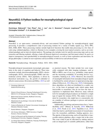
Chapter 03 p50-71.qxd4/1/069:46 AMPage 51Chapter 3 Neurophysiological Monitoring: A Tool for Neurosurgerywhen it is necessary to observe evoked EMGs. This technique may need to be modified if motor potentials arerecorded to predominantly total intravenous anesthesia.In many situations, halogenated hydrocarbon inhalationagents are desired to help control blood pressure. In our experience this problem is best approached by beginning theoperative procedure under balanced narcotic technique,and then once baseline responses have been obtained andcompared with the preoperative responses, introducing inhalation agents in a controlled fashion. It has also been ourexperience, particularly in children, that responses can bemaintained at an isoflurane level of 0.3 minimum alveolarconcentration (MAC), whereas many adults can maintaintheir responses at 0.5 MAC.Of the inhalation agents, isoflurane produces the weakesteffects on cortical activity. Thus, in those cases where a balanced narcotic technique can not be used, isoflurane is recommended as the anesthetic from a monitoring perspective. These effects are very individualized, and even lowlevels of inhalation agents may reduce the amplitudes ofcortically generated activity in some patients (Fig. 3–1).92The somatosensory short latency potentials (BSEPs) behavesimilarly to those from the auditory system (BAEPs) and areunaffected by most anesthetic manipulation.29,40,6551 Monitoring SystemsThe presently available systems supporting IOM allow thesimultaneous acquisition of evoked potentials, EEGs, andEMGs along with data from the anesthesiology monitoringunit,95 and have significantly increased the utility and sensitivity of IOM (Fig. 3–2).Neurophysiological signals are amplified using differentialamplifiers36 in which two input channels to the amplifier aredifferenced. This differencing has the effect of eliminatingidentical (in-phase) signal components that might be presentat each recording electrode (presumably noise), and retainingthe signals that are different (out-of-phase) and presumablyproduced by physiological generators. The effectiveness withwhich a differential amplifier rejects in-phase signals compared with its ability to amplify out-of-phase signals is calledthe common mode rejection ratio (CMRR). Differential amplifiers typically have CMRRs of greater that 10,000:1(80 dB). For efficient rejection of in-phase signals, it is extremely important that the electrode impedances of eachelectrode of a pair not only be as low as possible but as similar as possible because any inequality in electrode impedancewill produce amplitude differences in the in-phase activitythat will be amplified along with the desired signal.Figure 3–1 Bilateral median nerve evokedpotentials demonstrating effects of multiple anesthetic agents during carotid endarterectomy. Sequential recordings startat the top of the figure. Initial responses arewithin normal limits. Recordings start todeteriorate, and thiopental is given, reducing P30. When thiopental is turned off,recordings return rapidly, but the effect ofdesflurane can be seen. As desflurane levelsare decreased, recordings return to baselineat the bottom of the figure.
Chapter 03 p50-71.qxd524/1/069:46 AMPage 52IntroductionFigure 3–2 Example of median nerve evoked potentials (MSPs) andBAEPs being acquired simultaneously with the second channel (ch 4) ofBAEPs being digitally filtered. Channels 2, 3, and 4 are also being com-In evoked potential recording, the observed neuroelectricactivity, either from the scalp or propagating activity fromthe cord, is assumed to consist of a signal component representative of underlying activity evoked by the stimulus andrandom noise consisting of both physiological signals notrelevant to the study and environmental noise generated byubiquitous sources of electrical signals. Evoked potentialsare typically a fraction of the size of the spontaneous brainactivity appearing in the background EEG, and about onethousandth the size of the other physiological and extraneous potentials with which they are intermixed. The aim ofevoked potential recording is to acquire a large, clear response with the least possible noise contamination (i.e., thebest signal to noise ratio possible); thus the elimination ofunwanted signal components is essential. This eliminationis accomplished through the use of both analog and digitalfiltering techniques and signal averaging.After signal amplification, the most effective method forextracting a signal of interest from background noise is signal averaging. Signal averaging is in effect a cross-correlation between a point-process defined by the occurrence ofthe stimuli and the recorded evoked activity (i.e., an optimal filter).58 In averaging, the signal component at eachpoint is coherent and adds directly, whereas the background and noise components tend to be statistically independent and summate in a more or less root-mean-square(RMS) fashion.pared against baseline data. Waterfall displays of both modalities arealso shown with baseline responses at the bottom of each waterfall, andannotating comments are attached to the appropriate recording.Neurophysiological MeasuresNeurophysiological measures are available that provide afunctional map of nearly the entire neuroaxis. These measures include the EEG, an unstimulated measure of corticalfunction suitable for providing information concerning thedegree of cortical activation related to either metabolicprocesses (e.g., hypoxia) or pharmacological manipulation(e.g., pentobarbital-induced burst suppression to protectthe patient’s cortical function)75; the somatosensory andvisual cortical potentials (SEPs and VEPs), which provideadditional measures of cortical function specific to certainpathways and vasculature; the auditory and somatosensory brain stem potentials (BAEPs and BSEPs), which provide information about the brain stem function specific tocertain pathways86; compound nerve action potentials(CNAPs) providing information from both the spinal cord(SCAPs) and the peripheral nerves; and, finally, both continuous and evoked EMGs recorded from muscles [compound muscle action potentials (CMAPs)] innervated bythe various cranial and peripheral nerves, which providedirect information about the integrity of the cranial nerves,their underlying brain stem nuclei,51 the spinal cord, andperipheral nerves.All measured potentials may be characterized as eithernear-field or far-field potentials (NFPs or FFPs). These concepts express observed differences between types of potentials
Chapter 03 p50-71.qxd4/1/069:46 AMPage 53Chapter 3 Neurophysiological Monitoring: A Tool for Neurosurgeryand are meant to distinguish between two different manifestations of volume-conducted fields.54FFPs are recorded at some distance from the presumedgenerator, however, and the point at which a “near field”becomes a “far field” has never been clearly defined. A useful definition for the FFP is one that fails to decay in proportion to the square of the distance from the generator.ElectroencephalogramThe EEG is a valuable monitoring tool in almost all cerebrovascular procedures or tumor resections where significant risk for interruption of blood flow to the brain occurs.Cerebral blood flow alterations may occur during carotidendarterectomy, clipping or coiling of cerebral aneurysms,or repair of the internal carotid artery associated with tumor removal from the cavernous sinus. In most open cases,proximal and distal control of the feeding artery is required,potentially reducing blood flow to the brain. Associatedwith this decreased availability of blood may be hypoxiacaused by an inability of the collateral circulation to adequately supply blood to the brain (Fig. 3–3).The second most useful application of EEG monitoringhas been to help define the occurrence of embolic phenomena during these same procedures, which results in decreased blood flow and a potentially ischemic event, butwhich may be treated intraoperatively if recognized.12In all of these situations EEG monitoring can help identifythe presence of an insult, define the possibility for immedi-53ate therapy, and define the degree of burst suppression ifbarbiturate brain protection is instituted.The typical pattern seen in the EEG during cerebral hypoperfusion is a reduction or loss in high-frequency activityand the appearance of large-amplitude slow waves in therange of 1 to 4 Hz. There are situations where the EEG maybe acutely depressed upon injection of an anesthetic thatrapidly passes the blood–brain barrier. Such situations maybe found in high-dose opioid anesthesia, where fentanyl induces an immediate and marked reduction of fast frequencyactivity in the EEG, with an increase in low-frequency, highamplitude activity.33A simple but useful summary of possible changes is thatdecreased frequency with increased amplitude implies anischemic event to the cortex,106 widespread frequency slowing and decreased amplitude usually imply brain stem ischemia,87 whereas ischemic events affecting the thalamusand the internal capsule produce unremarkable changes inthe EEG106 but possibly significant changes in the SEPs.Somatosensory Evoked Potentials (Ascending SpinalCord Activity)SEPs are used during spinal surgery, vascular procedures,and cranial base procedures. For most cases we simultaneously stimulate the median or ulnar nerve at the wrist, andthe common peroneal nerve as it passes under the head ofthe fibula, or the posterior tibial nerve at the medial malleolus.Figure 3–3 Power spectrum of two channels of electroencephalography recordedduring temporary clipping of the left middlecerebral artery. Data are sequentially presented starting at the top of the figure. Thespectra in column 2 demonstrate changesassociated with cross-clamping and recoveryafter removal of the clip. Both channels ofdata reflect the generalized effect of thiopental on cortical activity and its underlyingmetabolic demand.
Chapter 03 p50-71.qxd544/1/069:46 AMPage 54IntroductionIn addition, dermatomal SEPs are of use in selected caseswhere concern exists about protecting particular nerveroots, such as during tethered cord release.SEPs are dependent on the stimulation of large afferentfibers of peripheral nerves. Following stimulation of peripheral nerves in the arms or the legs, SEPs can be reproduciblyrecorded over the spine and scalp. In the spinal cord, the SEPsare conducted primarily through the dorsal columns; however, extensive work has been done to clarify the generatorsfor the various components of the SEPs.24,25 In humans, lossof posterior column function is associated with abnormalityof the SEPs; however, extralemniscal pathways may alsomediate some mid- and long-latency scalp SEP components.In our experience, SEPs are extremely sensitive and specific to spinal cord injury whether it occurs in the dorsal orin the ventral pathways. This is confirmed in the literature,76 where a false-negative rate of 0.063% was found for51,263 spinal cases in which SEPs were the only modalitymonitored. Furthermore, the negative predictive value(i.e., the likelihood of normal spinal cord function in thepresence of stable SEPs) was 99.93%. This is a significantimprovement over the 0.72 to 1.4% incidence of spinal cordinjury reported for unmonitored cases.62Temperature changes significantly influence the SEPlatency. For each degree Celsius of local cooling, the nerveconduction velocity decreases by 2.5 msec. During longoperations, a drop in temperature around the nerve beingstimulated can result in a progressive increase in latenciesunrelated to surgical manipulation. Also, latencies may betransiently affected when the surgeon irrigates with physiological solution at cooler temperatures. Significant changesare also seen related to hypotension. Attention must be paidto these changes because spinal cord hypotension in patients with myelopathy may lead to an extension of themyelopathic lesion.Upper Extremity (Median and Ulnar Nerve) Evoked PotentialsMedian and ulnar nerve evoked potentials (MSPs and USPs)are both useful in assessing the brachial plexus, cervicalspinal cord, brain stem, and telencephalon. These potentialsare useful in preventing and reducing surgical morbidityduring procedures that pose potential harm to the cervicalcord and in assessing the level of hypoxia in cortical andspinal tissue.35 Only the MSPs are described in detail herebecause the USPs are usually identical except for their levelof entry into the spinal cord.Stereotypical MSPs are simultaneously recorded from theipsilateral Erb’s point referenced to the contralateral Erb’spoint, the neck (cervical CV7) and parietal (P3 or P4) (electrode locations are given in the International 10/20 system49)scalp with a reference to a frontal electrode (Fig. 3–4).At Erb’s point, the response is an NFP consisting of anapparently triphasic (positive-negative-positive) nerve actionpotential, reflecting the passage of a mixed nerve volley passing the brachial plexus. This component is usually labeled N11for the large negative-going component generated at 11 msec(all waves will be identified as N or P for their polarity subscripted by the latency of the wave). At the cervical C7recording site, the NFP consists of an N14 wave with an associated complex waveform. It has been postulated that thesewaves are generated in the dorsal roots, dorsal horn, posterior columns, and structures of the lower brain stem.27,28 Theresponse recorded from scalp electrodes placed (roughlyover the hand area of the sensory cortex) contralateral to thestimulated arm consists of a P15, N20, P30 complex. Some datasuggest that P15 is thalamic in origin,93 whereas N20 is generated in thalamocortical pathways, and P30 is generated inparietal cortex.2,37 After this wave, there is considerable disagreement as to the identity of the cortical underlying generators; however, it is clear that the parietal cortex is involvedin the generation of the N20/P30 complex and that the parietalassociation areas are involved in the generation of the laterwaves.Lower Extremity (Common Peroneal and Tibial Nerve)Evoked PotentialsIn the lower limbs, nerves used to elicit SEPs include thetibial and peroneal nerves. Occasionally the femoral nerve isalso used. Spinal potentials are most consistently obtainedby stimulation of the tibial nerve at the medial malleolus orperoneal nerve in the popliteal fossa.Recordings are made routinely at the lumbar spine andthe cerebral cortex (Pz/Fz). SEPs also can be recorded alongthe thoracic or cervical spine as clinically indicated. Stimulation of the posterior tibial nerve at the ankle evokes P32 andN42 potentials in the younger population,24 which graduallyincreases in latency until adult values of 40 and 48 msec, respectively, are obtained.108 Stimulation of the common peroneal nerve at the popliteal fossa produces waves that areslightly shorter in latency.25 The first useful component is anN20 wave, which is usually maximal over the T12 or L1 vertebra. At more rostral and caudal levels it may be possible torecord small “traveling” waves, representing the envelope ofaction potentials in the dorsal roots and sensory tracts of thespinal cord.Spinal SEPs are relatively easy to obtain in children, withthe amplitude and definition of the waves decreasing withincreasing age such that by the midteenage years these responses are more difficult to obtain. The response over themid and lower lumbar spine consists of an initially positivetriphasic potential. This reflects the NFP produced by a volley of action potentials ascending through the cauda equina(Fig. 3–5).Dermatomal ResponsesA disadvantage of SEPs produced by stimulation of largenerve trunks is that input to the spinal cord usually occursover more than one level. This problem can be addressed bydelivering the stimulus to small cutaneous nerves that arederived from a single dorsal root and which innervate the“signature area” of a particular dermatome.Pudendal nerve responses are a special case of dermatomal responses, particularly useful in patients withspina bifida or tethered cords. The pudendal nerve carriessensory fibers from the penis, urethra, anus, and pelvic floormuscles, and supplies motor innervation to the bulbocavernosus and pelvic floor muscles, the external urethral sphincter, and the external anal sphincter. Cortical responses to
Chapter 03 p50-71.qxd4/1/069:46 AMPage 55Chapter 3 Neurophysiological Monitoring: A Tool for Neurosurgery55Figure 3–4 Median nerve evoked potentials (MSPs)demonstrating the significant waves at different levels alongthe neuraxis from Erb’s point (ch 4) to the contralateral scalp(ch 1).electrical stimulation of the dorsal nerve of the penis, theurethra,41 and the urinary bladder4 have all been described.Pudendal nerve responses are similar in morphology to thetibial nerve SEP and are best recorded from the same area ofthe scalp.42nuclei; (3) P15 medial lemniscus; (4) N16, thalamus; and(5) later peaks representing the thalamocortical projections.It is believed that multiple parallel spinal cord pathways areactivated by suprathreshold stimulus intensities and thatthey contribute differentially to the various BSEP peaks.85Brain Stem Evoked ResponsesVentral Cord MonitoringBSEPs are monitored routinely during all procedures inwhich SEPs are recorded. We routinely record between electrode locations M1 (left mastoid) and Fz, similar to therecording locations for BAEPs. The waves and their generators27 are (1) P10, the brachial plexus; (2) P12, dorsal columnEven though the results of SEP monitoring in preventing bothmotor and sensory iatrogenic injury during spinal surgeryhave been excellent, they cannot provide complete coverageof the spinal cord, and several cases have been reported ofspinal injury going undetected using only SEP monitoring.19,59
Chapter 03 p50-71.qxd564/1/069:46 AMPage 56IntroductionFigure 3–5 Posterior tibial nerve evoked potentials demonstrating components at different levels of the neuroaxis from the lumbar regionbottom to the scalp top.Thus considerable attention has been paid to developing robust and reliable methods for monitoring the more ventralcorticospinal tracts.47 Two methods briefly reviewed here arecerebellar and motor potentials (evoked EMGs).Cerebellar Potentials (Ascending Activity)In addition to the ascending dorsal columns and spinothalamic tracts and descending ventral motor tracts, severalother ascending long tracts exist and are useful for IOMpurposes. These pathways include the ventral and lateralspinocerebellar and cuneocerebellar tracts, which providedense input to the cerebellum. Several studies have demonstrated that stimulation of the peripheral nerves commonlyutilized to generate SEPs also generates a cerebellar evokedpotential (CEP).45 Moreover, animal models have demon-strated that the evoked potential recorded from the cerebellum is generated via ascending ventral spinal cord tracts.44Thus this stimulation and recording technique provides anassay of the ventral spinal cord utilizing conventional techniques developed for SEP monitoring. These responses maybe obtained reliably and are reproducible from an additional electrode placed over the occiput and require thesame anesthetic technique used to record SEPs.Motor Evoked Potentials (Descending Activity)Motor evoked potentials (MEP) have been under serious investigation in the IOM field for 20 years.47,66 In general,stimulation has involved either cortical or spinal cord sites.Noninvasive stimulation has been investigated using eitherelectrical63 or magnetic9 stimulation of the motor cortex,89
Chapter 03 p50-71.qxd4/1/069:46 AMPage 57Chapter 3 Neurophysiological Monitoring: A Tool for Neurosurgeryindirect stimulation of the spinal cord via spinous processes,79or direct stimulation of the spinal cord100 by placing subduralor epidural stimulation electrodes. Various responses arerecorded distal to the operative site for these assorted stimulation techniques. Fig. 3–6 summarizes both the stimulating and the recording techniques currently available.105Recording sites include (1) spinal cord evoked potentials(SCEPs) using transcutaneous or direct (epidural or subdural) recording electrodes; (2) direct (D) and indirect (I)waves from the spinal cord using epidural recording sites;(3) CNAPs, referred to as NMEPs in Fig. 3–6 recorded fromperipheral nerves using transcutaneous or subdermal needle electrodes; and (4) CMAPs from muscle groups of theupper and lower extremities using transcutaneous or subdermal needle electrodes. CMAPs are referred to as myogenic potentials, whereas CNAPS have been termed neurogenic responses (Fig. 3–6).Transcranial StimulationThe motor cerebral cortex or brain stem can be activated byeither electrical or magnetic stimulation; however, onlyelectrical stimulators are currently approved by the Foodand Drug Administration (FDA) for transcranial stimulation.Scalp electrodes or electrode plates placed adjacent to thescalp or hard palate can be used to stimulate the cortex and57underlying tissues. Stimulation voltages are typically in therange of 100 to 250 V. SCEPs have been observed with between 100 and 4000 stimuli being averaged. CMAPs obtained following transcranial stimulation do not requireaveraging and provide a rapid assessment of motor pathwayfunction; however, these responses are very susceptible toanesthetic influence and have considerable intrinsic variability, making their interpretation difficult. CNAPs havebeen extremely unreliable, again due to anesthetic effects.Transcranial electrical stimulation through the intact scalphas been shown to result in a charge density that might resultin neuronal damage if applied directly to the cortex.78 However, electrical stimuli are attenuated by a factor of 30 due tothe high resistance of the intervening muscle, skull, andmeninges. Thus the charge density at the surface of the brainwith electrical stimulation is estimated to fall well within acceptable safe limits.3 One of the most important stimulationparameters for eliciting reliable transcranial Motor evokedpotentials (MEPs) is the interstimulus interval (ISI) of a burstof stimuli, applied at the above-referenced rate.101 It has beenfound that bursts of stimuli with an ISI between 2 and 5 msecproduce a maximal response by overcoming the depressant effects of general anesthesia.50,52 The significant parameters andmorphological fea- tures of CMAPs generated via transcranialstimulation are response threshold, onset latency, centralconduction time, and response size48 (Fig. 3–7).Figure 3–6 Various stimulating and recording sitesfor intraoperative motor evoked potential (MEP) monitoring. SCEPs are spinal cord evoked potentials. NMEPsare neurogenic motor evoked potentials, which arealso referred to as compound nerve action potentials(CNAPs). CMAPs are compound muscle action potentials. (Printed with permission from Kalkman CJ, UbagsLH. Motor evoked potential monitoring. Current Opinion in Anaesthesiology 1997; 10:327–332.)
Chapter 03 p50-71.qxd584/1/069:46 AMPage 58IntroductionFigure 3–7 Compound muscle action potentialsobtained by transcranial electrical stimulationduring lumbosacral instrumentation from the external anal sphincter muscle.Epidural spinal recordings of corticospinal tract activityfollowing transcranial stimulation produce a complex ofpositive and negative components composed of I and Dwaves48,,84 (Fig. 3–8).Indirect Spinal Cord StimulationIndirect stimulation of the spinal cord through vertebralbone has been used to produce descending neural activity recorded peripherally from mixed nerves (CNAP). 80The CNAPs are typically recorded at the popliteal fossa,with stimulation provided through a pair of electrodespositioned at adjacent spinal processes in the cervical region. Considerable controversy exists over the relativecontribution of the sensory and motor pathways to theseneurogenic spinal evoked potentials. Thus the term descending neurogenic evoked potential (DNEP), which describes the stimulation method and the direction of theneural volley along the spinal cord, but not necessarily itscomposition, has been adopted. Collision studies 56 haveraised serious doubts about the independent utility ofthis method because both modalities (SEPs and DNEPs)appear to be primarily mediated by the same neuralpathways, namely, the dorsal columns. 103 The consensus,based on these experiments, is that these responses arenot a pure motor response but are largely composed ofantidromic sensory activity and minimal orthodromicmotor activity.82Direct Cord StimulationConsiderable work has been done with direct stimulationover the midline of the posterior, rostral spinal cord andrecordings made either with epidural or subdural30 stripelectrodes or peripheral CMAPs.74 Epidural SCEPs recordedfrom the midline of the posterior spinal cord produced by adescending spinal cord volley of action potentials can varyin morphology depending on both the stimulating and therecording sites. The dominant waves seem to be an initialnegative spike (N1) followed by a slow negative component(N2). The conduction velocity of the N1 component has varied greatly, ranging from 47 to 90 m/s, whereas the averageconduction velocity of the N2 component has ranged from46 to 53 m/s.56,63
Chapter 03 p50-71.qxd4/1/069:46 AMPage 59Chapter 3 Neurophysiological Monitoring: A Tool for NeurosurgeryFigure 3–8 Typical direct (D) and indirect (I) waves recorded fromthe epidural space.Spinal Cord and Peripheral Nerve ElectromyographyThe EMG, a useful indicator of the integrity of descendingactivity in the spinal cord, is electrical activity produced inmuscle fibers below the skin and has a frequency contentranging from 15 to 150 Hz.26 The EMG is either spontaneous(e.g., anal sphincter activity produced by irritation of the S3to S5 roots during an untethering procedure involving thelower portion of the cauda equina) or evoked, of the typeproduced in selective rhyzotomy for the treatment of spasticity, for pedicle screw placement, or as already discussedfor spinal cord evaluation. Evoked EMGs have a considerablylarger amplitude ( 100 V) than sensory evoked potentialdata ( 0.2 V), and therefore these signals do not requireaveraging to extract them from the background noise.59lateral rectus muscle; the trigeminal nerve (V) through themasseter muscle; the trochlear nerve (IV) from the superioroblique muscle; and the oculomotor nerve (III) through themedial and inferior rectus and the inferior oblique muscles ofthe eye. When appropriate, the functioning of the glossophary
ment of the effects of surgical manipulations on the structure -function relationships of the patient's nervous system to facilitate the surgeon's decision making. Specific and sensi-tive measurements reflect the interactions between the sur-geon's intraoperative manipulations and the functioning of the patient's nervous system.