Transcription
10/25/2021Waldenstrom anddermatologyKate Mimken, RN, BSN, FNPOctober 28, 2021CONFIDENTIAL – Contains proprietary information.Not intended for external distribution.1Objectives To understand disease related cutaneous manifestations ofWaldenstrom’s To understand treatment related cutaneous manifestations ofWaldenstrom’s To understand what to do with either disease or treatment related skineffects2Kate Mimken, FNPCONFIDENTIAL – Contains proprietary information.Not intended for external distribution.21
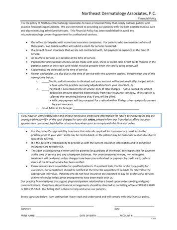
10/25/2021Quick overview of WM The IgM protein is the protein which is overproduced in WMo We test this through labs called quantitative immunoglobulins and the M-spike IgM is a large molecule and can infiltrate many things including the skin Because of the size of the molecule it can also cause other complicationswhich can lead to skin disorders which we will discuss in further detail There are 3 classifications of WM (IgM MGUS, smoldering, andsymptomatic) Various treatments that are necessary to treat symptomatic WM can alsolead to skin/hair/nail changes3Kate Mimken, FNPCONFIDENTIAL – Contains proprietary information.Not intended for external distribution.3IgM molecule compared to others4Kate Mimken, FNPCONFIDENTIAL – Contains proprietary information.Not intended for external distribution.42
10/25/2021IgM levels The level of IgM and/or the percentage of LPL (WM) cells in the bonemarrow varies tremendously between WM patients Some patients with very low IgM levels have lots of symptoms whileothers with very high levels may not have symptoms at all!5Kate Mimken, FNPCONFIDENTIAL – Contains proprietary information.Not intended for external distribution.5Various Dermatologic ConditionsTreatment RelatedDisease Related ( 5% patients w/ WM) Brittle/cracking nailsLPL cell infiltration Corkscrew hair changesHyperviscosity Skin infectionsPurpura Bruising/bleedingRaynaud's phenomenon Psoriasis post RituxanLivedo reticularis Neutrophilic dermatosis (Sweetssyndrome) and neutrophilic eccrinehidradenitisVasculitis6Chronic urticarial (Schnitzler'ssyndrome)CONFIDENTIAL – Contains proprietary information.Not intended for external distribution.Seiter, K., Ponce, D. (2021).63
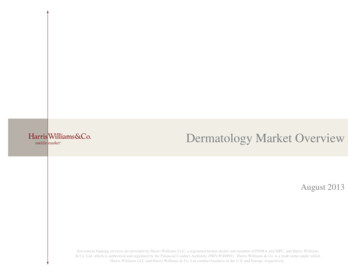
10/25/2021Disease Related Dermatologic manifestations with WM are a less common presentingsymptom upon diagnosis with only about 5% of people having this. There is neoplastic causes meaning the skin is infiltrated with the LPLcells and non-neoplastic meaning it is caused from complications of theIgM paraprotein. We will discuss these in the upcoming slides.7Kate Mimken, FNPCONFIDENTIAL – Contains proprietary information.Not intended for external distribution.7Disease Related (cont) Rarely the lymphoplasmacytic cells can infiltrate the tissue The rash can range from reddish-brown to purple w/ plaque like texture When IgM infiltrates the skin it is called cutaneous macroglobulinosis orbullous diseaseGressier L, Hotz C, Lelièvre J, et al8Kate Mimken, FNPCONFIDENTIAL – Contains proprietary information.Not intended for external distribution.84
10/25/2021Hyperviscosity Hyperviscosity with WM is common and can cause complication(bleeding, edema, headaches) This is when the blood becomes thick/sludge like from excess proteins inthe blood Because of bleeding risk use caution with brushing/flossing, blowing yournose The thicker blood can cause swelling in the legs (edema) which cancause the skin to blister or weep9Kate Mimken, FNPCONFIDENTIAL – Contains proprietary information.Not intended for external distribution.9Disease Related (cont)Cryoglobulinemia causes some of the dermatologic conditions associatedw/ WMThis is when blood proteins clump/precipitate in cold tempsTested through a blood samplePurpura- dark purple bruised looking spots on extremities This can cause pain at time is associated with vessel occlusion Can also be a result of hyperviscosityLehman, J. 201010CONFIDENTIAL – Contains proprietary information.Not intended for external distribution.105
10/25/2021Disease Related (cont)Raynaud's phenomenon occurs in cold temps from lack of blood flow Can cause acrocyanosis which is a blueish discoloration of the extremities, ears, and tip of nose Raynaud's occurs in cool or cold temperatures. This is when fingers become white, blue/purplefrom lack of blood flow, when blood flow return they become red. Keeping hands and/or feet warm with gloves, socks, hand or feet warmers to decrease pain andsymptoms associated with Raynaud's11Kate Mimken, FNPCONFIDENTIAL – Contains proprietary information.Not intended for external distribution.11Disease Related (cont)Cryoglobulinemia related conditions:Livedo reticularis this web-like rash can be benign and transient orpathologic/more consistent or permanentVasculitis inflammation/narrowing or blockage of blood vessels12Kate Mimken, FNPCONFIDENTIAL – Contains proprietary information.Not intended for external distribution.126
10/25/2021Schnitzler Syndrome Urticarial eruptions- neutrophil (WBC) infiltrates Associated with IgM protein- more common with IgM Kappa vs IgMlambda Often have constellation of other symptoms (fever, bone or joint pain,swollen lymph nodes, and neuropathy) Rare and underdiagnosed Treatment is directed at treating WM and a medication called anakinra(immunosuppressant to target interleukin 1) 13Tania Jain, Chetan P. Offord, Robert A. Kyle, David Dingli. (2013)Kate Mimken FNPCONFIDENTIAL – Contains proprietary information.Not intended for external distribution.13Neutrophilic dermatosis This is also called Sweets syndrome, named after Robert Douglas Sweet It is characterized by clinical, pathological, and lab findingso Clinical findings are fever, tender/red skin lesions, sometimes can affect areas outside the skino Labs show increased neutrophils (a portion of the white blood cell)o Biopsy (pathology) of the lesions shows infiltration of mature neutrophils in the upper layer of thedermis Some people can have this without have a cancer diagnosis, but oftentimes it is from an underlying malignancy Treatment requires systemic corticosteroids and sometimes topicalsteroid or injection into the skin lesion 14Al-Musalhi, B., Gerstein, W. (2016)Kate Mimken, FNPCONFIDENTIAL – Contains proprietary information.Not intended for external distribution.147
10/25/2021Neutrophilic eccrine hidradenitis This is a type of neutrophilic dermatosis This is a rash/skin eruption that is associated with chemotherapy most of the timeo When chemotherapy stops, the rash generally improves. It affects the eccrine (sweat glands) that make up most of the body Can appear infectious and some people have accompanying fevers. Definitively diagnosed by skin biopsy It is self limiting, but can be treated with steroids (cautiously)Crane JS, Krishnamurthy K. 2021Mills, L. DO, Steinmetz‐Rodriguez, C. DO, Folkes, A. DO, Shecter, R. DO, FAOCDCONFIDENTIAL – Contains proprietary information.Not intended for external distribution.1515Changes to skin when WM is managed There can be changes to skin when WM is managed, these are mostlyrelated to treatment side effects. Usually when specifically related to WM, meaning the skin lesion areinfiltrated with IgM protein, they usually improve with treatment and canflare when disease progresses You want to have a biopsy to determine the exact cause, because it couldbe related to other skin conditions or amyloidosis (another plasma celldisorder)16Kate Mimken, FNPCONFIDENTIAL – Contains proprietary information.Not intended for external distribution.168
10/25/2021Epidermal Growth Factor Receptor (EGFR) Epidermal growth factor is responsible for development/growth of newblood vessels Ibrutinib is intended to block BTK, but can also block the EGF receptorwhich can lead to unintended side effectso Rasho Bleeding/bruisingo Infections (skin as well as systemic)o Atrial fibrillationo High blood pressure (hypertension)CONFIDENTIAL – Contains proprietary information.Not intended for external distribution.1717Skin Infections Staphylococcus aureus- can be minor to very serious, requires oralantibioticso Often times redness, pain, swelling of the skin, feverso Called “cellulitis” Folliculitis- most minor type of staph infection at the base of a hair follicle,does not always require treatment, but can require an topical (applied toskin) antibiotic Panniculitis- inflammation of subcutaneous fat (adipose) tissueo May require corticosteroids and/or lower dose of ibrutinibSibaud, V., Beylot-Barry, M., Protin, C., Vigarios, E., Recher, C., & Ysebaert, L. (2020).Fabbro SK, Smith SM, Dubovsky JA, Gru AA, Jones JA.18Kate Mimken FNPCONFIDENTIAL – Contains proprietary information.Not intended for external distribution.189
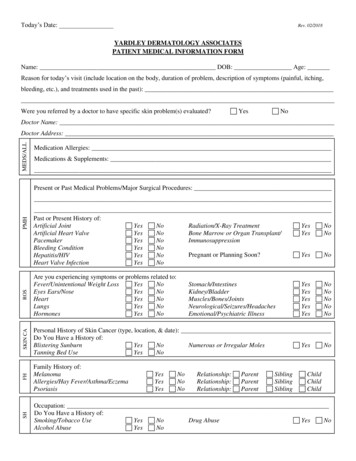
10/25/2021Skin Infections (cont) Herpes simplex and zoster (Shingles)o Prevention with medication or Shingrix vaccineo Post herpetic neuralgia can occur Aphthous ulcers/stomatitiso Generally not infectious or assoc. w/ neutropeniao Treat with corticosteroids (systemic and topical)o Pause ibrutinib and dose reduce19Kate Mimken FNPCONFIDENTIAL – Contains proprietary information.Not intended for external distribution.19Skin changes and what to do? Most common in the first year of treatment, but can occur at any time Cracked skin around fingers/toes, very brittle nails- Keep well moisturizedo Mane and Tail lotion, Working hands lotion, don’t use fragrance/alcohol based productso topical solutions such as hydrosoluble nail lacquer (Genadur) and polyureaurethane (Nuvail).o Biotin (2.5mg) -caution w/ thyroido Keep clean to prevent infections Rash Hair changes- some thinning and corkscrew like textureBitar, C., Farooqui, M. Z., Valdez, J., Saba, N. S., Soto, S., Bray, A., Marti, G., Wiestner, A., & Cowen, E. W. (2016). Hair and Nail Changes DuringLong-term Therapy With Ibrutinib for Chronic Lymphocytic Leukemia. JAMA dermatology, 152(6), 6.022520Kate Mimken FNPCONFIDENTIAL – Contains proprietary information.Not intended for external distribution.2010
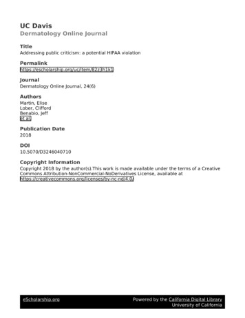
10/25/2021Skin/Nail changes21Kate Mimken, FNPCONFIDENTIAL – Contains proprietary information.Not intended for external distribution.21Skin changes (cont) Psoriasis post Rituxano Rare cases (1.04 in 1,000)- reported anytime during treatmento Generally resolves once Rituxan stops, sometimes requiring topical or systemic steroidso Cause is unknown, but could be from depletion of B-cell causing activation of T-cells, maybefrom impaired response to infection, or from Rituxan induced auto-immune changeso Alahmari, H.S., Alhowaish, N.Y., Omair, M.A. (2019)22Kate Mimken FNPCONFIDENTIAL – Contains proprietary information.Not intended for external distribution.2211
10/25/2021Managing Toxicities Always discuss with your provider There are many interventions as we mentioned above and sometimesthey require treatment breaks or dose reductions to minimize side effects23Kate Mimken FNPCONFIDENTIAL – Contains proprietary information.Not intended for external distribution.23Thank YouKate Mimken, RN, BSN, FNPCONFIDENTIAL – Contains proprietary information.Not intended for external distribution.2412
10/25/2021References Alahmari, H.S., Alhowaish, N.Y., Omair, M.A. "Rituximab-Induced Psoriasis in a Patient with Granulomatosis with Polyangitis Treated withAdalimumab", Case Reports in Rheumatology, vol. 2019, Article ID 5450863, 3 pages, 2019. https://doi.org/10.1155/2019/5450863 Alegría-Landa, V, Prieto-Torres, L, Santonja, C, Córdoba, R, Manso, R, Requena, L, Rodríguez-Pinilla, SM. MYD88 L265P mutation in cutaneousinvolvement by Waldenström macroglobulinemia. J Cutan Pathol. 2017;44:625–631. https://doi.org/10.1111/cup.12944 Al-Musalhi, B., & Gerstein, W. (2016). Monoclonal Gammopathy of Undetermined Significance and Neutrophilic Dermatosis. Oman medicaljournal, 31(5), 394–395. https://doi.org/10.5001/omj.2016.79 Bitar, C., Farooqui, M. Z., Valdez, J., Saba, N. S., Soto, S., Bray, A., Marti, G., Wiestner, A., & Cowen, E. W. (2016). Hair and Nail Changes DuringLong-term Therapy With Ibrutinib for Chronic Lymphocytic Leukemia. JAMA dermatology, 152(6), 6.0225 Crane JS, Krishnamurthy K. Neutrophilic Eccrine Hidradenitis. [Updated 2021 Jul 18]. In: StatPearls [Internet]. Treasure Island (FL): StatPearlsPublishing; 2021 Jan-. Available from: https://www.ncbi.nlm.nih.gov/books/NBK448175/ Fabbro SK, Smith SM, Dubovsky JA, Gru AA, Jones JA. Panniculitis in Patients Undergoing Treatment With the Bruton Tyrosine Kinase InhibitorIbrutinib for Lymphoid Leukemias. JAMA Oncol. 2015;1(5):684–686. doi:10.1001/jamaoncol.2015.0457 Gressier L, Hotz C, Lelièvre J, et al. Cutaneous Macroglobulinosis: A Report of 2 Cases. Arch Dermatol. 9.359 Jensen, A. B., Stausbøl-Grøn, B., Riber-Hansen, R., & d'Amore, F. Ibrutinib-Associated Skin Toxicity: A Case of Maculopapular Rash in a 79-YearOld Caucasian Male Patient with Relapsed Waldenstrom's Macroglobulinemia and Review of the Literature. Dermatology reports, 2017, 9(1), 6976.https://doi.org/10.4081/dr.2017.697625Kate Mimken FNPCONFIDENTIAL – Contains proprietary information.Not intended for external distribution.25References (cont) Lehman, J. Waldenstrom’s Macroglobulinemia and the Skin. IWMF Torch, 11(1). 2010, 14. Mills, L. DO, Steinmetz-Rodriguez, C. DO, Folkes, A. DO, Shecter, R. DO, FAOCD. Neutrophilic Eccrine Hidradenitis: AnUnusual Case and a Review of the Literature. Retrieved esmgr/jaocd/contents/volume38/38-10.pdf. October 10, 2021. Oscoz-Jaime S, Agulló-Pérez AD, Llanos-Chavarri C, Yanguas-Bayona JI. Infiltración cutánea por macroglobulinemia deWaldenström. Actas Dermosifiliogr. 2018;109:75–78. Seiter,K., Ponce, D. (2021). Waldenstrom Macroglobulinemia Clinical Manifestations. Medscape. Retrieved clinical#b3 Sibaud, V., Beylot-Barry, M., Protin, C., Vigarios, E., Recher, C., & Ysebaert, L. (2020). Dermatological Toxicities of Bruton'sTyrosine Kinase Inhibitors. American journal of clinical dermatology, 21(6), 799–812. https://doi.org/10.1007/s40257-020-00535x Tania Jain, Chetan P. Offord, Robert A. Kyle, David Dingli. Schnitzler syndrome: an under-diagnosed clinical entity.Haematologica 2013;98(10):1581-1585; https://doi.org/10.3324/haematol.2013.084830. Vigarios, E., Beylot-Barry, M., Jegou, M-H., Overic, L., Ysevaert, L., Sibaud, V. British Journal of Haematology. 2017. 185 (4),784-788. DOI: https://doi.org/10.1111/bjh.1562026Kate Mimken FNPCONFIDENTIAL – Contains proprietary information.Not intended for external distribution.2613
This is when blood proteins clump/precipitate in cold temps Tested through a blood sample Purpura-dark purple bruised looking spots on extremities This can cause pain at time is associated with vessel occlusion Can also be a result of hyperviscosity Lehman, J. 2010 Disease Related (cont) 9 10 10/25/2021 6 11