Transcription
mineralsArticleOn the Color and Genesis of Prase (Green Quartz)and Amethyst from the Island of Serifos,Cyclades, GreeceStephan Klemme 1, *, Jasper Berndt 1 , Constantinos Mavrogonatos 2Ioannis Baziotis 3 , Panagiotis Voudouris 2 and Stamatios Xydous 3123*, Stamatis Flemetakis 1 ,Institut für Mineralogie, Westfälische Wilhelms Universität Münster, Corrensstrasse 24,48149 Münster, Germany; jberndt@uni-muenster.de (J.B.); stam.flemetakis@uni-muenster.de (S.F.)Faculty of Geology and Geoenvironment, National and Kapodistrian University of Athens,15784 Athens, Greece; kmavrogon@geol.uoa.gr (C.M.); voudouris@geol.uoa.gr (P.V.)Laboratory of Mineralogy and Geology, Agricultural University of Athens, 11855 Athens, Greece;ibaziotis@aua.gr (I.B.); stxydous@aua.gr (S.X.)Correspondence: stephan.klemme@uni-muenster.de; Tel.: 49-(0)251-8333047Received: 10 September 2018; Accepted: 24 October 2018; Published: 26 October 2018 Abstract: The color of quartz and other minerals can be either caused by defects in the crystal structureor by finely dispersed inclusions of other minerals within the crystals. In order to investigate themineral chemistry and genesis of the famous prase (green quartz) and amethyst association fromSerifos Island, Greece, we used electron microprobe analyses and oxygen isotope measurements ofquartz. We show that the color of these green quartz crystals is caused by small and acicular amphiboleinclusions. Our data also shows that there are two generations of amphibole inclusions within thegreen quartz crystals, which indicate that the fluid, from which both amphiboles and quartz havecrystallized, must have had a change in its chemical composition during the crystallization process.The electron microprobe data also suggests that traces of iron may be responsible for the amethystcoloration. Both quartz varieties are characterized by isotopic compositions that suggest mixing ofmagmatic and meteoric/marine fluids. The contribution of meteoric fluid is more significant in thefinal stages and reflects amethyst precipitation under more oxidizing conditions.Keywords: green quartz; prase; amethyst; color; amphibole; actinolite; skarn; Serifos; Greece1. IntroductionColored varieties of quartz (SiO2 ) are ubiquitous in many igneous, metamorphic and sedimentaryrocks [1–3]. They are well-loved collectors’ items and comprise a number of semi-precious gems suchas violet amethyst, yellow citrine, pinky rose quartz, and dark smoky quartz. Color in quartz is causedeither by electronic defects of the crystal structure, transition metals or other elements incorporated inthe crystal structure. Furthermore, color in quartz, and in other minerals, can also be caused by small,often nano-sized, well-dispersed mineral inclusions [3]. Blue quartz, pink rose quartz or jasper areknown to be colored because of small inclusions of other minerals. Whilst it was thought for a longtime that the color of rose quartz was caused by small rutile (TiO2 ) inclusions, an electron microscopystudy [4] showed that the rose quartz crystals from Montana, USA, contained small inclusions ofdumortierite [(Al,Fe3 )7 (BO3 )(SiO4 )3 O3 ]. Furthermore, there is evidence that other rose quartz typesare colored by the incorporation of Mn, Ti or P in the quartz structure [3].Compared to yellow, violet or pink quartz, green varieties of quartz are rare. There are somedescriptions of natural green quartz which are commonly referred to as prasiolite [5,6]. However, most greenprasiolite is prepared from natural amethyst by heating to 300–600 C [7,8]. Some early studies found thatMinerals 2018, 8, 487; s
Minerals 2018, 8, 4872 of 13the color of green prasiolite quartz was caused by Fe2 on an interstitial site in octahedral coordinationwithin the quartz structure [2,9] but more recent studies using Mössbauer Spectroscopy questioned theseresults and concluded that the green color of prasiolite was caused by Fe3 [6]. Detailed studies illustratehow color changes from amethyst to prasiolite during heating [3,7,8]. Rare natural examples of prasiolitehave been reported from Reno, Nevada, USA [10] and Thunder Bay, Ontario, Canada [11,12].Another green variety of quartz is commonly referred to as chrysoprase. The color of chrysoprasevaries from olive-green to pale sea-green [3]. As far as we know, such chrysoprase has been describedfrom Australian serpentinites [13] and jadeite-rich veins in lower Silesia [14]. Rossman concludesin his review [3] that the color of chrysoprase is caused by “fine-grained nickel compounds in thesilicate matrix rather from substitutional Ni in the silica itself”. Possible Ni-minerals include bunsenite(NiO) or other complex hydrous Ni-silicates [3]. Recent studies of chrysoprase samples from Szklary(Poland) and Sarykul Boldy (Kazakhstan) showed that the “apple-green” color is caused by nano-scaledinclusions of Ni-kerolite and minor pimelite but they found no evidence for bunsenite inclusions [15].Finally, Yang et al., [16] reported green quartzites (also known as “Guizhou Jade”) from the Quinglongantimony deposit, in the Guizhou province, China, and claimed that green coloration is due to thepresence of Cr3 - and V3 -bearing dickite inclusions.Amethyst, on the other hand, is a relatively common variety of quartz that forms in a widevariety of environments [17]. It is well known as a gemstone since antiquity, as it is mentioned byTheofrastus, the disciples of Aristotle (4th century B.C.). Amethyst comes in an attractive violetcolor with reddish or bluish tint, that was originally interpreted as a result of the presence of Fe3 ,which turns into Fe4 after irradiation and substitutes for Si4 in a deformed tetrahedral position [3].However, recent studies, questioned this result and by using modern analytical techniques, like EPR,Mössbauer and synchrotron X-ray absorption spectroscopy attributed the coloration of amethyst inthe presence of Fe3 [6,18–20].Amethyst is not very common in skarn deposits. When present, it is usually only of mineralogicalinterest, as it rarely comes in gem quality in this geological environment [21]. Amethyst has been describedfrom the Fe-rich skarn deposits in Angara-Ilim in Russia [21,22]. Collector specimens of amethyst havebeen reported from the Dashkesan skarn deposit in Azerbaijan [21]. Sceptre-shaped amethyst crystals arealso found in the skarns of Denny mountain, Washington, USA [21] and finally, amethyst crystals havealso been found in Hässellkulla, Sweden, in the Örebro Fe-rich skarn deposit [21,23,24].Here we will present some results of an electron microprobe and oxygen isotope study on greenquartz crystals and associated amethyst from the skarn of Serifos Island in Greece, which comprises, toour knowledge, the only locality that produces specimens with both quartz varieties. These green quartzand amethyst crystals are well known to collectors and fetch rather high prices during auctions [25,26].However, not much is known about the origin of these quartz crystals or the origin of the green color inSerifos quartz. Similar-looking green quartz crystals are available for sale from internet mineral dealers andare sourced from Dalnegorsk, Russia [27]. As to the color, there are, to our knowledge, no peer-reviewedarticles that investigated the nature of the green Serifos or similar green quartz crystals in any detail.Hyrsl and Niedermayr [28] reported actinolite from XRD analyses of prase from Serifos, but presentedno further analytical data. Similarly, Maneta and Voudouris [29] reported actinolite inclusions to beresponsible for the green color of quartz from Serifos, and Voudouris and Katerinopoulos [30] describedsimilar green quartz crystals from the skarn occurrences at the Xanthi and Kresti/Drama areas in northernGreece, but without any detailed analytical data. Some recent published studies suggested inclusionsof hedenbergite as potential candidates that may be responsible for the color of green quartz crystals inSerifos quartz [27,31].2. Materials and MethodsWe analyzed representative cm-sized prismatic crystals of prase (green quartz), and amethyst.To characterize both the quartz varieties and to search for possible mineral inclusions, we used opticalmicroscopy and electron microprobe analyses (EMPA). Electron microprobe analyses on prase and its
Minerals 2018, 8, 4873 of 13inclusions were performed with a JEOL 8530F field emission microprobe at the University of Münster(Germany). This microprobe enables us to take high-resolution back-scattered electron images andalso to analyze small crystals with high accuracy. Analytical conditions for amphiboles were 15 kVaccelerating voltage, 15 nA beam current, 5 µm spot size, and counting times of 10 s for peak and 5 s forthe background signal except for Na and K (5 s for peak and 2 s for background). Prior to quantitativeanalyses all elements were standardized on matrix matched natural (Na, Mg, K, Si, Al, Ca, Fe, Mn)and synthetic (Ti, Cr) reference materials. Although quartz is known to be a robust mineral in regardsto structure, hardness and weathering, it is nevertheless very sensitive to electron irradiation. Thus,special care must be taken analyzing quartz and its trace elements like Al, Fe, Ti, and K. We followeda recently published analytical protocol [32] using a beam current of 80 nA at 15 kV, 60 s peak and 30 sbackground counting time with a probe diameter of 15 µm. A blank quartz sample was measuredalong with the unknowns in order to check for “true-zero” concentrations. The average detectionlimits (3σ) for Al, K, Ti, and Fe is in the range of 0.011 wt % oxides. The phi-rho-z correction wasapplied to all data. To monitor accuracy and precision over the course of this study micro-analyticalreference materials were analyzed and obtained results match published values within error.Amethyst in polished sections embedded in resin, was analyzed using a JEOL JXA 8900Superprobe equipped with four wavelength-dispersive spectrometers (WDS) and one energy-dispersivespectrometer (EDS) at the Agricultural University of Athens (Greece). All analyses were performed withan accelerating voltage of 15 kV, 15 nA beam current, slightly defocused beam (2 µm), 20 s countingtime on peak position and 10 s for each background. Natural mineral standards used were jadeite (Na),quartz (Si), corundum (Al), diopside (Ca), olivine (Mg), fayalite (Fe), spessartine (Mn), microcline (K),ilmenite (Ti), chromite (Cr), apatite (P) and Ni-oxide (Ni).Stable isotope analyses were performed at the Stable Isotope and Atmospheric Laboratories,Department of Geology, Royal Holloway, University of London (UK). The oxygen isotope compositionof quartz was obtained using a CO2 laser fluorination system similar to that described by Mattey [33].Each mineral separate is weighed at 1.7 mg 10%. These were loaded into the 16-holes of a nickelsample tray, which was inserted into the reaction chamber and then evacuated. The oxygen wasreleased by a 30 W Synrad CO2 laser in the presence of BrF5 reagent. The yield of oxygen was measuredas a calibrated pressure based on the estimated or known oxygen content of the mineral being analyzed.Low yields result in low δ18 O values for all mineral phases, so accurate yield calculations are essential.Yields of 90% are required for most minerals to give satisfactory δ18 O values. The oxygen gas wasmeasured using a VG Isotech (now GV Instruments, Wythenshawe, UK) Optima dual inlet isotoperatio mass spectrometer (IRMS). All values are reported relative to the Vienna Standard Mean OceanWater (V-SMOW). The data are calibrated to a quartz standard (Q BLC) with a known δ18 O value of 8.8‰ V-SMOW from previous measurements at the University of Paris-6 (France). This has beenfurther calibrated for the RHUL laser line by comparison with NBS-28 quartz. Each 16-hole traycontained up to 12 sample unknowns and 4 of the Q BLC standard. For each quartz sample a smallconstant daily correction, normally less than 0.3‰, was applied to the data based on the average valuefor the standard. Overall, the precision of the RHUL system based on standard and sample replicatesis better than 0.1‰.3. Geological SettingSerifos Island is located in the NW edge of the Cyclades Island complex. In the Cyclades area,three major tectono-metamorphic units have been distinguished, namely the Cycladic ContinentalBasement Unit (CCBU), the Cycladic Blueschists Unit (CBU) and the Upper Cycladic Unit (UCU) [34–37].Serifos is occupied by a metamorphic sequence which comprises [38–40]: (a) a basal gneiss,mylonitic schists and marbles unit, with a maximum thickness of about 200m, that has been interpretedas member of the CCBU; (b) an intermediate rock unit made of amphibolites and overlying greenschistswith rare marble intercalations, occupying the northern part of the Island and containing relics ofglaucophane, that are typical of the CBU [41]; (c) and finally, an uppermost unit with marbles,
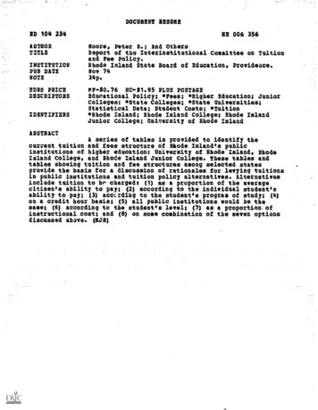
Minerals 2018, 8, x FOR PEER REVIEW4 of 13Serifos is occupied by a metamorphic sequence which comprises [38–40]: (a) a basal gneiss,mylonitic schists and marbles unit, with a maximum thickness of about 200m, that has beeninterpretedandMinerals2018, 8,as487member of the CCBU; (b) an intermediate rock unit made of amphibolites4 of13overlying greenschists with rare marble intercalations, occupying the northern part of the Island andcontaining relics of glaucophane, that are typical of the CBU [41]; (c) and finally, an uppermost unitankeritisedprotocataclasticshales and minorserpentinite[31].Contacts [31].betweenthe rockunits thearewith marbles,ankeritised protocataclasticshalesand lopasdetachmentrock units are marked by tectonic structures (Figure 1), namely the Megalo Livadi and Kavosfaults,whichbelong tofaults,the WestCycladicDetachmentSystem,(WCDS[42]) andare characterizedbyKyklopasdetachmentwhichbelongto the WestCycladicDetachmentSystem,(WCDS, [42])top-to-theSSWkinematicsonthefootwalls.and are characterized by top-to-the SSW kinematics on the footwalls.(a) etheNWedgeCycladesIslandcomplexin Aegeanthe AegeanFigure 1. (a)NWedgeof ofthetheCycladesIslandcomplexin theSea,Sea, Greece.(b) Simplifiedgeologicalmapof Serifos(modifiedafter Ducouxet al.,[31]).BlackGreece.(b) Simplifiedgeologicalmap ofSerifosIslandIsland(modifiedafter Ducouxet al. [31]).Blacksquaressquares indicatestudyareas of Avessalos(South)and Neroutsikaindicatethe studytheareasof Avessalos(South) andNeroutsika(North). (North).The above-mentionedareareintrudedby edrockrockunitsunitsintrudedbyI-type,an e-grainedequigranular,which displays two variants [43]: the inner part of the intrusion comprises a fine-grainedunfoliatedrock,while therock,outerwhilepart isthecomposedof iatedouter partcomposed of ite,micrograniteandcharacterized by large biotite flakes and mafic enclaves. Many dykes and sills of granodiorite,rhyodacite/daciteare scattered aroundintrusion[44].the intrusion [44].microgranite and rhyodacite/daciteare thescatteredaroundA number of skarns have formed close to the granodiorite intrusion [39–41,43] and have beenlong known for their Fe-rich mineralisation, that was exploited until the 1960’s. Mines are widespreadover the Island and were targeted at magnetite-rich skarn zones or in hematite/limonite bariteorebodies in marbles [31]. These skarns are also the general area where the unique Serifos green quartz,
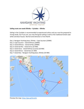
Minerals 2018, 8, x FOR PEER REVIEW5 of 13A number of skarns have formed close to the granodiorite intrusion [39–41,43] and have beenlong known for their Fe-rich mineralisation, that was exploited until the 1960’s. Mines arewidespreadover the Island and were targeted at magnetite-rich skarn zones orMinerals2018, 8, 4875 of in13hematite/limonite barite orebodies in marbles [31]. These skarns are also the general area where theunique Serifos green quartz, mostly known as prase (or prasem), is found (Figure 2). Except for themostlyknownas prase(ora prasem),is found(Figure2). Exceptfor canthe alsowell-knownwell-knowngreenquartz,great varietyof otheraestheticmineralsbe foundgreenin ].Serifos [25].Figure 2. Field and raphs:photographs: (a);(a);TheTheAvessalosAvessalos locationlocation (view towardsSW), n(viewtowardsNE)whereuniqueits prase; (b) The Neroutsika location (view towards NE) from wherefromuniquecombinationscombinationsof prase withrecovered(Figurewithin2g); (c)Geode withinhedenbergiticofprase with amethystwere amethystrecovered were(Figure2g); (c) Geodehedenbergiticskarn,filled withskarn, ;(d,e)aesthetic s,Avessaloslocality;(d,e) aestheticcrystalscombinationscombinationsfrom scepter,Avessaloslocality;negativescepter,formedby anupperpartofaamethystinelocality;(f) negativeformedby an(f)upperpart ofamethystinequartzthatgrowsatopbasal part ofprase,locality;bi-coloreda basal prasepart and anupperconsistingamethyst partquartzAvessalosthat growsatop a(g)basalpart ofcrystalprase,consistingAvessalosoflocality;(g) bi-coloredcrystalof aofgemprasequality,NeroutsikaFigures andspecimenand 3e–fare courtesyof C. Mavrogonatosbasalpartand an locality.upper amethystpartof gem3dquality,Neroutsikalocality.Figures andandP. Voudourisspecimen3d and respectively.3e–f are courtesy of C. Mavrogonatos and P. Voudouris respectively.Amongilvaite, aa rareAmong them,them, ilvaite,rare sorosilicatesorosilicate mineralmineral isis mainlymainly foundfound inin KoundourosKoundouros area,area, whichwhich sfromthislocality,areconsideredtobeamonglocated a few kilometers north from Avessalos. Samples from this locality, are considered to bethebesttheof itskind.amongbestof its kind.Andraditic garnets, often zoned are found in euhedral translucent crystals on top of hedenbergite ingeodes with varying dimensions in the areas of Avessalos and Agia Marina. Finally, tabular translucent
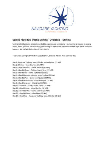
Minerals 2018, 8, 4876 of 13barite and rhombohedral calcite have been found in exceptional crystals in places close to the Fe-richmineralization [25,30].The green quartz from Serifos is usually translucent, with a characteristic green color in differentintensities. Typical are cigar-shaped crystals which extend from a fine-grained matrix consisting ofhedenbergite. Often smaller crystals sprout from the prism of larger crystals, resulting in very aestheticaggregates. Green quartz is extracted from a few localities in the SW part of Serifos Island. The mostimportant of these are the localities of Avessalos (Figures 1 and 2 and Neroutsika (Figures 1 and 2b).Our samples are from the western part of Serifos (Figure 1), within amphibolite facies rocks and theskarn mineralization [25,26,29,30].Minor green quartz occurrences can also be found in Agia Marina and Koundouros. The studiedsamples come from the well-known locality of Avessalos (Figure 2a,c–f), from where the best greenquartz samples have been found. Crystals from this site are usually lustrous with vivid green color,in contrast to other sites, from where green quartz is usually not translucent, but with varying intensityof its green color. Green quartz is often found in geodes between brecciated, up to tens of meters-sizedblocks of skarnified amphibolites [25,26,29,30]. Brecciation of the amphibolites has been caused by theactivation of the WCDS and created pathways through which hydrothermal fluids arising from thegranodiorite created the widespread skarns on the Island [31].Empty spaces between the more-or-less skarnified amphibolite blocks, commonly occur intriangular shapes that may vary significantly in size (Figure 2c), sometimes reaching a width ofa few meters across. These spaces are filled by hedenbergite, followed by ilvaite, prase amethyst,hematite and carbonate (calcite) deposition towards the centre of the open spaces. Inside these geodes,the prase (green quartz) crystals develop in a variety of crystals shapes. Figure 2d–g displays someaesthetic aggregates with sharply terminated crystals, sceptres and reverse sceptres of prase combinedwith amethyst from both Avessalos and Neroutsika areas. In many cases, prase is accompanied bycalcite and iron-roses composed of hematite [25,26]. Its crystal shape is usually spindle-like in thebasal part and evolves into hexagonal on the top of the crystal. Prismatic green quartz crystals arealso found, as well as bi-colored, rocket-like shapes, with amethyst in the base and green quartz ontop. The sceptres found in the area are normal or inverse with green quartz in the base and an oftentrigonal amethystine termination (Figure 2f–g). An unusual type of crystals encountered in the areais the “interrupted” prase or amethyst crystals which form when platy calcite crystals are depositedtogether with quartz in successive layers [21,26].4. Results4.1. Mineral ChemistryDetailed microscopy and back-scattered electron (BSE) images reveal that the Serifos green quartzcontains numerous small inclusions. We find two general types of mineral inclusions. The first typecomprises myriads of acicular amphibole crystals which are often 10–100 µm long, but their thicknessis limited to below a few µm. These thin actinolite needles are practically found everywhere andseem to be randomly oriented. We did not observe any obvious relationship between amphiboleorientation and the crystallographic orientation of the quartz crystals. Some of the needles (Figure 3a,b)are clearly curved, a fact that supports random orientation of the amphibole needles in relation to thehost quartz crystals. With our electron microprobe we could only identify the needles as actinoliticamphibole but accurate analysis was not possible, due to the small size of the inclusions. We, therefore,assume that the chemical compositions of these thin amphibole needles are identical to the rims ofthe larger amphibole inclusions, a fact that also seems to be supported by electron microprobe maps(Figure 3e–f).The second type consists of larger inclusions (Figure 3a–c) with sizes that reach a few tens of µm inlength (commonly between 10 and 60 µm), and that always display zonation. EPMA analyses revealedthat these inclusions are Ca-Fe-rich actinolitic amphiboles with compositions very close to the end-member
Minerals 2018, 8, 4877 of 13ferro-actinolite. These minerals contain about 5 wt % MgO, almost no Na or K, and about 2 wt % Al2 O3 .All the studied larger actinolite inclusions contain a core, which is significantly lighter in BSE images.Microprobe analyses (Table 1) reveal that these cores are also actinolite, with some differences comparedto the rims. MnO content is almost doubled compared to the rim (0.56 and 0.25 wt % respectively) andFeO is slightly higher. Subsequently, the cores contain lower MgO and (3.8 wt %) and much less Al2 O3(0.3 wt %).Table 1. Electron microprobe measurements of amphibole inclusions, prase and amethyst fromSerifos Island.Amphibolen.o.a.SiO2TiO2Al2 O3Cr2 O3FeOMnOMgOCaONa2OK2OP2 O5Total3 (core)PraseAmethyst11146 (rim)wt %2σwt %2σwt %2σwt 0.010.110.010.030.010.010.010.010.010.02Fe2 O3Structural formulaeSiAlVIM1-3siteAlVITiFe3 MgFe2 MnΣM1-3M4siteMgFe2 MnCaNaΣ M4A siteNaKΣAΣ .03Fe3 MnMgCaNaK0.0000.0000.00020.000-4.925.00P-0.000Σ .880.081.960.000.020.0215.00.000.040.0415.0n.a. not analysed; (-) below detection, n.o.a. number of analyses.
Minerals 2018, 8, 487Minerals 2018, 8, x FOR PEER REVIEW8 of 138 of 13Figure 3.3. Back-scattered electron (BSE) images and quantitative electron microprobe elementelement maps.maps.(a): thin, nm-sized needles of amphibole together with a larger amphibole. The latter larger ithwithno clearlydefinededges,edges,as theasrapidcontains amphiboleno clearlydefinedthegrowthof the crystalstopped.Furthermore,it seems thatthis amphibolealready hasdeterminedrapid growthof the hascrystalhas stopped.Furthermore,it seemsthat this hasamphibolealreadytheouter edgethe crystalit wouldhavegrown haveinto ifgrowngrowthhadnot beenhadinterrupted.determinedtheofouteredge ofwhichthe crystalwhichit wouldintoif growthnot beenTheorigin of Thethe observedis unknown;(c)isSimilarlarger(c)amphibolewith aamphibolefringed edgealsoainterrupted.origin oftexturesthe observedtexturesunknown;Similar largerwithindicatingrapidgrowth;(d) high-resolutionof the fine-graineddark blebsare emptyfringed edgealsoindicatingrapid growth; image(d) high-resolutionimagematrix.of the Thefine-grainedmatrix.Theandinterpretedas fluidthat wereduring samplepreparation;Quantitativedarkareblebsare emptyandinclusionsare interpretedas openedfluid inclusionsthat wereopened (e)duringsampleelectronmicroprobemap of Fe.The coresof the largercontainmore FeOpreparation;(e) Quantitativeelectronmicroprobemapamphibolesof Fe. The coresof significantlythe larger t-handsideindicateschemicalcomposition(wt% FeO).contain significantly more FeO (Table 1) than the rims. The color bar on the right-hand side indicatesNotethat compositionthe very thin(wt.%needle-shaped(blueishin thisactinolite crystalsare toosmallto bechemicalFeO). Notethat theveryFigure)thin needle-shaped(blueishin thisFigure)analyzedthat theareFeO-content(f)thatQuantitativeelectron microprobemap of Mn.actinolite socrystalstoo smallistounderestimated;be analyzed sothe FeO-contentis underestimated;(f)Thecores Mn thanthe rimofamphibole.actinolite,indicating two different generations of amphibole.
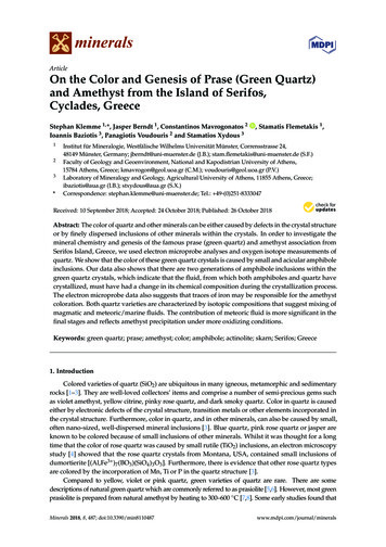
Minerals 2018, 8, 4879 of 13The CaO content is quite similar in both rim and core with values around 11.5 wt %. Finally,traces of Na2 O and K2 O were detected in both rim and core actinolites. Values for both elementsincrease towards the rims of the crystals, reaching values of up to 0.26 and 0.19 wt % respectively.Average chemical formulae for the compositions from the cores and the rims of the crystals correspondto [Ca1.95 Na0.01 K0.02 ]1.98 [Mg0.89 Fe2 3.87 Mn0.08 Al0.07 Ti0.01 ]4.92 [Si]8.08 O22 (OH)2 and [Ca1.88 Na0.08 K0.04 ]2[Mg1.15 Fe2 3.55 Fe3 0.13 Mn0.03 Al0.13 ]4.99 [Si7.85 Al0.15 ]8 O22 (OH)2 , respectively. Analytical data fromprase and amethyst yielded traces of Al2 O3 , FeO, MgO, MnO, CaO, Na2 O, K2 O and P2 O5 . The highestFeO and Al2 O3 contents were found in prase (up to 0.26 and 0.024 wt % respectively). Amethyst Fe2 O3content reaches up to 0.020 wt % (calculated from the total FeO of the analyses).4.2. Quartz Oxygen IsotopesThe quartz oxygen isotopic compositions were analyzed in hand-picked prase and amethystcrystals. The compositions of the fluid in equilibrium with the prase and amethyst crystals werecalculated following Sharp et al. [45], using temperatures of 350 C for prase and 250 C for amethyst,based on the respective mineral assemblages of Salemink [43].The studied prase samples yielded isotopic δ18 OQz values of 12.72 and 12.74‰, which correspond tocalculated δ18 OFl values of 7.32 and 7.14‰ (Table 2). Amethyst samples yielded δ18 OQz values of 9.94and 9.85‰, which correspond to calculated δ18 OFl values of 1.04 and 0.95‰ respectively. The isotopicsignature of prase is comparable to those of quartz samples from different assemblages of the skarn zone,but amethyst displays significantly lighter isotopic signature (Figure 4).Table 2. Oxygen isotope compositions of quartz from the skarn of Serifos Island [δ values in‰ relativeto Standard Mean Ocean Water (SMOW)]. The oxygen isotope values in the fluid (δ18 OFl ) in equilibriumwith quartz have been calculated according to Sharp et al., [45].SampleVarietyAssociated Mineralsδ18 OQzT ( C)δ18 OFlSR2aSR2bSR1aSR1b20–11 *27–30 *26–49 *26–48 *26–87 *137 *55 *55-(1) *56 *26–35 *ML-2.2 *ML-1 *27-7B uartzacachemhemepepMt hemMt hemEp acAc ccivivivmtAc mtAc mtPy hem 713.314.76.86.06.77.87.87.55.2Samples marked with (*) are from Salemink [43]. Abbreviations: ac actinolite; hem hematite; ep epidote; mt magnetite; iv ilvaite; cc calcite; py pyrite.
Minerals 2018, 8, 487Minerals 2018, 8, x FOR PEER REVIEW10 of 1310 of 13FigureO valuesof quartzfromthetheskarnskarn ofof SerifosSerifos Island,Valuesfor formagmaticwaterwaterFigure4. δ184.Oδ18valuesof quartzfromIsland,Greece.Greece.Valuesmagmaticand andesiticvolcanicvaporfromTaylorΤaylor[46][46] andand Giggenbachrespectively.and 7][47]respectively.5. Discussion5. Discussionor pr
Serifos Island, Greece, we used electron microprobe analyses and oxygen isotope measurements of quartz. We show that the color of these green quartz crystals is caused by small and acicular amphibole inclusions. Our data also shows that there are two generations of amphibole inclusions within the