Transcription
Hou et al. BMC Surgery 2014, ESEARCH ARTICLEOpen AccessComparison of single and two-tunnel techniquesduring open treatment of acromioclavicular jointdisruptionZhiyong Hou1*, Jove Graham2, Yingze Zhang1, Kent Strohecker2, Daniel Feldmann2, Thomas R Bowen2,Wei Chen1 and Wade Smith3AbstractBackground: Coracoclavicular (CC) ligament reconstruction with semitendinosus tendon (ST) grafts has becomemore popular and has achieved relatively good results; however optimal reconstruction technique, single-tunnel ortwo-tunnel, still remains controversial. This paper is to compare the clinical and radiographic data of allogenous STgrafting with single- or two-tunnel reconstruction techniques of the AC joint.Methods: The outcomes of 21 consecutive patients who underwent anatomical reduction and ST grafting for ACjoint separation were reviewed retrospectively. Patients were divided into two groups: single-tunnel group (11) andtwo-tunnel group (10). All patients were evaluated clinically and radiographically using a modified UCLA rating scale.Results: The majority of separations (18 of 21) were Rockwood type V, with one each in type III, IV and VI categories.The overall mean follow-up time was 16 months, and at the time of the latest follow-up, the overall mean UCLA ratingscore was 14.1 (range 8–20).The percentage of good-to-excellent outcomes was significantly higher for patients with the two-tunnel techniquethan for those with the one-tunnel technique (70% vs. 18%, respectively, p 0.03). Within the single-tunnel group, therewas no statistically significant difference in percentage of good-to-excellent outcomes between patients with vs.without tightrope augmentation (17% vs 20%, p 0.99). Similarly, within the two-tunnel group, there was nosignificant difference in the percentage of good-to-excellent outcomes between the graft only and augmentgroups (67% vs. 75%, p 0.99).Conclusion: Anatomical reduction of the AC joint and reconstruction CC ligaments are crucial for optimal jointstability and function. Two-tunnel CC reconstruction with an allogenous ST graft provides superior significantlybetter radiographic and clinical results compared to the single-tunnel reconstruction technique.Keywords: Acromioclavicular joint, Single-tunnel, Two-tunnel, Reconstruction, AugmentationBackgroundAcromioclavicular (AC) joint injuries are among themost commonly occurring problems in the young andactive patient population. Higher-grade AC joint injuries(Rockwood types III through VI) represent failure of thecoracoclavicular (CC) ligament complex, which is formedby the conoid and trapezoid ligaments. This complex hasbeen termed the primary suspensory structure of the* Correspondence: drzyhou@gmail.com1Department of Orthopaedic Surgery, Third Hospital of Hebei MedicalUniversity, Shijiazhuang, Hebei 050051, ChinaFull list of author information is available at the end of the articleupper limb [1,2]. In the literature, the incidence oftraumatic AC joint separation varies from 3 to 4 per100,000 people with 25-52% of these occurring duringsporting activities, and they are also one of the mostcommon shoulder injuries seen in orthopaedic traumatology [2-5]. For certain Rockwood type III AC jointseparations and all type IV, V, and VI injuries, surgicaltreatment has been recommended to prevent disablingpain, weakness, and deformity [6-8]. Although more than60 surgical techniques have been reported, the frequencyof failure to maintain reduction after surgical treatmentremains high [9,10]. 2014 Hou et al.; licensee BioMed Central Ltd. This is an Open Access article distributed under the terms of the CreativeCommons Attribution License (http://creativecommons.org/licenses/by/2.0), which permits unrestricted use, distribution, andreproduction in any medium, provided the original work is properly credited. The Creative Commons Public DomainDedication waiver ) applies to the data made available in this article,unless otherwise stated.
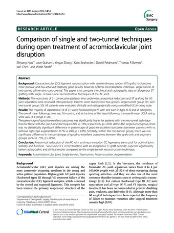
Hou et al. BMC Surgery 2014, ecently, CC ligament reconstruction with tendon graftshas become more popular and has achieved relativelygood results [11,12]. Biomechanical studies focusing on ananatomic reconstruction of the CC ligament complexusing tendon grafts have reported promising potentialfor this technique [13-15]. Semitendinosus tendon (ST)grafting and anatomic reconstruction can be imitated,providing stability to the clavicle that is very close tothat provided by the intact ligaments [13]. However, optimal reconstruction technique, single-tunnel or two-tunnel,still remains controversial. Anatomical two-tunnel reconstruction with tendon grafts or synthetic materials seemsappealing because it has been shown by biomechanicalstudies to restore the original two ligaments (the conoidand trapezoid) and to produce an ultimate failure loadthat is equivalent to that of native CC ligaments [13-15].However, it is technically difficult and theoretically increases the risk of fracture [16].The purpose of this retrospective study was to analyzethe clinical and radiographic data of allogenous ST tendongrafting with single- or two-tunnel reconstruction techniques of the CC ligaments. We hypothesize that anatomicreconstruction of the AC joint disruption using two-tunnelreconstruction technique results in a satisfying clinicalfunction and provides stable fixation.MethodsBetween June 2003 and January 2009, twenty-three patientsunderwent open operation for AC joint reconstructionwith ST allograft at our institution. In the earlier studyperiod before 2007, we mostly used single-tunnel technique, and after 2007 mostly the two-tunnel technique.For analysis we divided patients into two groups: singletunnel group and two-tunnel group. Patient data werecollected retrospectively, including gender, age at thetime of surgery, injury mechanism, classification accordingto Rockwood, and surgical technique. Patients with atleast 12 months of clinical follow-up were included in thisstudy. Patients were excluded if they had a previous shoulder injury, arthritis, or an associated neurological deficiton the side of injury.The procedure was performed with the patient in thebeach chair position under general anesthesia in combination with an interscalene block. An anterior deltopectoral approach was utilized with saber incision, TheAC joint, the lateral end of the clavicle, and the coracoidprocess were exposed. Subperiosteal detachment of thedeltotrapezial fascia from the clavicle was performed.The distal end of the clavicle was resected 8 to 10 mmusing an oscillating saw. For the single-tunnel technique,a 6-mm drill hole was made about 1.5-2 cm medial tothe remaining end of the clavicle superior to inferior ina 300 posterior to anterior angle. A ST allograft was prepared by placing a whipstitch (Arthrex #2 FiberwirePage 2 of 7suture, Naples, FL, USA) on either end. After reducingthe distal clavicle down to the acromion anatomically, theST graft was introduced around the base of the coracoidand then both ends of the graft up through the claviclehole. The graft was then mechanically tensioned and a5.5 mm Bio-tenodesis screw was placed down through thecenter of the ST graft fixing it to the clavicle. The freeends of the graft were then passed underneath the clavicleand tied to themselves for additional fixation (Figure 1).If using a tightrope augment (Arthrex Fiberwire No. 5,Naples, FL, USA), a guide was used to place a pin froma point medial to the lateral tunnel, to the base of thecoracoid. A 4.5 mm reamer was then used to create atunnel through the clavicle and coracoid. The tight ropedevice was placed through the clavicular and then coracoid tunnel and endobutton secured against inferior cortexof coracoid. The tight-rope was then tied after fixation ofthe graft. Later in the series, a single clavicular tunnel wasutilized for both the graft and tight-rope. The graft wasplaced around the coracoid and through the claviculartunnel and tightrope device (Figure 2).For the two-tunnel technique, the same delto-pectoralapproach was used. Two holes were drilled in the clavicleto reconstruct each of the two CC ligaments, trapezoidand conoid ligaments. The lateral tunnel is created as inthe single-tunnel technique. The medial tunnel is located4.5 cm medial to the AC joint. A 5.5 mm tunnel is reamedlike the medial tunnel. A single ST graft was prepared andlooped under the coracoid. The lateral free end wasbrought up through the lateral tunnel, and the medial freeend through the medial tunnel. The AC joint is reduced,and the grafts fixed into the tunnels with 5 mm biotenodesis screws and the graft tied to itself (Figure 3). If usingtightrope augment, a guide pin is placed between the twograft tunnels, from midline, through the clavicle and baseof coracoid. A 4.5 mm tunnel is reamed over the guidewire and the Tight-rope device placed through the clavicleand coracoid and secured to the inferior cortex of theFigure 1 ST allograft reconstruction of the AC joint withsingle-tunnel technique.
Hou et al. BMC Surgery 2014, age 3 of 7Figure 2 ST Allograft with tightrope augment reconstruction ofthe CC joint with single-tunnel technique.Figure 4 ST Allograft with tightrope augment reconstruction ofthe CC joint with two-tunnel technique.coracoid. The device is tightened and tied after graftfixation (Figure 4). After reconstruction, attention wasdirected to repair of the deltotrapezial interval. Thiswas performed in a pants-over-vest fashion using #1 or #2non-absorbable sutures in an interrupted fashion. Alayered closure was then performed. A drain was notutilized.All patients were placed in a sling immobilizer post-opfor 4 to 6 weeks. Gentle pendulums and Codman’s werebegun post-op day 1. At 4 weeks therapy was begun withpassive motion and cuff isometrics. Resistive programstarted at 8 weeks. Patients were generally allowed to returnto manual work and athletics at 4 to 6 months dependingon level of rehabilitation. Contact sports not prior to sixmonths. All patients were evaluated clinically and radiographically using a modified UCLA rating scale [5,17],which reflects three parts: maintenance of reduction,objective evaluation of the patient’s function, and complications secondary to operation. In the radiologicalevaluation, the roentgenographic rating was determinedby the degree of displacement of the AC joint, whichwas evaluated by measuring the relation between theacromion and the clavicle on the anteroposterior viewfor vertical displacement (reduced 4 points, subluxed 2points, dislocated 0 points). In the physical evaluation,range of motion (ROM), pain, weakness, and complicationswere recorded. Finally, patients were asked their overallsatisfaction with the postoperative result, with 0 points fordissatisfaction or unsure and 2 points for satisfaction.Table 1 shows the relative weight given to each categoryof the rating scale and describes the criteria by which apatient was assigned an overall final result of excellent,good, fair, or poor.Percentages of good-to-excellent outcomes and maintenance of reduction (reduced or subluxed) were comparedbetween the two reconstruction procedures (single vs.two-tunnel), and between augmentation techniques(with vs. without tightrope). Because of the relativelysmall sample sizes, Fisher’s exact test was used in placeof chi-square testing at a significance level of p 0.05.All analysis was performed using SAS statistical software(SAS 9.2, Cary, NC). Waiver of patient consent was grantedby Institutional Review Board of Geisinger Medical Centerfor retrospective chart review.Figure 3 ST allograft reconstruction of the CC joint withtwo-tunnel technique.ResultsFrom the initial 23 patients who were surgically treated,two patients were lost to follow up and were excluded.Table 2 summarizes the demographics and injury characteristics of the 21 patients remaining in the study.The majority of fractures (18 of 21) were Rockwoodtype V, with one fracture each in type III, IV and VIcategories. Most of those patients had received primaryunsuccessful conservative care and switched to operative management, and one patient underwent a failedWeaver-Dunn procedure.The overall mean follow-up time was 16 months, andat the time of the latest follow-up, the overall mean UCLArating score was 14.1 (range 8–20). Eleven (52%) patients
Hou et al. BMC Surgery 2014, age 4 of 7Table 1 The modification of the UCLA rating scale8.17CategoryPointsTable 2 Demographic and injury characteristics, bysingle-tunnel and two-tunnel groupMaintenance of ocation0FemaleMean age (range), yearsRange of motionSingle-tunnel (11)Two-tunnel (10)695137 (20–55)42 (20–63)Full2Side of FractureImproved from preoperative1Right58No change from preoperative0Left62Mechanism of InjuryStrength2Sporting66Improved from preoperative1Traffic accident42Unimproved from preoperative0Fall12NormalRockwood ClassificationPain4C310With strenuous activity3C401With moderate activity2C5108With mild activity1C601All the time0Mean length of follow up(range), months16 (12–38)15 (12–40)NoneWeaknessNone2With strenuous activity1All the time0Change in occupationSame or more strenuous2Less fected outcome0Patient satisfactionYes2No or unsure0Results: excellent, 18–20; good, 15–17; fair, 12–14; poor, 11.rated the outcome as good to excellent, 3 (14%) rated it asfair, and 7 (33%) rated it as poor. Three of 21 patientsunderwent additional revision surgery for the failed CCligament repair or reconstruction.Of the 21 patients, eleven patients underwent allogenous ST grafting with single-tunnel reconstructiontechnique, and 6 of these received tightrope augmentation. Ten patients underwent allogenous ST grafting with two-tunnel reconstruction technique: fourof these received one ST graft plus one tightrope graft(“ST-tightrope”), while the other six received two ST grafts(“ST-ST”).Table 3 summarizes the UCLA rating scale scores atlast follow-up for the two groups (single- and two-tunnel),subdivided by augmentation type. The percentage ofgood-to-excellent outcomes was significantly higher forpatients with the two-tunnel technique than for thosewith the one-tunnel technique (70% vs. 18%, respectively,p 0.03). Within the single-tunnel group, there was nostatistically significant difference in percentage of goodto-excellent outcomes between patients with vs. withouttightrope augmentation (17% vs 20%, p 0.99). Similarly,within the two-tunnel group, there was no significantdifference in the percentage of good-to-excellent outcomes between ST-tightrope and ST-ST patients (75%vs. 67%, p 0.99).We noted that complications were observed in threeof the 21 patients: two patients in the two-tunnel grouphad infection, and one patient in the single-tunnel grouphad a coracoid fracture. Calcification of the CC ligamentoccurred in one case, but it did not appear to causesymptoms, and was therefore not considered a complication. No patient had neurovascular or post-traumaticarthritis of the injured AC joint.DiscussionOur data demonstrated that allogenous ST grafting withtwo-tunnel reconstruction technique of the AC jointyielded excellent or good clinical outcomes more frequently compared to single-tunnel reconstruction technique. These results also suggest that the materials used
Hou et al. BMC Surgery 2014, age 5 of 7Table 3 Number of patients receiving single-tunnel vs. two-tunnel techniques, subdivided by augmentation type, withclinical outcome results based on modification of the UCLA rating scaleUCLA rating scaleTwo-tunnel (n 10)*Single-tunnel (n 11)With augmentWithout 2111Poor3301Total65461 (17%)1 (20%)3 (75%)4 (67%)N(%) with excellent or good *Two-tunnel group had significantly higher percentage of good-to-excellent outcomes than single-tunnel group, p 0.03. No significant difference between with vs. without augmentation for single-tunnel group, p 0.99.#No significant difference between ST-tightrope vs. ST-ST for two-tunnel group, p 0.99.for augmentation in the two-tunnel reconstructiontechnique do not impact the clinical result. In thistechnique, one ST allograft combined with one tightrope graft construction can provide similar outcomesto using ST allograft in both tunnels. We also saw no significant differences between patients with and withouttightrope augment in the single-tunnel technique group.Based on well established anatomical ligament reconstruction in the knee injury, reconstructing the CCligament using tendon graft for AC joint injury has become more popular because the construct is more physiologic, does not require implant removal and preserves theCA ligament [18,19]. ST tendon grafts are most commonused for this procedure, which can be either autografts orallografts, and have achieved relatively good results[11-13,20,21]. The harvesting of an autogenous tendonmay not result in long-term functional impairment butmay still cause some morbidity associated with the donorsite, and also create a second operative site during ACjoint surgery [22]. Nicholas et al. [12] achieved excellentoutcomes after fresh-frozen ST allograft reconstruction ofthe CC ligament; patients reported significant pain relief,return of normal strength and function, negligible loss ofmotion, and no loss of reduction on postoperative radiographs. Based on this information, the substitution of allograft material has become a routine procedure in ourinstitution. The current surgical technique for the CC ligament reconstruction can be graft tendon passed thoughthe clavicle with single tunnel or two tunnels technique[16,23], looped around the base of the coracoids [24],passed through a transosseous tunnel in the coracoids[25], or fixed to the base of coracoid using an anchor technique [6]. The CC ligament is stabilized by 2 sets of ligamentous structures: the conoid and trapezoid. Single-tunnelor two-tunnel reconstruction still remains controversial.Mazzocca et al. considered that each CC ligament hasa separate function, and so each must be considered inreconstructive procedures [26]. Anatomical two-tunnelreconstruction with tendon grafts has yielded goodresults because it restores the original 2 ligaments andproduces an ultimate strength that is equivalent to thatof native CC ligaments [14,15,23]. However, two-tunneltechniques are technically difficult, with increased riskof fracture, and sometimes are not possible in patientswith a small clavicle [13,16]. This technique should beperformed by an experienced arthroscopist [23]. Yoo et al.[16] reported that single-tunnel reconstruction has someadvantages over two-tunnel techniques. They reconstructedCC ligaments in 21 patients using a single-tunnel ST autograft and achieved superior clinical result. 17 (81%) ofthe 21 patients maintained complete reduction, and only 1patient (reportedly a manual laborer) had complete reduction loss. In our cohort, there was a statistically significantdifference in percentages of good-to-excellent UCLA scoresbetween the single-tunnel and two-tunnel groups. Thetwo-tunnel group had better scores, with the caveat that weobserved two cases of infection in the two-tunnel groupwhich may be related to the greater length and complexity of this procedure as compared to the singletunnel technique.Anatomical two-tunnel reconstruction with ST tendongrafts or synthetic materials provided similar results. Thetightrope system, consisting of one round clavicle titaniumbutton and one long coracoid titanium button connectedby non-absorbable sutures (No. 5 Ethibond suture), hasbeen initially utilized for repair of acute syndesmosis disruptions. The application has been extended and previouslydescribed for AC joint dislocations [27,28]. It can be usedas a single graft device or an augment for the other tendongraft construction. Two-tunnel reconstruction techniquehas been shown by biomechanical studies to restore thestrength of the original two ligaments (the conoid andtrapezoid) and result in significantly higher stability in thesuperoinferior as well as the anteroposterior plane whencompared with the native CC ligaments [11,14,15,29].Grafting materials for the two-tunnel technique useare variable, and may include two tendon grafts, twotightrope grafts, or one tendon with one tightrope
Hou et al. BMC Surgery 2014, rafts. Salzmann et al. [23] reported on 23 consecutivepatients with the acute AC joint disruption who underwent two-tunnel anatomical reconstruction of CC ligaments using two flip-button tightropes. This procedureyielded satisfactory clinical function and provided a stablefixation at intermediate-term follow-up. In our two-tunnelgroup, most patients had good-to-excellent UCLA scoresat last followup, and this result did not vary between thecases treated with one ST graft and one tightrope graftversus those treated with two ST grafts.Augmentation has been shown to be beneficial duringCC ligament reconstructions by biomechanical studies[30,31]. An effective augmentation must have biomechanical properties enabling it to shield the repair or reconstruction from excessive tensile force, ideally allowingearly rehabilitation. It seems desirable for an augmentation to possess strength and stiffness similar to those ofthe intact CC ligament complex, thus protecting againstphysiologic loads while allowing for physiologic motionbetween the clavicle and coracoid. Tienen et al. [32] hadgood results with using an open modified Weaver-Dunntechnique and AC joint augmentation with absorbable,braided suture in 21 paptients. The tightrope augmentation was initially described for acute AC joint dislocationand represented an excellent biological augmentationtechnique by Hernegger [27]. Scheibel et al. [33] alsoreported using a gracilis tendon reconstruction augmentedwith a tightrope achieved good and excellent results andmaintained good reduction for acute AC joint dislocationswith one year follow up. Recently, Yoo et al. [16] also reported a superior result by using the tightrope augmenttechnique to protect the ST graft though the same tunnelduring the healing period. They considered the tightropeaugment was really important factor for their successfulsurgical procedure and good outcomes. However, in ourone-tunnel group, although the sample size was small, wesaw no significant difference between patients treated withand without tightrope augmentation. Both of them had ahigher re-dislocation rate and achieved the inferior resultscomparing to the two-tunnel group. From our results, wecannot definitively state that tightrope augmentation is notimportant and effective for the CC complex reconstruction,but our results do provide strong evidence that the reconstruction technique (specifically the choice betweenone or two tunnels) largely impacts the radiographicand clinical outcomes.The principal limitations of this study are the relativesmall sample size who met our inclusion criteria and thefact that we did not have preoperative functional scores.Thus, our conclusions are focused on the substantial difference in success rates we saw between the single-tunneland two-tunnel groups (18% vs. 70%), and we have limitedability to assess and compare other aspects of the procedures. In addition, because this was an observationalPage 6 of 7study, our data did not permit an accurate assessmentof the time to functional recovery. The two-tunneltechnique became a standard technique at our institutionat a later date than the single-tunnel technique, and so itis possible that surgeon experience may have played a rolein the different outcomes among groups. However, we donot believe this confounding factor would be substantialenough to explain the large difference in the two groupsthat we observed.ConclusionAnatomical reduction the AC joint and biomechanicalreconstruction CC ligaments are crucial for the optimaljoint stability and function. Two-tunnel CC reconstructionwith an allogenous ST graft provides superior radiographicand clinical results compared to single-tunnel reconstruction technique.AbbreviationsCC ligament: Coracoclavicular ligament; ST tendon: Semitendinosus tendon;AC joint: Acromioclavicular joint; UCLA shoulder rating scale: University ofCalifornia at Los Angeles shoulder rating scale; ROM: Range of motion.Competing interestsThe authors declare that they have no competing interests.Authors’ contributionsZH and WS designed research; JG, KS and WC analyzed data and performedstatistical analysis. All authors read and approved the final manuscript.Author details1Department of Orthopaedic Surgery, Third Hospital of Hebei MedicalUniversity, Shijiazhuang, Hebei 050051, China. 2Department of OrthopaedicSurgery, Geisinger Medical Center, Danville, PA 17822, USA. 3MountainOrthopaedic Trauma Surgeons at Swedish, 701 East Hampden Avenue Suite515, Englewood, CO 80113, USA.Received: 27 July 2013 Accepted: 11 August 2014Published: 15 August 2014References1. Bosworth BM: Acromioclavicular separation: New method of repair. SurgGynecol Obstet 1941, 73:866–871.2. Rockwood CA, Williams GR, Young DC: Injuries to the AcromioclavicularJoint. In Fractures in Adults, Volume 2. Fourthth edition. Edited by RockwoodCA, Green DP, Bucholz RW. Philadelphia: Lippincott-Raven Pub Publishers;1996:1341–1413.3. Horn JS: The traumatic anatomy and treatment of acuteacromioclavicular dislocation. J Bone Joint Surg Br 1954, 36:194–1201.4. Lemos MJ: The evaluation and treatment of the injured acromioclavicularjoint in athletes. Am J Sports Med 1998, 26:137–144.5. Lizaur A, Marco L, Cebrian R: Acute dislocation of the acromioclavicularjoint. Traumatic anatomy and the importance of deltoid and trapezius.J Bone Joint Surg Br 1994, 76:602–606.6. Bannister GC, Wallace WA, Stableforth PG, Hutson MA: The management ofacute acromioclavicular dislocation. A randomised prospectivecontrolled trial. J Bone Joint Surg Br 1989, 71:848–1850.7. Fremerey RW, Lobenhoffer P, Ramacker K, Gerich T, Skutek M, Bosch U:Acute acromioclavicular joint dislocation–operative or conservativetherapy? Unfallchirurg 2001, 104:294–299.8. Kumar S, Sethi A, Jain AK: Surgical treatment of completeacromioclavicular dislocation using the coracoacromial ligament andcoracoclavicular fixation: report of a technique in 14 patients. J OrthopTrauma 1995, 9:507–510.
Hou et al. BMC Surgery 2014, 6.27.28.29.30.31.Warren-Smith CD, Ward MW: Operation for acromioclavicular dislocationA review of 29 cases treated by one method. J Bone Joint Surg Br 1987,69:715–718.Weaver JK, Dunn HK: Treatment of acromioclavicular injuries, especiallycomplete acromioclavicular separation. J Bone Joint Surg Am 1972,54:1187–1194.Jones HP, Lemos MJ, Schepsis AA: Salvage of failed acromioclavicacromioclavicular joint reconstruction using autogenous semitendinosustendon from the knee: surgical technique and case report. Am J SportsMed 2001, 29:234–237.Nicholas SJ, Lee SJ, Mullaney MJ, Tyler TF, McHugh MP: Clinical outcomesof coracoclavicular ligament reconstructions using tendon grafts. Am JSports Med 2007, 35:1912–1917.Costic RS, Labriola JE, Rodosky MW, Debski RE: Biomechanical rationale fordevelopment of anatomical reconstructions of coracoclavicularligaments after complete acromioclavicular joint dislocations. Am J SportsMed 2004, 32:1929–1936.Lee SJ, Nicholas SJ, Akizuki KH, McHugh MP, Kremenic IJ, Ben-Avi S:Reconstruction of the coracoclavicular ligaments with tendon grafts: acomparative biomechanical study. Am J Sports Med 2003, 31:648–655.Mazzocca AD, Santangelo SA, Johnson ST, Rios CG, Dumonski ML, ArcieroRA: A biomechanical evaluation of an anatomical coracoclavicularligament reconstruction. Am J Sports Med 2006, 34:236–246.Yoo JC, Ahn JH, Yoon JR, Yang JH: Clinical results of single-tunnelcoracoclavicular ligament reconstruction using autogenous semitendinosustendon. Am J Sports Med 2010, 38:950–957.Guy DK, Wirth MA, Griffin JL, Rockwood CA Jr: Reconstruction of chronicand complete dislocations of the acromioclavicular joint. Clin OrthopRelat Res 1998, 347:138–149.Fu FH, Shen W, Starman JS, Okeke N, Irrgang JJ: Primary anatomicdouble-bundle anterior cruciate ligament reconstruction: a preliminary2-year prospective study. Am J Sports Med 2008, 36:1263–1274.Stannard JP, Riley RS, Sheils TM, McGwin G Jr, Volgas DA: Anatomicreconstruction of the posterior cruciate ligament after multiligamentknee injuries: a combination of the tibial-inlay and two-femoral-tunneltechniques. Am J Sports Med 2003, 31:196–202.LaPrade RF, Hilger B: Coracoclavicular ligament reconstruction using asemitendinosus graft for failed acromioclavicular separation surgery.Arthroscopy 2005, 21:1277.Tauber M, Gordon K, Koller H, Fox M, Resch H: Semitendinosus tendongraft versus a modified Weaver-Dunn procedure for acromioclavicularjoint reconstruction in chronic cases: a prospective comparative study.Am J Sports Med 2009, 37:181–190.Yoo JC, Choi NH, Kim SY, Lim TK: Distal clavicle tunnel widening aftercoracoclavicular ligament reconstruction with semitendinous tendon: acase report. J Shoulder Elbow Surg 2006, 15:256–259.Salzmann GM, Walz L, Buchmann S, Glabgly P, Venjakob A, Imhoff AB:Arthroscopically assisted 2-bundle anatomical reduction of acuteacromioclavicular joint separations. Am J Sports Med 2010, 38(6):1179–1187.Hessmann M, Gotzen L, Gehling H: Acromioclavicular reconstructionaugmented with polydioxanonsulphate bands. Surgical technique andresults. Am J Sports Med 1995, 23:552–556.Wolf EM, Pennington WT: Arthroscopic recons
begun post-op day 1. At 4 weeks therapy was begun with passive motion and cuff isometrics. Resistive program started at 8 weeks. Patients were generally allowed to return to manual work and athletics at 4 to 6 months depending on level of rehabilitation. Contact sports not prior to six months. All patients were evaluated clinically and radio-