Transcription
Hindawi Publishing CorporationEvidence-Based Complementary and Alternative MedicineVolume 2012, Article ID 971896, 11 pagesdoi:10.1155/2012/971896Research ArticleThe Physical Demands of the Tree (Vriksasana) and One-LegBalance (Utthita Hasta Padangusthasana) Poses Performed bySeniors: A Biomechanical ExaminationSean S.-Y. Yu,1 Man-Ying Wang,1 Sachithra Samarawickrame,1 Rami Hashish,1Leslie Kazadi,1 Gail A. Greendale,2 and George J. Salem11 Divisionof Biokinesiology and Physical Therapy, University of Southern California (USC), 1540 E. Alcazar Street, Los Angeles,CA 90033, USA2 Division of Geriatrics, Geffen School of Medicine at the University of California, Los Angeles (UCLA),924 Westwood Boulevard, Suite 200, Los Angeles, CA 90024, USACorrespondence should be addressed to George J. Salem, gsalem@usc.eduReceived 3 May 2012; Accepted 23 July 2012Academic Editor: Andreas MichalsenCopyright 2012 Sean S.-Y. Yu et al. This is an open access article distributed under the Creative Commons Attribution License,which permits unrestricted use, distribution, and reproduction in any medium, provided the original work is properly cited.Yoga is considered especially suitable for seniors because poses can be modified to accommodate practitioners’ capabilities andlimitations. In this study, biomechanical assessments on healthy seniors (n 20; 70.1 3.8 yr) were used to quantify the physicaldemands, (net joint moments of force [JMOFs] and muscular activation in the lower extremities) associated with the performanceof 3 variations (introductory, intermediate, advanced) of 2 classical Hatha yoga poses – Tree and One-Leg Balance (OLB). ANOVAand Cohen’s-d were used to contrast the postural variations statistically. The advanced (single-limb, without additional support)versions were hypothesized to generate the greatest demands, followed by the intermediate (single-limb [Tree] and bilaterallimb [OLB] with support) and introductory (bilateral-limb) versions. Our findings, however, suggest that common, long-heldconceptions about pose modifications can be counter-intuitive. There was no difference between the intermediate and advancedTree variations regarding hip and knee JMOFs in both the sagittal and frontal planes (P 0.13–0.98). Similarly, OLB introductoryand intermediate variations induced sagittal JMOFs that were in the opposite direction of the classic advanced pose version at thehip and knee (P .001; d 0.98–2.36). These biomechanical insights provide evidence that may be used by instructors, cliniciansand therapists when selecting pose modifications for their yoga participants.1. IntroductionYoga is a popular exercise activity for older adults, withsenior participation in the US estimated at greater than900,000 participants—an increase of 90% between years2007 to 2010 [1]. Likely contributing to this rise in participation are anecdotal and lay-journal reports that yogaincreases joint range of motion, strength, and balance,improves emotional and spiritual wellness, and that it isrelatively safe. Moreover, yoga appears to be especiallysuited for seniors because programs and individual posturescan be modified and tailored to accommodate a varietyof participant physical capabilities and limitations [2–4].Unfortunately, well-designed, randomized controlled trialsexamining yoga in seniors are rare, and little is knownregarding the physical demands, program efficacy, and safetyof programs designed for older adult participants. Moreover,seniors have diminished strength and balance, reduced jointrange of motion, and a greater prevalence of spinal-canalstenosis, osteoarthritis, and hyperkyphosis [5–7]; therefore,yoga programs designed without using evidenced-basedprescription may place elders at risk for musculoskeletaland neurological pain and injury (e.g., strains, sprains, andimpingements).In the present study, we provide some of this evidence bycharacterizing the physical demands associated with the performance of 3 common variations (we termed these introductory, intermediate, and advanced) of 2 classical Hatha
2poses, Tree (Vriksasana) and One-Leg Balance (Utthita HastaPadangusthasana; OLB). The physical demands of yoga canbe quantified by estimating the net joint moments of force(JMOFs) and muscular activation patterns generated duringperformance of the individual poses and their modifications.While performing a pose, external reaction forces (e.g.,ground reaction forces; GRFs) acting on body segments produce external JMOFs about the joints. Acting in the oppositedirection, these external JMOFs must be met by internalJMOFs generated via muscular and ligamentous constraintsin order to maintain the position of the body’s center of massand/or prevent collapse of the limbs. Although increasedmuscle loading due to JMOFs may stimulate beneficial adaptational responses (e.g., strength and endurance), excessivelyhigh JMOFs may lead to the detrimental loading of articular,ligamentous, and capsular structures and exacerbate existingjoint pathology (e.g., osteoarthritis; OA) [8, 9]. For example,a pose that induces a high medial (abductor) JMOF at theknee is likely to increase the stress on the medial collateralligament and raise the joint reaction forces across the lateralcondyles. Conversely, high lateral knee (adductor) JMOFs,produced during functional activities (e.g., walking), havebeen shown to predict OA progression [10, 11].Information regarding the JMOFs and muscle activationpatterns associated with yoga poses may be used by instructors, clinicians, and therapists to select the most appropriatepose versions, targeted at each participant’s experience, physical capabilities, and injury history. This information mayalso be used to determine when it is appropriate to “advance”participants by prescribing the more demanding pose variations. For the present investigation, we hypothesized that for,both the Tree and OLB poses, the “introductory” versions(with bilateral-limb and wall support (Tree); bilateral-limband block support (OLB)) would generate the smallestphysical demands, the “intermediate” versions (with singlelimb and wall support (Tree) and bilateral-limb and chairsupport (OLB)) would generate intermediate demands, andthe most “advanced” versions (with unilateral-limb supportalone (Tree and OLB)) would generate the greatest physicaldemands across all lower extremity (LE) joints and planesof motion. Our a priori hypotheses are based on the accumulative experience of the research team, which includeda geriatrician, biomechanist, yoga instructor with extensiveclinical-trial and senior-student training experience, and aphysical therapist [12].2. Subjects and Methods2.1. Subjects. Participants in this study were part of theYoga Empowers Senior Study (YESS) [12]. The YESS studysample consisted of 6 men and 14 women, aged 70.7years ( 3.8 years). Mailing lists, physician referrals, flyers,and newspaper advertisements were used to recruit theparticipants. Initial eligibility (e.g., age) was evaluated byphone, and then additional inclusion and exclusion criteriawere assessed in person. The following were exclusions:active angina; uncontrolled hypertension (SBP 160 or/andDBP 90); high resting heart rate (greater than 90)Evidence-Based Complementary and Alternative Medicineor respiratory rate (greater than 24); unstable asthma orexacerbated chronic obstructive pulmonary disease; cervicalspine instability or other significant neck injury; rheumatoidarthritis; unstable ankle, knee, hip, shoulder, elbow, orwrist joints; hemiparesis or paraparesis; movement disorders;peripheral neuropathy; stroke with residual deficit; severevision or hearing problems; walker or wheelchair use; notable to attend in-person classes; has not had checkupby regular provider within 12 months (if not taking anyprescription medications) or in the past 6 months (if anyregular medicines taken). Participants also had to executethe following safety tests stably and independently: transitionfrom standing to recumbent on the floor and reverse; liftboth arms to shoulder level; stand with feet side-by-side for30 seconds; stand with feet hip-width apart for 60 seconds.2.2. Study Design. The YESS study was a single armed prepost intervention study of 2 specified series of Hatha yogapostures, conducted in ambulatory, community-dwellingadults aged 65 years or greater. The Yoga intervention wasdelivered 2 days per week, 1 hour per session, for 32weeks. We used Hatha yoga, which teaches asanas (postures)and pranayama (breathing). The intervention consisted of2 sets of postures, Series I and Series II, designed to beprogressive (i.e., to advance in difficulty) and to train majormuscle groups that are integral to conducting activities ofdaily living. These included the shoulder/upper extremity(necessary for reaching and for carrying light loads); trunkstabilizers (for balance); hip/lower extremity (for static anddynamic balance, locomotion, and transfers (e.g., sit tostand)). We used modified versions of standard asanas,tailored in a manner that we believed to be suitable forindependent, ambulatory seniors. Specific postures wereselected by the research team based upon our combinedknowledge of yoga, biomechanics, physical therapy, andmovement science. Reproducibility was also a specific goal;we therefore modeled the YESS program after the AshtangaSchool, which uses a standard set of opening and closingpostures, and a variable middle series which progressesin difficulty. Similarly, YESS used opening and closingsequences and 2 ordered, progressive middle sequences,termed Series I (first half of the study) and Series II (secondhalf of the study). Using this standardized approach makesthe intervention transparent to both the research and theteaching communities.This paper focuses on the biomechanical analysis of 2poses, Tree (Vriksasana) and OLB (Utthita Hasta Padangusthasana) (see the following for description of biomechanical data collection and analysis). Three versions ofeach of these poses were instructed across the 2 seriesof the YESS intervention. The poses were “progressed” indifficulty by reducing the limb support from a bilateral stance(introductory versions) to single-limb stance (advancedversions) and by reducing or removing additional supportingstructures (wall, chair, blocks). By the end of the study,the participants were practiced at performing all 3 versionsof each pose. The object of the current analysis was toexamine the biomechanics of the 3 progressive versions ofeach pose; therefore, we used biomechanical data from the
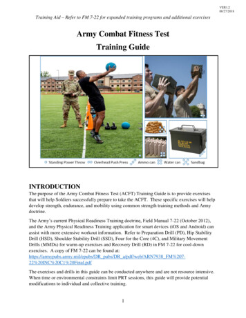
Evidence-Based Complementary and Alternative Medicine(a)3(b)(c)Figure 1: The variations of Tree pose, performed while the participant is instrumented for biomechanical analysis. (a) TreeWF: Tree posewith hands on wall and contralateral foot on the ground; (b) TreeW: Tree pose with hands on wall; (c) Tree: free-standing, single-supportTree pose.final biomechanics study visit, during which biomechanicsdata on all 3 versions of each pose were obtained.The introductory version of Tree pose consisted ofstanding on the dominant limb (the limb with which onewould kick a ball; we refer to this as the stance limb) andplacing the contralateral foot on the floor, with toes andforefoot in contact with the floor and the heel resting justabove the contralateral ankle. The participant’s hands lightlytouched a wall (TreeWF, Figure 1(a)). In the intermediateversion of Tree, participants placed their contralateral footon the medial aspect of the support-limb shank, just abovethe ankle (Figure 1(b)) and again used the wall for support(TreeW). The advanced version of Tree approached theclassic posture; it was the same as version 2, but the wallsupport was not used (in the classical version of Tree, thecontralateral foot is placed on the mid-to upper thigh) (Tree,Figure 1(c)).OLB was also a single-limb-stance pose in which theparticipants stood on their dominant limb. The introductoryOLB variation included the use of 2 or 3 standard yoga blocks(OLBb; block height 20 or 30 cm depending on participantheight). Here the subjects placed their contralateral foot onthe blocks with their knee flexed to 90 (Figure 2(a)). Theintermediate version of OLB included the use of a chair(OLBc). Here, the participants flexed their contralateral hipand rested their extended limb on the seat of a standardfolding chair (seat height 44.5 cm; Figure 2(b)). Theadvanced version of OLB was a free standing pose with thecontralateral limb lifted anteriorly with a flexed hip (OLB).They also lifted their arms above their head by flexing theirshoulders to approximately 180 (Figure 2(c)).2.3. Biomechanical Assessments. Biomechanical analysis wasperformed at the USC Musculoskeletal BiomechanicsResearch Laboratory using standard techniques [13]. Wholebody kinematic data were collected using an eleven-cameramotion capture system at 60 Hz (Qualisys Tracking Systemwith Oqus 5 cameras; Qualisys, Gothenburg, Sweden).Reflective markers were placed on a head band and overthe following anatomical landmarks of the lower and upperextremities bilaterally: first and fifth metatarsal heads, malleoli, femoral epicondyles, greater trochanters, acromions,greater tubercles, humeral epicondyles, radial and ulnarstyloid processes, and third metacarpal heads. Markers werealso attached to the spinous process of the 7th cervicalvertebrae (C7), jugular notch, L5/S1, bilateral iliac crests, andbilateral posterior superior iliac spines, in order to definethe trunk and pelvis. Based on these markers, a total of 15body segments were modeled, including the upper arms,forearms, hands, head, trunk, pelvis, thighs, shanks, and feet.Noncollinear tracking marker plates were placed on each ofthese segments to track segmental position during the poses,using previously documented procedures [14, 15].Once instrumented, the subjects performed the posesequences, while guided by their instructor. The sequenceof the poses was the same as when it was carried out in theregular yoga classes. A firm but portable clear plexiglass wall,which permitted capture of the markers, was positioned forwall support in the lab visits. For each pose, the participantwas instructed to begin in a starting position, move smoothlyinto the pose, hold the pose while taking one full breath, andthen return back to the original position. Simultaneously,the instructor also performed each pose in order to providevisual cueing. Once the participant had moved into thepose position, the instructor provided a verbal cue to theresearch associate to initiate the 3-second data collection.Two successful trials of each pose version were collected, andall 3 seconds of each pose were used for the analyses.
4Evidence-Based Complementary and Alternative Medicine(a)(b)(c)Figure 2: The variations of One-Leg Balance (OLB) pose, performed while the participant is instrumented for biomechanical analysis. (a)OLBb: double-support OLB pose with contralateral foot on the yoga blocks and knee flexed; (b) OLBc: double-support OLB pose withcontralateral foot on a chair and knee extended; (c) OLB: free-standing, single-support OLB pose.The subjects also completed 2 successful walking trialsat their self-selected “comfortable speed.” The walking trialsprovided a reference condition [16–18]. That is, walking is awell-studied, stereotypical activity and the JMOFs measuredduring comfortable-pace walking could be compared to theJMOF’s generated by the 2 yoga poses.GRFs were measured from a force platform at 1560 Hz(AMTI, Watertown, MA). Qualisys Track Manager Software(Qualisys, Gothenburg, Sweden) and Visual 3D (C-motion,Rockville, MD) were used to process the raw coordinate dataand compute segmental kinematics and kinetics. Trajectorydata was filtered with a fourth-order zero lag Butterworth12 Hz low-pass filter. In Visual 3D, the head was modeledas a sphere, the torso and pelvis as cylinders, and theupper and lower extremity segments as frusta of cones. Thelocal coordinate systems of body segments were derivedfrom the standing calibration trial. Joint kinematics werecomputed based upon Euler angles with the following orderof rotations: flexion/extension, abduction/adduction, internal/external rotation. The principle moments of inertia weredetermined from the subject’s total body weight, segmentgeometry, and anthropometric data. Using standard inversedynamics techniques, along with the International Society ofBiomechanics recommended coordinate systems, net JMOFsin the sagittal and frontal planes, for the ankle, knee, andhip, were calculated from the inertial properties, segmentalkinematics, and GRFs [19, 20]. JMOFs were normalizedto each subject’s bodyweight in kg. Additionally, a supportmoment, calculated as the sum of the ankle, knee, and hipsagittal plane JMOFs, was determined for each pose [17,21].These instrumentation and data-processing techniqueshave previously been used in our laboratory to assess exerciseperformance with high reliability (Cronbach’s alpha 0.98;[22]).Surface electromyographic (EMG) signals of the lowergluteus medius, hamstrings, vastus lateralis, and gastrocnemius muscles were collected on the subjects’ dominantlimb at 1560 Hz using active surface electrodes (Motion LabSystems, Baton Rouge, LA). Standard procedures, includingpreparation of the skin and electrode placement, wereemployed [23]. The obtained EMG signals were amplified( 1000), notch filtered at 60 Hz, and band-pass filteredat 20–500 Hz. A root mean square smoothing algorithm[24] with a 75 ms constant window was used to smooththe EMG data over the 3-second data collection periodcorresponding to the epoch of kinematic and kinetic data.EMG processing and smoothing were performed usingMATLAB (MathWorks, Natick, MA). The study protocol wasapproved by the Institutional Review Boards at the Universityof Southern California (USC) and University of California atLos Angeles (UCLA), and all subjects provided their writtenconsent to participate.2.4. Statistical Analysis. The JMOFs of the stance limb ankle,knee, and hip joints, in the sagittal and frontal plane,were the primary dependent variables. These were averagedover the collected period and both repetitions. Secondarydependent variables included the EMG activity, which wasalso averaged across the 2 trials. Repeated measures ANOVA(1 group 3 tasks) were used as omnibus tests to identifysignificant differences in the JMOFs and EMG results foreach dependent variable within each pose group (Tree andOLB). Tukey’s post hoc tests were used to examine thepairwise comparisons between tasks in each group when theANOVA tests were significant. Additionally, Cohen’s d effectsizes (small d 0.2; medium d 0.5; large d 0.8) arereported for all statistically significant post hoc comparisons[25]. Statistical analysis was conducted via PASW Statistics
Evidence-Based Complementary and Alternative Medicine5Table 1: Lower extremity peak net joint moments of force duringthe stance phase of gait at a self-selected walking speed.1.2 HipKneeAnkleSagittal planeExtensorFlexor0.81 0.060.54 0.030.63 0.040.30 0.031.33 0.04‡0.29 0.06†Frontal planeAbductor0.87 0.040.38 0.030.09 0.01Adductor0.08 0.010.09 0.010.19 0.02Mean standard error.‡ Plantar flexor moment.† Dorsiflexor moment.1Support moment (Nm/kg)Moments (Nm/kg) 0.8 0.60.40.218 (IBM SPSS Statistics, Armonk, NY), and significance levelwas set at P 0.05.0Tree3. Results3.1. Self-Selected Walking JMOFs. The average peak JMOFsacross the self-selected walking trials are presented in Table 1.These JMOFs generated during walking provide a metricagainst which the pose JMOFs can be compared.OLBYoga poseVariationsTreeTreeWTreeWFOLBOLBcOLBbFigure 3: Magnitude of support JMOF during variations of Treeand OLB. The whiskers represent standard errors ( P 0.05).3.2. Tree Support JMOF. The repeated measures ANOVAtest identified a significant difference in the support JMOFamong the 3 Tree variations (F2,38 36.12; P 0.001).Pairwise comparisons (post hoc analysis) revealed that thesupport JMOF during the classical Tree pose (the advancedversion) was 30% (P 0.001, d 0.65) greater than Treewith wall support (TreeW, the intermediate version) and103% greater than Tree with toes touching the floor andwall support (TreeWF, the introductory version) (P 0.001,d 1.76; Figure 3). Additionally, the TreeW support JMOFwas 57% greater than TreeWF (P 0.001, d 0.83).Tree versions in gastrocnemius activation level (F2,38 68.29;P 0.001). Average gastrocnemius activation during theclassical Tree was 123% greater than Tree with wall support(TreeW; P 0.001, d 1.29) and 324% greater thanTree with toes touching the floor and wall support (TreeWF;P 0.001, d 1.94). Gastrocnemius activation duringTreeW was 90% greater than that during TreeWF (P 0.052,d 0.91).3.2.1. Tree Sagittal Plane JMOFs and EMG. Figure 4 illustrates comparisons of the JMOF at the hip, knee, and ankle inthe sagittal plane across Tree variations. Repeated measuresANOVA identified significant differences in hip flexor andankle plantar flexor JMOFs (F2,38 13.36 and 29.40, resp.;P 0.001), but not the knee extensor JMOF (F2,38 0.795; P 0.46). Pairwise comparisons revealed that theaverage hip flexor JMOF generated during TreeWF was 188%greater than classical Tree (P 0.001, d 0.70) and 268%greater than TreeW (P 0.001, d 0.83). There wasnot a statistically significant difference between the 2 singlesupport Tree versions (Tree and TreeW; P 0.88). Regardingthe ankle plantar flexor JMOF, the pairwise comparisondemonstrated that the traditional Tree induced a JMOF thatwas 31% greater than TreeW (P 0.001, d 0.92) and 66%greater than TreeWF (P 0.001, d 1.77); while TreeWwas 27% greater than the introductory TreeWF (P 0.01,d 0.75).Surface EMG signals from the gastrocnemius muscleswere used to support the sagittal-plane JMOFs at the ankle(Table 2). Significant differences were identified across the3.2.2. Tree Frontal Plane JMOFs and EMG. In the frontalplane, the overall statistical analyses identified significantdifferences across the 3 Tree variations at each LE joint(F2,38 50.05, 36.44, and 19.03 for, hip, knee, andankle, resp.; P 0.001). Pairwise comparisons revealedthat the 2 single-support Tree poses (Tree and TreeW)engendered similar hip abductor JMOFs (P 0.98) whichwere approximately 55% greater than the JMOF engenderedduring the introductory version TreeWF (P 0.001, d 1.90). Complementing the frontal-plane JMOF findings, theclassical Tree and TreeW poses generated higher averagegluteus medius EMG signals (85% and 54%, resp.) thanTreeWF. However, statistical significance in the EMG levelwas only found in the difference between the classical Treeand TreeWF (P 0.007, d 0.60), and only a trend wasidentified between TreeW and TreeWF (P 0.12). Gluteusmedius activation also did not differ between the traditionalTree and TreeW (P 0.46). Similar to findings related tothe hip joint, the classical Tree and TreeW produced kneeabductor JMOFs which were 54% and 71% greater than theJMOF generated by the TreeWF, respectively (P 0.001,
Evidence-Based Complementary and Alternative MedicineJMOF ( : extensor/plantar flexor; : dorsiflexor) (Nm/kg)6Table 2: Average EMG activity of lower extremity muscle groupsduring Tree poses. 0.2EMG (mV)0 0.2TreeWTreeGluteus Medius 17.35 3.26Hamstrings11.96 2.63Vastus lateralis 62.22 15.0226.64 4.4624.70 7.3677.37 20.4632.15 7.1348.05 10.60101.47 23.7124.33 3.7546.21 6.63103.12 12.31GastrocnemiusTreeWFMean standard error. 0.4 0.6 0.8 1HipKneeJointAnkleVariationsTreeTreeWTreeWFFigure 4: Mean JMOFs in the sagittal plane during Tree variations.The whiskers represent standard errors ( P 0.05).d 1.12 and 1.43; Figure 5). All 3 Tree variations producedankle invertor JMOFs. The classical Tree ankle invertorJMOF was 40% greater than that generated during TreeW(P 0.006, d 0.39) and 118% greater than that generatedduring the introductory version, TreeWF (P 0.001, d 0.87).3.3. OLB Support JMOF. Similar to the Tree findings, therepeated measures ANOVA test identified that the supportJMOF differed statistically among the 3 OLB variations(F2,38 97.00; P 0.001). Pairwise comparisons revealedthat the support JMOF during the classical OLB pose (theadvanced version) was 106% greater than OLB with foot ona chair (OLBc, the intermediate version; P 0.001, d 2.48)and 137% greater than OLB with foot on blocks (OLBb, theintroductory version; P 0.001, d 2.90). There was nota significant difference in support JMOF between OLBc andOLBb (P 0.36).3.3.1. OLB Sagittal Plane JMOFs and EMG. Comparisons ofJMOFs at the hip, knee, and ankle in the sagittal plane areillustrated in Figure 6, and EMG data for the OLB versionsare presented in Table 3. Repeated measures ANOVAs identified statistical differences among OLB variations in sagittalplane JMOFs at all 3 LE joints (F2,38 92.55, 36.10, and54.69 for hip, knee, and ankle, resp.; P 0.001). At thehip, an extensor JMOF was generated during the traditionalOLB; whereas a flexor JMOF was generated during the OLBbwhich was 135% greater than the flexor JMOF generatedduring the OLBc (P 0.02, d 0.72). At the knee,Table 3: Average EMG activity of lower extremity muscle groupsduring OLB poses.EMG (mV)OLBbGluteus medius 19.34 2.95OLBcOLB30.95 4.6744.28 6.38HamstringsVastus lateralis21.49 8.2978.20 15.0329.47 6.95106.89 20.4094.78 13.5586.92 12.50Gastrocnemius41.37 6.3249.48 5.16117.72 12.33Mean standard error.the classical OLB generated a flexor JMOF, whereas similarextensor JMOFs were generated with the 2 double-supportOLB variations (OLBb and OLBc) (P 0.74). At the ankle,pairwise comparisons revealed that the plantar flexor JMOFassociated with the traditional OLB was 85% greater thanOLBb (P 0.001; d 2.17) and 91% greater than OLBc(P 0.001; d 2.34). The plantar flexor JMOF did notdiffer between OLBb and OLBc variations (P 0.94).These ankle plantar-flexor results were supported by thegastrocnemius EMG data. The classical OLB pose inducedsignificantly greater gastrocnemius muscle activation thanthe intermediate version of OLB with foot on a chair (OLBc;138%; P 0.001, d 1.61) and the introductory version ofOLB with foot on blocks (OLBb; 185%; P 0.001, d 1.74).No significant EMG difference was found between the OLBcand OLBb (P 0.605).3.3.2. OLB Frontal Plane JMOFs and EMG. In the frontalplane, the JMOFs generated at each LE joint differedstatistically among the 3 OLB variations (F2,38 80.66,21.92, and 11.94 for hip, knee, and ankle, resp.; P 0.001).The traditional OLB induced a hip abductor JMOF whichwas 17% greater than that induced during the intermediateversion, OLBc (P 0.001, d 0.82), and 53% greater thanthat produced by the introductory version, OLBb (P 0.001,d 2.24) (Figure 7). A higher gluteus medius activationwas also induced by the traditional, single-support OLB posecompared to OLBc (43%, P 0.003, d 0.53) and OLBb(129%, P 0.001, d 1.12). Complementing the significantdifferences (31%) found in the hip abductor JMOF betweenthe 2 double-support OLB variations (OLBc and OLBb; P 0.001, d 1.22), gluteus medius activation during OLBcwas 60% greater than that during OLBb (P 0.009, d 0.66). At the knee, the pairwise comparison revealed thatthe advanced OLB and intermediate OLBc poses producedabductor JMOFs which were 76% (d 0.91) and 55% (d 0.75) greater than that produced during the introductory
Evidence-Based Complementary and Alternative Medicine7 1.2JMOF (abductor/invertor) (Nm/kg) 10.8 0.6 0.4 0.2 0HipKneeJointAnkleVariationsTreeTreeWTreeWF 1.2 0.2JMOF (abductor/invertor) (Nm/kg)JMOF ( : extensor/plantar flexor; : dorsiflexor) (Nm/kg)Figure 5: Mean JMOFs in the frontal plane during Tree variations. The whiskers represent standard errors ( P 0.05).0 0.2 0.4 0.61 0.8 0.60.4 0.2 0.8 ntAnkleVariationsOLBOLBcOLBbFigure 6: Mean JMOFs in the sagittal plane during OLB variations.The whiskers represent standard errors ( P 0.05).Figure 7: Mean JMOFs in the frontal plane during OLB variations.The whiskers represent standard errors ( P 0.05).version, OLBb (P 0.001). There was not a difference inknee abductor JMOF between traditional OLB and OLBc(P 0.21). At the ankle, the invertor JMOF produced duringthe traditional OLB pose was 23% greater than that producedduring OLBc (P 0.007, d 0.46) and 38% greater thanthat produced during OLBb (P 0.001, d 0.68). Nodifference was observed between OLBb and OLBc (P 0.27).
84. DiscussionIn this study, we quantified the JMOFs and average EMGactivity associated with 3 versions of yoga poses that aretraditionally done standing on one leg: Tree and OneLeg Balance. The 3 versions of each pose progressivelyincreased the difficulty level of performing each versionfrom introductory, through intermediate, to advanced. Byconducting biomechanical tests during the performance ofeach version of Tree and One-Leg Balance, we formallyevaluated whether the poses were indeed becoming morephysically demanding as hypothesized.Our findings, which were not always intuitive, demonstrated that, within each pose, the different variationsengendered significantly different JMOFs and EMG activity,in some but not all joints, planes of motion, and musclegroups. Thus, our general hypothesis that the introductoryposes would generate the smallest physical demands, theintermediate versions would generate intermediate demands,and the most advanced versions would generate the greatestphysical demands, in a uniform fashion that is across all LEjoints and planes of motion, was not upheld. Moreover, wewere surprised to find that within a given pose type (e.g.,One-Leg Balance) some pose variations generated JMOFs inopposite directions (i.e., hip flexor versus hip extensor) andthus
and therapists when selecting pose modifications for their yoga participants. 1.Introduction Yoga is a popular exercise activity for older adults, with senior participation in the US estimated at greater than 900,000 participants—an increase of 90% between years 2007 to 2010 [1]. Likely contributing to this rise in par-