Transcription
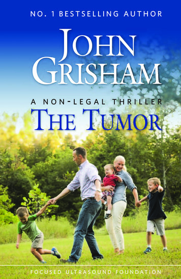
NO. 1 BESTSELLING AUTHORJOHNGRISHAMA NON-LEGALTHRILLERTHE TUMORF O C U S E D U LT R A S O U N D F O U N D AT I O N
THE TUMORA NON-LEGALTHRILLERJohn GrishamFocused Ultrasound Foundation . Charlottesville VA
Focused Ultrasound Foundation1230 Cedars Court, Suite 206Charlottesville VA 22903fusfoundation.orgText: 2015–2020 John GrishamMedical Illustrations: 2015–2020 Anatomical Justice, LLC,pp. 16, 17, 18 (fig. 6 & 7)All rights reserved under International andPan-American Copyright Convention.ISBN 978-1-4951-7940-22JOHN GRISHAM
4Dear Reader6 Chapter 1The Patient9 Chapter 2The Tumor15 Chapter 3The Treatment24 Chapter 4The End29 Chapter 5The Alternative34 Chapter 6The End—Revised Version37 Chapter 7The Present44Focused Ultrasound Foundation46The AskTHE TUMOR Focused Ultrasound Foundation3
Dear ReaderMy knowledge of medicine and medicalresearch is quite limited. When I wasa student, I drifted away from scienceand math, preferring instead subjectsI considered less demanding. I eventuallymade it to law school and became alawyer. After a brief career suing people(never a doctor, though), I stumbledupon fiction and wrote a couple ofbooks. Others followed, and I happilyshuttered the law office. Because thebooks have done well, I have been luckyenough to dabble in philanthropy. Once you get the reputation of beinggenerous, a lot of opportunities present themselves.Seven years ago, my friend and neighbor, Neal Kassell, gave a PowerPointpresentation on focused ultrasound therapy. Neal is a prominent neurosurgeonwho’s spent his career drilling through skulls and making repairs to brains.During the PowerPoint, Neal, with great enthusiasm, explained thatfocused ultrasound therapy could one day alleviate the need for conventionalbrain surgery. Tumors would be destroyed using beams of ultrasoundenergy, and afterward the patient would walk out of the operating roomand go home. Not only would the treatment be non-invasive, painless,quick, and relatively inexpensive, it could also save the patient’s life.Focused ultrasound therapy is still in its early stages, still experimental, butthere is enough research to date to be very optimistic.4JOHN GRISHAM
The brain is just the starting point. Tumors in the breast, prostate, pancreas,liver, kidneys, and bones could be treated on an out-patient basis. Neal loves touse the example of a man with prostate cancer undergoing focused ultrasoundtherapy, then driving himself back to the office for a few hours. Later, he goeshome to celebrate his wedding anniversary with his wife. They share a champagnetoast to growing old together.This is not science fiction. Around the world, more than 70,000 men withprostate cancer have been treated with focused ultrasound. Nearly 100,000women with uterine fibroids (benign tumors of the uterus) have been treated,thus avoiding hysterectomies and infertility. Clinical trials for tumors of thebrain, breast, pancreas, and liver, as well as Parkinson’s disease and arthritis,are inching forward at over 360 research sites around the world.Though focused ultrasound technology is in its infancy, there is great enthusiasmfor its potential to improve quality of life and decrease cost of care. For thispotential, to be fully demonstrated, additional laboratory research and clinicaltrials are necessary.But progress is too slow. There are barriers from regulators, insurance companies,even many in the medical field.I have found no other cause, issue, non-profit, or charity that can potentiallysave so many lives. One day in the not-too-distant future, you or someoneyou love will be diagnosed with a tumor. After the shock, you will think offocused ultrasound.Let’s hope it’s available.THE TUMOR Focused Ultrasound Foundation5
Chapter 1The PatientMeet Paul, a 35-year-old banker with a lovely wife, Karen, and three smallchildren. They enjoy a nice life in the suburbs with lots of friends andthe usual activities—backyard cookouts, swim parties, tee-ball, church onSundays. They are active and enjoy great health. Paul’s parents are intheir 60s and also very healthy. Paul gets a complete physical once a year,jogs twenty miles a week, plays golf and tennis at a nearby club, andavoids extra pounds. He has an occasional beer, doesn’t smoke, and takesno medication.6JOHN GRISHAM
Chapter 2The TumorBut Paul has a problem. He has a tumor in the right frontal lobe of hisbrain, about the size of a hen’s egg.Looking back, the first symptom was a gradual decrease in his ability toconcentrate at work. Naturally curious and active, he noticed anuncharacteristic tendency to procrastinate. At times he felt listless and tired.Then the headaches arrived, and with a fury. He blamed them on stressand took lots of ibuprofen. As he drove to work one morning, his visionbecame so blurred he stopped the car. Karen began to notice mood swingsand a loss of patience with the kids. He grew more irritable, both athome and at the office. His boss chastised him for barking at a coworker.He quarreled with Karen over his dour moods and crankiness. She knewsomething was changing with her husband and urged him to see a doctor.He refused.On a Wednesday morning, as Paul is in the bathroom shaving, Karen hearsa loud thump. She finds him on the floor, shaking in a full-blown grandmal seizure. She calls 911, and as she waits the seizure stops and hegradually awakens. He is confused, disoriented—doesn’t recognize Karenand doesn’t know where he is. The rescue squad arrives. Paul is loadedinto an ambulance and taken to the hospital. In the emergency room,he is still drowsy and confused and complains of weakness on his left side.Upon examination, his left hand is very weak and he has difficulty lifting hisleft arm and leg. An MR scan reveals the tumor.He is admitted to the hospital and started on anticonvulsant medicationto prevent further seizures, as well as steroids to decrease the swelling inhis brain around the tumor. Paul and Karen are not shown the MR scan.A neurosurgeon is consulted.THE TUMOR Focused Ultrasound Foundation9
By Thursday morning, the confusion and disorientation are gone, as is theweakness in his left side. He feels much better, briefly, but things willchange. When the neurosurgeon arrives early that morning for the initialconsultation, he produces the MR scan (opposite). As they stare at it,Paul and Karen are too stunned to speak. The doctor explains that Paulindeed has a tumor in his brain and it appears to be the type known asa glioma. Surgery is needed to remove as much of it as possible and toobtain tissue to determine the type of tumor.They talk about the operation. The doctor covers the risks. For complicationslike death and paralysis, the risks are very small. The most likely complicationwill be a weakness on the left side. The surgery will take about three hours,and if all goes well, Paul can expect to go home in three days.10JOHN GRISHAM
MR scan of Paul’s tumorThe neurosurgeon explains that gliomas are graded one through four, withone and two being benign. Three and four are malignant. Grade four,the most catastrophic, is called a glioblastoma. The life expectancy fora grade four diagnosis is short. Regardless of treatment—surgery, chemo,radiation—the average length of survival is about one year. Left untreatedbut managed with pain medication only, the patient can expect to liveseveral months. About 22,000 Americans are diagnosed each year withglioblastomas; 15,000 die within 12 months. The lucky ones, about onein ten, live for five years.Based on the MR scan, the neurosurgeon thinks the odds are about 50–50that the tumor is benign.THE TUMOR Focused Ultrasound Foundation11
He recommends surgery at the earliest convenient time and it is scheduledfor the following Monday. After the doctor leaves, Paul and Karen attemptto come to grips with what’s happening. Should they get another opinion?It seems senseless when staring at the MR scan. There is no doubt aboutthe tumor. They like their neurosurgeon and a quick search online proveshe’s one of the best. They are in the finest hospital in the city. Surgery isneeded sooner rather than later. There is no time to waste.Needless to say, the weekend is long and agonizing. Karen gives the badnews to the family but not to their children. She refuses to believe thetumor is malignant and is convinced the surgery will go well.She spends hours online gathering frightening and depressing informationabout brain tumors. Ted Kennedy, Susan Hayward, Beau Biden, LeeAtwater, George Gershwin, Lou Rawls, Bobby Van, Pete Rozelle, WilmaRudolph—they are just a few of those who died from a glioblastoma.On average, they survived about 12 months after being diagnosed.OppositeWell-known people who died from aglioblastoma. On average, they surviveda year after diagnosis.top, left to rightLee Atwater, 40Wilma Rudolph, 54middle, left to rightGeorge Gershwin, 38Edward “Ted” M. Kennedy, 77bottom, left to rightJoseph Robinette “Beau” Biden, III, 46Susan Hayward, 5712JOHN GRISHAM
Chapter 3The TreatmentAt 6 a.m. on Monday, Paul and Karen are in his hospital room, wide-awake,fearful but trying to appear brave as they wait for the day to unfold.At 6:30, Paul is prepped for surgery and two orderlies arrive with a gurneyfor the short ride to the operating room. Paul hugs his wife, who’sovercome with emotion. After he’s gone, she is led by a nurse to the waitingroom. His parents are already there. The nurse tells them the surgeryshould be over around noon. As they settle in for a long morning, theroom begins to fill with other anxious families.Paul is rolled into the operating room and put to sleep with general anesthesia.THE TUMOR Focused Ultrasound Foundation15
His head is shaved and his skull fixed in a three-prong headrest toimmobilize it (fig. 1).A question-mark-shaped incisionis made from the midpoint ofhis forehead just below thehairline to a point in front of hisright ear.Burr holes are made in the skull,and a power saw is used to fashiona bone flap more or less like thetop of a cookie jar (fig. 2).The dura mater—a membranebetween the inner table of theskull and the brain—is cut (fig. 3).The surface of the frontal lobe isdiscolored and distorted because1of the tumor.The tumor is localized with an intraoperative navigation device tominimize damage to the motor cortex—the portion of the braincontrolling movement of the left side of the body. Under themagnification of an operating microscope, an incision is made into thebrain, and just beneath this the abnormal tissue is identified.A portion is cut out and sent to the pathology lab for a preliminarydiagnosis (fig. 4). The tumor is removed by suction and thebleeding is controlled with coagulation (fig. 5).16JOHN GRISHAM
2345THE TUMOR Focused Ultrasound Foundation17
67The surgeons are able to remove everything that appears abnormal.They then suture the dura mater, secure the bone flap with screws (fig. 6),and staple the skin flap (fig. 7).After three hours of surgery, they are confident things went as well aspossible. Paul is taken to recovery.The surgeon goes to the waiting room and meets with Karen and Paul’sparents. He reports that everything went as expected: the tumor wasremoved, there were no complications, and they should be able to see Paul18JOHN GRISHAM
in about an hour. The initial results of the biopsy are not good—it looks likea glioblastoma, but it will take several days for the final report.Meanwhile, Paul is waking up,and there are problems. He hasprofound weakness in the left sideof his body. He cannot lift his leftarm or leg off the bed. He has onlya flicker of motion in his fingers.The surgeon immediately ordersa CT scan to make sure theweakness is not caused byhemorrhaging or a blood clot inhis brain from the surgery. It is not.The CT scan is unremarkable.Early Monday evening, Paul andKaren meet with the neurosurgeon,who has a preliminary pathologyreport; the final one is a fewdays away. While the tumor wassuccessfully removed, it is the typePost-operative MR scanof tumor that will likely recur.The next day another MR scan confirms that there was total removal of allvisible tumor, a rare bit of good news (above).Chemotherapy and radiation will be necessary to slow its regrowth. As for theweakness in Paul’s left side, the doctor says it is undoubtedly the result ofsurgical manipulations and should get better.By Tuesday morning, the weakness has improved slightly. Paul is able to lifthis arm but movements of his fingers are slow and his grip is weak. He is ableto stand but can walk only with assistance. Karen stays by his side as the hoursdrag on. He wants to discuss what’s on his mind: death, life insurance, his lastTHE TUMOR Focused Ultrasound Foundation19
will and testament, their savings, her future, the kids’ futures. Karen, though,is simply not ready for this. She doggedly maintains a veneer of optimism.To Paul, it seems more like denial. She tells him that a lot of friends are eagerto stop by for a visit, but he says no. He looks awful, feels worse, and wantsto see no one, not even his own children.Early Wednesday morning, one week after his seizure, they meet again withthe neurosurgeon. The pathology report confirms their worst fears:glioblastoma, grade four. Although the tumor has been removed, it leftbehind microscopic portions that extend into the normal brain and cannot besurgically removed. These remnants of the tumor will almost certainly regrow,and must be treated with radiation and chemotherapy. When the tumorreturns, there will be the likelihood of more surgery.With as much professional sympathy as possible, the doctor tells them that,according to statistics, Paul can expect to live 12 to 14 months. Occasionallya patient will live 5 to 10 years, but that’s uncommon. He offers his usual,“Hope for a miracle, but plan for the average.”“Plan for the average,” Paul repeats after the doctor leaves. Karen pulls theshades and turns off the lights. They sit in the darkness, holding hands, asthe monitors beep occasionally. When they speak, they discuss the best wayto tell the children.Thursday morning, the radiation oncologist stops by. He describesradiation therapy and explains that it will be administered five days a weekfor the next six weeks. Among other side effects, Paul will lose his hairand his face will swell and become disfigured from the steroids he’ll begiven. Later, the neuro-oncologist stops by and they discuss chemotherapy,which has its own set of unpleasant side effects.On Friday, Karen brings their children to the hospital to visit. She and Paulhave decided to wait a few months before delivering the bad news. They stillbelieve in miracles and are praying a lot. After the kids leave, Paul’s parentsOpposite, radiation therapy treatmentTHE TUMOR Focused Ultrasound Foundation21
visit him. With Karen home with the children, he does not pull punches.He’ll be lucky if he’s still living a year from now.Later, alone and in a dark room, Paul opens his laptop and pulls up acalendar for the next 12 months. It’s all there, all planned: the school year,their upcoming vacation, the holidays and birthdays, a golfing trip withhis friends, several business trips, his parents’ 40th anniversary. Would he beable to enjoy any of it? Would he even be alive? “Plan for the average”means he should be able to make it to Christmas. What does a father ofthree young children do to celebrate his last Christmas?Paul thinks about the next 12 months and asks himself many questions.There are no answers.Later that afternoon, he is transferred to a rehabilitation facility to address theweakness in his left side. He cannot raise his left hand to his face, nor can hewalk without a cane. Ten days after surgery, he is discharged and taken home.He is instructed to return to the rehab facility three times per week. His leftside continues to improve.Being at home lifts his spirits. Friends arrange meals and there is a steadyflow of traffic to the house. He tries to eat but his appetite is gone. Twoweeks after surgery, Karen drives him to the office where he’s greeted likea hero. He is determined to work at least half a day until he regains hisstrength, and he assures his colleagues he’ll be back. Paul begins radiationtherapy Monday through Friday, five days per week.His hair falls out rapidly from the radiation and, worse, his face begins toswell from the steroids (opposite). The moon face seems to grow each day.He looks terrible. He is constantly fatigued, and his thinking becomes slowand dull from the damage to his brain caused by the radiation.22JOHN GRISHAM
Before surgery and radiation therapyAfter surgery and radiation therapyHe gives up on the idea of going to the office. His boss promises to coverfor him and keep the paychecks coming. The group medical policy covers80 percent of the expenses.His lawyer drafts a new will, not that one is really needed. Paul and Karenown everything jointly; upon his death, it’s all hers anyway. She certainlygets the kids. His life insurance policy is for only 250,000. They haveabout 40,000 in savings. With three children under the age of eight,the future is anything but secure. Karen secretly begins checking outemployment opportunities on the internet. Their minister stops by everyother day for a devotion and prayer.THE TUMOR Focused Ultrasound Foundation23
Chapter 4The EndSix months later, the weakness in Paul’s left side increases dramatically.He cannot grasp objects with his left hand. He drags his left foot whenwalking and cannot move around without assistance. He notices he cannotconcentrate for more than a few seconds. His short-term memory is shot.An MR scan shows the tumor is back and growing rapidly (below).It also reveals damage in his braincompatible with the effects of radiation.His neurosurgeon offers the optionof another surgery to remove therecurrent tumor.Paul and Karen discuss this for severaldays. A second operation is morelikely to damage the brain. There is nocertainty that the tumor will not recuragain, and again. They are losinghope, and their thoughts of miraclesare fading rapidly. Paul could almostMR showing recurrent tumorthrow in the towel, opt for a few finalweeks with pain medication, andsuffer as little as possible to the end. Karen, though, still believes in luck.The second operation is similar to the first. The visible portions of thetumor are removed, and Paul’s skull is put back together. When he awakens,though, the weakness in his left side is much worse. He is transferred toa rehabilitation center. After three weeks of intense therapy, his conditiondoes not improve. He can no longer stand without assistance, nor walkwithout a walker. His left hand is essentially useless. He is discharged home,24JOHN GRISHAM
where he arrives in a wheelchair. The chemotherapy has been ineffectiveand is terminated. Paul takes steroids in an effort to reduce swelling inhis brain.At this point, Paul begins saying goodbye to his family and friends.He bravely accepts the fact that his days are numbered, and he wishes tosay farewell on his terms. As bad as he looks, he knows that things willonly get worse.In a heart-wrenching scene, he and Karen finally tell the children that theirfather is about to leave them.The steroids are not working and are cut off. He’s left with only somepowerful narcotics to deaden the horrible headaches, which occur withincreasing severity.THE TUMOR Focused Ultrasound Foundation25
Paul prays for a quick and painless end, but this doesn’t happen. He slowlydeteriorates and becomes increasingly confused and disoriented. He losesalmost all consciousness and his ability to move. He is bedridden andrequires around-the-clock care for feeding and bathing. Karen sleepwalksthrough the days and nights, thoroughly drained, but trying gamely toshield his condition from the children as much as possible. Eight monthsafter his seizure, Paul has completely checked out, but his heart still managesto beat. Karen finally begins praying for a merciful end.Nine months after the first surgery, he passes away, at the age of 36.The total cost of his treatment and care isapproximately 300,000.26JOHN GRISHAM
Chapter 5The AlternativePaul was born in 1980, ten years too early. Had he been born in 1990 anddiagnosed with a brain tumor at the age of 35, in 2025, his story could berewritten as follows:That same Wednesday morning, Karen hears a crash in the bathroom,and she finds Paul on the floor in a grand mal seizure. He’s taken to theER and admitted to the hospital. An MR scan is performed with molecularimaging, a more advanced scan than was available ten years earlier.Based on the scan, the neurosurgeon, with virtual certainty, makes a diagnosisof a glioblastoma and explains the prognosis and the treatment options,including focused ultrasound therapy. The size and location of Paul’s tumormake it amenable to treatment with focused ultrasound therapy, whichis what the neurosurgeon recommends. He explains that the tumor in allprobability cannot be cured and will return, but it can be controlled withrepeated treatment, giving Paul additional years with a high quality of life.Opposite, the focused ultrasound braintransducer fits over the head andemits beams of energy that penetratethe skull to target a tumor.THE TUMOR Focused Ultrasound Foundation29
12Early Friday morning, Paul and Karen walk into the focused ultrasoundtherapy suite (fig. 1). He changes into a gown, takes a light sedative, and ispositioned on his back on a table.His head is securely fixed in a hemispheric focused ultrasound brain transducer(fig. 2). The transducer is capable of transmitting more than 1,000 intersectingbeams of ultrasound energy through the scalp and skull to the tumor witha high degree of accuracy and without damaging the adjacent normal tissue.After the transducer is in place, Paul is inserted into the bore of the MR machine.30JOHN GRISHAM
3 45In the adjacent control room, the surgeon manually outlines the tumor,which is to be destroyed by the focused ultrasound beams (fig. 4). Paul isawake and feels no discomfort. Karen stays by his side, holding his hand andchatting with him. The treatment begins. The surgeon is in constant voicecontact with Paul and Karen (fig. 3). He uses continuous images from theMR scan to guide the precise point where the ultrasound is focused and tocontrol the delivery of its energy to the target (fig. 5).THE TUMOR Focused Ultrasound Foundation31
Before focused ultrasound therapyAfter focused ultrasound therapyIt takes about an hour to kill all of the tumor that can be seen on theMR scan. At the completion of the procedure, another scan is performed toconfirm the entire tumor was treated.While this is happening, powerful chemotherapy agents enclosed inmicroscopic nanoparticles are injected intravenously. These circulate withthe blood in every tissue and organ in the body, but the chemotherapydrugs are inactive because they are trapped inside the nanoparticles.After eliminating all of the tumor that can be seen on the MR scan, thesurgeon then refocuses the ultrasound to the surrounding brain to activatethe nanoparticles, which release their pharmacological payload in theprecise area around the tumor where residual microscopic extensions of thetumor have infiltrated. This allows very high concentrations of the drugsto be delivered focally to the brain while minimizing systemic side effects.The remainder of the chemo-laden nanoparticles will be excreted.32JOHN GRISHAM
Less than two hours after the treatment began, Paul gets off the table andwalks to the recovery room for observation. There are no complications,and he is discharged home Friday afternoon. He feels fine. The onlyaftershock is some residual clumsiness in his left hand from his initialseizure, which is decreasing. On Saturday afternoon, he and Karen and thekids walk down the street for a block party. They have yet to tell theirfamilies and friends about the tumor and the treatment. On Sunday, theentire family goes to church.On Monday, Paul is at the office before 8 a.m., eager to catch up after threedays off. He explains to his colleagues that he was in the hospital for “tests,”but everything is fine. He looks and feels like himself. The weakness in hisleft side continues to fade away.A month after the ultrasound procedure, he undergoes another MR scan.The scan reveals that the ablated tissue is being absorbed harmlessly byPaul’s body. The mass is not regrowing due to the efficacy of the focusedchemotherapy treatment that targeted the microscopic extensions of themalignant tumor. The deadened tissue continues to shrink.Three years later, Paul again notices difficulty using his left hand. His leftfoot occasionally drags. He does not hesitate and calls his doctor immediately.An MR scan is done and reveals that the tumor is back. The following day,as an outpatient, he undergoes another focused ultrasound procedure.The weakness goes away after a month. Four years later, seven years after hisinitial diagnosis, the tumor is growing again, and the procedure is done forthe third time.Cost to date: approximately 75,000Savings: about 225,000 and one life prolongedTHE TUMOR Focused Ultrasound Foundation33
Chapter 6The End—Revised VersionThe tumor will eventually take Paul’s life. However, focused ultrasoundtherapy could transform a fatal condition into one that is chronic butmanageable. In contrast to the best current treatment circa 2015, thefuturistic ultrasound therapy depicted here circa 2025 could potentiallybe accomplished on an outpatient basis without multiple days ofhospitalization, without surgery and its attendant risks of infection andcomplications like blood clots and brain damage, without the harmfuleffects of radiation, and with minimal side effects of chemotherapy due tofocused drug delivery. The net result could be a dramatic improvement inthe quality and longevity of countless lives, and decreased cost of treatment.How many years will it add? At this time, the answer is uncertain. Clinicaltrials for brain tumors have just begun, and the patients selected for thesetrials represent the most desperate of cases. Much more research is needed.Researchers believe, with caution, that five additional years are realistic.Perhaps ten.Who wouldn’t bargain for ten more years, especially with a high qualityof life?With time, research, and improved technology, neurosurgeons are hopefulthat a guy like Paul can live to the age of 45 or even 50, long enough to seehis children mature.34JOHN GRISHAM
Chapter 7The PresentFocused ultrasound is a new, revolutionary, groundbreaking, non-invasivetherapeutic technology that has the potential to transform the treatment ofa variety of serious medical disorders in addition to brain tumors, improveoutcomes, and decrease the cost of care. It could become an alternative to,or complement for, traditional surgery, radiation therapy, and drug delivery.Focused ultrasound could result in fewer complications, such as damage tonormal tissue, infection, hemorrhage, and pain, as well as shorten recoverytimes. By providing safer and more effective therapy, it could reduce death,disability, and suffering for millions of people around the world.Focused ultrasound utilizes intersecting beams of high-frequency soundconcentrated accurately and precisely on tissue deep in the body, much assunlight passing through a magnifying glass can be focused to burn a holein a leaf.THE TUMOR Focused Ultrasound Foundation37
Table 1Effects at focal pointVariety of effects,variety of disorders Thermal ablationprecise heating and destructionof tissue Focal drug deliverydelivery of very highconcentrations of drugs preciselywhere they are needed Blood-brain barrier openingtemporary access of drugs tothe brain Immunomodulationstimulation of immune responseallowing body to fight cancer Neuromodulationreversible stimulation orinhibition of cells in the brainand nervous system Radiation sensitizationsensitizing tumors to effects ofradiation allowing use of lowerdose to kill cancer cells Stem cell deliveryspecific “homing” of stem cellsto targeted tissue38JOHN GRISHAM
At the point where the beams converge, the ultrasound energy induces avariety of biological effects while surrounding structures and tissues remainundamaged (table 1).Magnetic resonance or ultrasound imaging is used to identify, guide, andcontrol the treatment in real time.In the theoretical example used in this all too common story, focusedultrasound treatment of the malignant brain tumor controlled but did noteradicate it. This alternative treatment provided years of high quality oflife to a patient with an ultimately fatal condition. The treatment of braintumors is not the only area of medicine in which focused ultrasoundtherapy shows promising results. There are many more applications,including uterine fibroids, prostate cancer, and essential tremor, wherefocused ultrasound treatment could potentially cure the disease.Today, focused ultrasound is in various stages of development for treatingover 120 diseases and conditions, including epilepsy, Alzheimer’s andParkinson’s diseases, and tumors of the brain, liver, pancreas, and lung(table 2). But despite the progress so far, much work remains to be donebefore focused ultrasound can be widely used to treat large numbersof patients.Unfortunately, it often takes decades for a new therapeutic technologylike focused ultrasound to become widely adopted as a mainstreamstandard of care.THE TUMOR Focused Ultrasound Foundation39
Table 2Status of progress by diseasePre-clinicalResearchClinical TrialsOutside ertensionAtrial fibrillationPeripheral artery diseaseVaricose veinsArteriovenous malformationsCardiac hypertrophyCongestive heart failureDeep vein thrombosisEssential tremorParkinson’s diseaseDepressionNeuropathic painOCDALS/Lo
liver, kidneys, and bones could be treated on an out-patient basis. Neal loves to use the example of a man with prostate cancer undergoing focused ultrasound therapy, then driving himself back to the office for a few hours. Later, he goes home to celebrate his wedding anniversary with his wife. They share a champagne toast to growing old together.