Transcription
VOLUME 21, NUMBER 12012200 University Ave. E.St. Paul, MN tOrganizationU.S. PostageP A I DTwin Cities, MNPermit No. 5388CHANGE SERVICEREQUESTEDPediatric Back Pain:When to Sit Up and Take NoteA Pediatric Perspective focuses onspecialized topics in pediatrics, orthopedics,neurology, neurosurgery and rehabilitationmedicine.To subscribe or unsubscribe fromA Pediatric Perspective, please send anemail to Publications@gillettechildrens.com.Back pain is uncommon among children who are under 10 years old, but the incidenceof back pain increases for adolescents. A 2005 study of 7542 European teenagersstates, “A total of 1180 (20.5%) teenagers reported one or more episodes of low backpain (LBP), of whom 900 (76.3%) had consulted a health provider.”1 Not surprisingly,athletes have a higher incidence of back pain than nonathletes.Copyright 2012. Gillette Children’s SpecialtyHealthcare. All rights reserved.For primary care providers, the key questions are—when is back pain the result ofoveruse or muscle strain? When is the pain symptomatic of more serious pathology,such as a herniated disc, spondylolysis, scoliosis, Scheuermann’s disease, osteomyelitis,discitis, leukemia, tumors, or ankylosing spondylitis? What follows is a practical guideto evaluating pediatric back pain, with recommendations about which cases should bereferred to an orthopedic specialist and which cases can be managed in the primarycare setting.To make a referral, call 651-325-2200 or855-325-2200 (toll-free).Start With the Basics – A Thorough HistoryTenner Guillaume, M.D., is an orthopedic surgeonwho specializes in spine surgery. His professionalNEWS & NOTESinterests include management of pediatriccongenital and idiopathic scoliosis as well asHas A New Lookisthmic spondylolisthesis. He received his medicaldegree from the University of Minnesota MedicalSchool. He completed an internship and residencyat the University of California – San FranciscoMedical Center and a spine surgery fellowshipthrough the Twin Cities Spine Center inMinneapolis. Guillaume has presented research,posters and abstracts and has professionalpublications. He is a member of the AmericanVisit www.gillettechildrens.org/MedicalStaffBios to learn more aboutGillette’s services and specialists.Clinical EducationFind videos and professional presentations.Go here to see a video of TennerGuillaume, M.D., conducting a back painhistory and physical.Our redesigned newsletter includes these new features: “Key Insights,” a summary on P. 1 Links and a QR code for quick access to relevant resources on the Gillette website.Although the newsletter has a new design, the editorial focus remains on providing apractical, in-depth discussion of clinical topics. We hope the newsletter’s new design willbe PCOMING CONFERENCESAcademy of Orthopaedic Surgeons and the NorthFor orthopedic surgeons, pediatric rehabilitation medicine specialists and allied practitionersAmerican Spine Society. He is a candidate for22nd Annual ConferenceClinical Gait Analysis: A Focus on InterpretationMay 21 – 23, 2012membership in the Scoliosis Research Societyand the Pediatric Orthopedic Society ofNorth America.Back Issues ofA Pediatric pectiveFor pediatric neurologists, pediatric neurosurgeons, pediatric rehabilitation medicine specialists and allied practitioners3rd Biennial Pediatric Neurosciences ConferenceMoving Forward in the Treatment of Pediatric Neurological DisordersMay 30 – June 1, 2012The answers to the following questions will provide nearly all of the informationneeded to differentiate a benign issue requiring conservative management from amore serious condition. The answers will also help you determine whether to simplytreat the symptoms or whether a complete radiographic study is necessary.1. Characterize the back pain.Is the pain acute? The result of trauma? Unremitting or more subtle? Has it grownprogressively worse? Is the pain recurrent, related to activity or worse at night? Isthe pain associated with constitutional symptoms, such as unintentional weight loss,fevers, chills or malaise?2. Determine the location of the pain.Establish whether the pain is lumbar, thoracic or cervical and whether it is focal,diffuse, radiating or radicular. The more focal the pain, the easier it will be to potentiallyidentify an underlying cause. Nonfocal, diffuse pain (“My whole back hurts” or “I can’tput my finger on it because it hurts everywhere”) is less likely to result in the discoveryof a focal underlying pathology with radiographic work-up. Back pain is surprisingly commonamong adolescents and youngathletes, but cases require furtherinvestigation to identify causes andtreatment recommendations.A thorough history and a comprehensive physical exam for back painprovide most of the informationneeded for a differential diagnosis.For initial screening, begin with AP,lateral or oblique view radiographs. AnMRI is needed only if there is evidenceof a serious condition or if the patienthas not improved after six weeks.See the guide on P. 2 for moredetails about which imaging studiesto request.Younger patients (ages 10 and under)who complain of back pain are moreworrisome—a pathologic abnormalityis more likely.Inside 3. Consider the patient’s age and check neurological signs.Is the patient 10 or younger? The younger the patient, the more worrisome the backpain, because a pathologic abnormality is more likely to be the cause. Also, ask abouttransient paraparesis, paralysis, numbness or paresthesias, and determine if bowel orbladder function is affected. Neurological impairment signals a more complex condition.4. Listen for these reassuring signs.Ask if the patient has missed school or sports because of the pain, whether the painhas improved over time, and if the pain responds to nonsteroidal anti-inflammatorydrugs (NSAIDs) or acetaminophen. Additionally, discuss whether the pain is intermittent or activity-related, generalized or focal.2012KEY INSIGHTSby Tenner Guillaume, M.D.Editor-in-Chief – Steven Koop, M.D.Editor – Ellen ShrinerDesigners – Becky Wright, Kim GoodnessPhotographers – Anna Bittner,Paul DeMarchiTenner Guillaume, M.D.Pediatric Spine SurgeonVOLUME 21, NUMBER 1 Guide to Imaging Studies forPediatric Back Pain, P. 2Differential DiagnosisOverview, P. 3Case Study - SuspectedScoliosis, P. 3 (inside flap)www.gillettechildrens.org
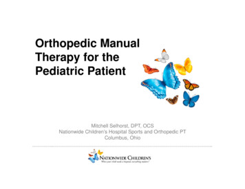
If the pain is decreasing, responds well to NSAIDs, is generalized or is activity-related, it is more likely to be the result ofmuscle strain or injury. Similarly, if the patient participates innormal activities, (i.e., pain does not prevent him or her fromparticipating), the back pain is less likely to be serious. Theexception would be a highly competitive athlete or dancerwho might be motivated to compete despite significant pain.An absence of all of the “red flags” listed below is also reassuring.5. Be aware of these red flags.Associated findings like those mentioned below are definitelycause for concern. Pursue additional testing if the pain is: Associated with constitutional symptoms like fever, malaise,night sweats, unintentional weight loss, easy bleeding orbruising Worse at night or if it wakes the patient from sleep Associated with neurologic symptoms Acute, unremitting or focal pain that is not responsive toNSAIDs or acetaminophen Constant (not activity-related)Complete a Comprehensive Exam for Back PainEvaluate patients while they are standing, walking, bendingat the waist and lying on the exam table. Include these exams:Standing and walking – Are the shoulders level? Does thepatient have any obvious abnormalities of the spine, hips orstance? Does the patient limp?Appearance – Look for rash, bruising or ecchymoses, whichare potential symptoms of leukemia. Check for cutaneousmanifestations of dysraphism: hairy patches, dimples and deepRefer patients suspected of having leukemia to a pediatriconcologist. If patients have any of these conditions, referthem to a pediatric spine specialist: Vertebral fractures Spondylolysis Discitis Scheuermann’s disease Apophyseal ring fracture Vertebral osteomyelitis Osteoid osteoma Disc herniation Spondylolisthesis Idiopathic scoliosis Ankylosing spondylitissinuses at the base of the spine (possible neural tube defect);café au lait spots (possible neurofibromatosis); heart-shapedbuttocks (possible high-grade spondylolisthesis); kyphosis.Palpation and inspection – Palpate to determine the locationand nature of the pain: focal, diffuse, in the spinous process orparaspinal. Have the patient do an Adam’s forward bend test,and inspect the spine for any deformity, such as scoliosisor kyphosis.Range of motion – Is the pain worse with spine flexion?Extension?Neurologic exam – Test sensory and motor responses. Checkthese reflexes: biceps (C5); triceps (C6); brachioradialis (C7);patellar (L3, L4); Achilles (S1); upgoing Babinski; Hoffman’s testof the upper extremity; and clonus. Also look for unilateral orbilateral weakness, asymmetric abdominal reflexes, foot drop, orloss of fine motor skills in the hands.Guide to Imaging Studies for Pediatric Back Painmetabolic activity or bone turnover. MRIs are currently the preferred test of choice among orthopedic surgeons and radiologists.Radiographs For trauma or a history that suggests the pain is not muscular,start with AP and lateral radiographs, rather than an MRI. Request a standing view (or seated view, if the patient cannotstand). For lumbosacral involvement, avoid pelvic shielding. Request an oblique view radiograph for suspected spondylolysis. Request flexion/extension view radiographs for suspectedspondylolisthesis.MRI Reserve MRIs for cases in which the back pain has lasted morethan six weeks and is not responding to NSAIDs or therapy. Consider an MRI if the patient’s history and physical reveal focalback pain or radicular pain. Consider an MRI if there are obvious red flags during historytaking that suggest possible underlying infection or neoplasm. Request an MRI if the preliminary radiographs point to a seriouscondition (acute stress reactions or fractures, spondylolysis,spinal neoplasms, discitis and so forth). If you are likely to refer the patient to a specialist, the specialistcan order an appropriate MRI.CT ScanReserve for cases that fail nonoperative management or forvisualization of bony changes that have resulted from fracture,tumor or infection. This can largely be left to the specialist to orderif it is indicated.Bone Scan/Scintigraphy/SPECT ScanBecause these tests are highly sensitive and nonspecific, they areused much less commonly today. They will identify areas of highInterpreting Imaging Results – What to Look ForOverview – What to Request and When2Refer Red Flag Issues ImmediatelyAnterior/Posterior Radiographs Check overall alignment – Any scoliosis? Pedicles – Normal cascade? Widening at any level? Clearlyidentifiable at all levels? Any effacement or cortical destruction? Spinous processes – Widening between spinous processes?Well-aligned? No offset in coronal plane?Lateral Radiographs Overall alignment – Lumbar lordosis, thoracic kyphosis,cervicothoracic lordosis? Posterior vertebral body cortical line – Is it aligned? Is there anterior or posterior offset of a vertabra (possiblespondylolisthesis)? Interspinous widening? Cervical spine posterior laminar line?Lumbar Oblique RadiographsLook for spondylolysis (the infamous “Scotty dog”).Flexion/Extension RadiographsCheck for atlantoaxial instability (cervical) or spondylolisthesis(lumbar).MRIExamine images for acute stress reactions or fractures,apophyseal ring fractures, cord abnormalities, discitis, herniatednucleus pulposus (HNP), juvenile degenerative disc disease,spinal neoplasms, spondylolysis.CT ScanExamine bony anatomy for suspected cases of vertebral, burst orpedicle fractures or of osteoid osteoma.Special tests – Include these additional exams, if you suspect: Spondylolysis – check extension in single leg stance andpopliteal anglesIf the patient has pain while standing on the right leg andextending the spine, but does not have pain when standing onthe left leg and extending the spine, presume a unilateral parsstress fracture/spondylolysis on the right. If the pain occursbilaterally, presume that the pars stress fracture/spondylolysisis bilateral. Herniated nucleus pulposus or apophyseal ring avulsion –straight leg raiseConsider the test positive if any degree of passive elevationreproduces pain radiating down the affected leg. The straightleg raise specifically tests nerve roots that contribute to thesciatic nerve (L4, L5, S1). For suspected femoral nerve rootinvolvement (L1, L2, L3), perform a femoral stretch test,because those nerve roots contribute to the femoral nerve. Cervical radiculopathy – Spurling’s testTurning the patient’s head in the direction of the suspectePrevalence of nonspecific low back pain in schoolchildren aged between 13 and 15years. Masiero S, Carraro E, Celia A, Sarto D, Ermani M., Acta Paediatr.2008;97(2):212HistoryPhysical ck pain, trauma.Boys girls. Parent may note “poorposture.” Pain at apex of kyphosis orlower lumbar spine. Aching pain, doesnot radiate or wake patient at night.Most patients do not complain of backpain, but apex curves left with spinalcord abnormalities.Loss of spine mobility, back pain, boys girls. Pain worse in morning.HistoryThe patient denied any history of back pain. However, he hada history of anxiety and insomnia and takes melatonin andsertraline. He had never missed school or extracurricularactivities because of back pain.PhysicalDuring the examination, he was alert, oriented and in no acutedistress. He had normal lordotic posture and mild evidence ofpositive sagittal imbalance. He had no evidence of shoulderheight imbalance. His gait appeared normal with no evidenceof ataxic or antalgic gait. He was able to toe walk, heel walk, andperform tandem gait maneuvers without significant difficulty.Skin examination of his back demonstrated no cutaneousmanifestations of underlying spinal dysraphism. During theAdam’s forward bend test, he had no evidence of any clearthoracic, thoracolumbar, or lumbar prominences. He did,however, have evidence of increased thoracic kyphosis bothin the erect position and even more so when bending forward.He had an acute thoracic kyphosis with an apex at themidthoracic spine. When he stood erect, he was able to retracthis scapulae somewhat to straighten his posture. Althoughthere was some evidence of correction of his kyphosis, someof the kyphosis seemed structural. He had no pain withlumbar extension.1Focal tenderness when palpated.Pain is worse with erect posture.Acute traumatic onset of pain. Symp- Positive single leg raise. Othertoms mimic a herniated nucleus pulpo- neurologic signs are presentsus with lower extremity radiation.infrequently.DiscitisMost common cause of back pain inFever, malaise. Pain is worse whenpatients who are 5 or younger.standing or with forward flexion.Back pain, limping, refusal to walk,Gowers’ sign is present. Patient isabdominal pain.systemically ill.VertebralBack pain, refusal to walk, fever, night Fever, malaise. Pain is worse whenOsteomyelitissweats.standing or with forward flexion.Gowers’ sign is present. Patient issystemically ill.LeukemiaBack pain (6 – 25 percent of patientsFever, ecchymoses, possibly focalpresent with it initially), pallor, fatigue, tenderness with palpation.malaise, anorexia, fever, bruising,abnormal bleeding.Osteoid Osteoma Most common benign spinal tumorPatient often stands with a list,in children. Severe back pain, worse at decreased spine range of motion.night, relieved by NSAIDs.Disc Herniation Back pain with radiation into legs.Straight leg test is positive for 85Pain worse with valsalva (sneezing,percent of patients.coughing, or straining).Absent reflexes, weakness andsensory changes rare in children.Spondylolysis or Patient is gymnast, offensive lineman, Pain with extension and single legSpondylolisthesis ballet dancer or diver. Low back pain is stance extension. Tight hamexacerbated by hyperextension.strings, increased popliteal angle.Scheuermann’sDiseaseThis tall, thin, 13-year-old male wasreferred by his primary care physicianfor a scoliosis evaluation. Originally, thepatient’s dermatologist noticed someprominence during a physical exam.The patient’s primary care physicianrequested radiographs, which indicateda 14-degree thoracolumbar curve.Follow In Clinic or Refer?The history and physical exam will help determine the severityand acuity of the patient’s back pain. If there are no red flagissues, send the patient to be evaluated by a physical therapistwho provides care for children and follow up with the patientin clinic. If the history and physical uncover red flag issues,request appropriate radiographs and lab tests. See Page 2for a guide to imaging studies. If screening radiographs pointto a serious orthopedic condition, request an MRI or referthe patient to an orthopedic specialist who will get thenecessary ealRing FractureSuspected Scolioiscervical radiculopathy while tipping and extending the neckin the same direction will cause foraminal compression. Thatcompression should reproduce the patient’s experience ofradiculopathy and pain radiating down the affected upperextremity.Kyphosis, particularly on forwardbending.Rigid kyphosis on extension orover bolster.Positive Adam’s forward bend test.May have focal tenderness overrib prominence.Loss of lumbar flexibility onAdam’s forward bend.Increased kyphosis, positiveFABER test.Imaging Studies Radiographs – AP and lateral views. CT scan – Evaluate specific anatomy of fracture. Radiograph – Look for osseous fragment posterior tovertebral body. CT scan – Identify size and location of bony fragment. Radiograph – Check for narrowing of disc space, softtissue swelling. MRI – Use to localize infection and determine softtissue involvement. Radiograph – Check for bony destruction, corticalscalloping. MRI with IV contrast – Determine extent of bony andsoft tissue involvement, possible abscess. Radiograph – Look for generalized osteopenia,compression fracture. CBC with differential, peripheral smear, erythrocytesedimentation rate. MRI, CT scans, SPECT scan.The examination of his bilateral lower extremities showed thathis motor strength, neurological signs and reflexes were allwithin normal limits. He was noted to have a popliteal angleon the right of approximately 75 degrees and on the left ofapproximately 70 degrees. His hamstrings were extremely tight.That tightness was associated with some spasm and pain whenhis knees were extended passively.Imaging Studies Radiograph – AP and lateral. MRI – Visualize HNP, rule out tumors or epiduralabscesses.Lateral radiographs were repeated, which showed thoracichyperkyphosis that measured from T1 to T12 at approximately70 degrees. He also had findings consistent with Scheuermann’sdisease from T6 to T9. He had anterior wedging of greater than10 degrees at each of these levels and more than 30 degrees ofkyphosis across these segments. There were also end plate irregularities suggestive of Schmorl’s nodes. This was consistent witha diagnosis of Scheuermann’s disease. His C7 plumb line wasnoted to fall posterior to the superior posterior corner of the S1vertebral body, suggesting somewhat negative sagittal balance. Radiograph – Lateral lumbar spine and oblique views MRI – Look for increased pedicular signal intensityon T2/STIR. CT or SPECT scan – For better bony definition(recalcitrant or pre-op). Radiograph – Three contiguous vertebrae with 5degrees of anterior wedging, intervertebral disc spacenarrowing, Schmorl’s nodes. Radiograph – AP and lateral to define extent ofscoliosis. MRI – Specialist will order this if deemed appropriate. Radiograph – “Bamboo spine,” sacroiliac (SI) jointsclerosis. MRI – SI joint increased T2 signal intensity. Labs – Human leukocyte antiger (B27) is positive.Treatment3Given the patient’s tight hamstrings and thoracic hyperkyphosis,we recommended physical therapy: hamstring stretching, corestrengthening exercises, and postural exercises with a focuson thoracic extension. We recommended monitoring him andrepeating PA and lateral spine films in six months. There was noindication for bracing. Should the PA film again demonstrate noevidence of scoliosis, we will discontinue PA films altogether andonly obtain lateral films on follow-up.
If the pain is decreasing, responds well to NSAIDs, is generalized or is activity-related, it is more likely to be the result ofmuscle strain or injury. Similarly, if the patient participates innormal activities, (i.e., pain does not prevent him or her fromparticipating), the back pain is less likely to be serious. Theexception would be a highly competitive athlete or dancerwho might be motivated to compete despite significant pain.An absence of all of the “red flags” listed below is also reassuring.5. Be aware of these red flags.Associated findings like those mentioned below are definitelycause for concern. Pursue additional testing if the pain is: Associated with constitutional symptoms like fever, malaise,night sweats, unintentional weight loss, easy bleeding orbruising Worse at night or if it wakes the patient from sleep Associated with neurologic symptoms Acute, unremitting or focal pain that is not responsive toNSAIDs or acetaminophen Constant (not activity-related)Complete a Comprehensive Exam for Back PainEvaluate patients while they are standing, walking, bendingat the waist and lying on the exam table. Include these exams:Standing and walking – Are the shoulders level? Does thepatient have any obvious abnormalities of the spine, hips orstance? Does the patient limp?Appearance – Look for rash, bruising or ecchymoses, whichare potential symptoms of leukemia. Check for cutaneousmanifestations of dysraphism: hairy patches, dimples and deepRefer patients suspected of having leukemia to a pediatriconcologist. If patients have any of these conditions, referthem to a pediatric spine specialist: Vertebral fractures Spondylolysis Discitis Scheuermann’s disease Apophyseal ring fracture Vertebral osteomyelitis Osteoid osteoma Disc herniation Spondylolisthesis Idiopathic scoliosis Ankylosing spondylitissinuses at the base of the spine (possible neural tube defect);café au lait spots (possible neurofibromatosis); heart-shapedbuttocks (possible high-grade spondylolisthesis); kyphosis.Palpation and inspection – Palpate to determine the locationand nature of the pain: focal, diffuse, in the spinous process orparaspinal. Have the patient do an Adam’s forward bend test,and inspect the spine for any deformity, such as scoliosisor kyphosis.Range of motion – Is the pain worse with spine flexion?Extension?Neurologic exam – Test sensory and motor responses. Checkthese reflexes: biceps (C5); triceps (C6); brachioradialis (C7);patellar (L3, L4); Achilles (S1); upgoing Babinski; Hoffman’s testof the upper extremity; and clonus. Also look for unilateral orbilateral weakness, asymmetric abdominal reflexes, foot drop, orloss of fine motor skills in the hands.Guide to Imaging Studies for Pediatric Back Painmetabolic activity or bone turnover. MRIs are currently the preferred test of choice among orthopedic surgeons and radiologists.Radiographs For trauma or a history that suggests the pain is not muscular,start with AP and lateral radiographs, rather than an MRI. Request a standing view (or seated view, if the patient cannotstand). For lumbosacral involvement, avoid pelvic shielding. Request an oblique view radiograph for suspected spondylolysis. Request flexion/extension view radiographs for suspectedspondylolisthesis.MRI Reserve MRIs for cases in which the back pain has lasted morethan six weeks and is not responding to NSAIDs or therapy. Consider an MRI if the patient’s history and physical reveal focalback pain or radicular pain. Consider an MRI if there are obvious red flags during historytaking that suggest possible underlying infection or neoplasm. Request an MRI if the preliminary radiographs point to a seriouscondition (acute stress reactions or fractures, spondylolysis,spinal neoplasms, discitis and so forth). If you are likely to refer the patient to a specialist, the specialistcan order an appropriate MRI.CT ScanReserve for cases that fail nonoperative management or forvisualization of bony changes that have resulted from fracture,tumor or infection. This can largely be left to the specialist to orderif it is indicated.Bone Scan/Scintigraphy/SPECT ScanBecause these tests are highly sensitive and nonspecific, they areused much less commonly today. They will identify areas of highInterpreting Imaging Results – What to Look ForOverview – What to Request and When2Refer Red Flag Issues ImmediatelyAnterior/Posterior Radiographs Check overall alignment – Any scoliosis? Pedicles – Normal cascade? Widening at any level? Clearlyidentifiable at all levels? Any effacement or cortical destruction? Spinous processes – Widening between spinous processes?Well-aligned? No offset in coronal plane?Lateral Radiographs Overall alignment – Lumbar lordosis, thoracic kyphosis,cervicothoracic lordosis? Posterior vertebral body cortical line – Is it aligned? Is there anterior or posterior offset of a vertabra (possiblespondylolisthesis)? Interspinous widening? Cervical spine posterior laminar line?Lumbar Oblique RadiographsLook for spondylolysis (the infamous “Scotty dog”).Flexion/Extension RadiographsCheck for atlantoaxial instability (cervical) or spondylolisthesis(lumbar).MRIExamine images for acute stress reactions or fractures,apophyseal ring fractures, cord abnormalities, discitis, herniatednucleus pulposus (HNP), juvenile degenerative disc disease,spinal neoplasms, spondylolysis.CT ScanExamine bony anatomy for suspected cases of vertebral, burst orpedicle fractures or of osteoid osteoma.Special tests – Include these additional exams, if you suspect: Spondylolysis – check extension in single leg stance andpopliteal anglesIf the patient has pain while standing on the right leg andextending the spine, but does not have pain when standing onthe left leg and extending the spine, presume a unilateral parsstress fracture/spondylolysis on the right. If the pain occursbilaterally, presume that the pars stress fracture/spondylolysisis bilateral. Herniated nucleus pulposus or apophyseal ring avulsion –straight leg raiseConsider the test positive if any degree of passive elevationreproduces pain radiating down the affected leg. The straightleg raise specifically tests nerve roots that contribute to thesciatic nerve (L4, L5, S1). For suspected femoral nerve rootinvolvement (L1, L2, L3), perform a femoral stretch test,because those nerve roots contribute to the femoral nerve. Cervical radiculopathy – Spurling’s testTurning the patient’s head in the direction of the suspectePrevalence of nonspecific low back pain in schoolchildren aged between 13 and 15years. Masiero S, Carraro E, Celia A, Sarto D, Ermani M., Acta Paediatr.2008;97(2):212HistoryPhysical ck pain, trauma.Boys girls. Parent may note “poorposture.” Pain at apex of kyphosis orlower lumbar spine. Aching pain, doesnot radiate or wake patient at night.Most patients do not complain of backpain, but apex curves left with spinalcord abnormalities.Loss of spine mobility, back pain, boys girls. Pain worse in morning.HistoryThe patient denied any history of back pain. However, he hada history of anxiety and insomnia and takes melatonin andsertraline. He had never missed school or extracurricularactivities because of back pain.PhysicalDuring the examination, he was alert, oriented and in no acutedistress. He had normal lordotic posture and mild evidence ofpositive sagittal imbalance. He had no evidence of shoulderheight imbalance. His gait appeared normal with no evidenceof ataxic or antalgic gait. He was able to toe walk, heel walk, andperform tandem gait maneuvers without significant difficulty.Skin examination of his back demonstrated no cutaneousmanifestations of underlying spinal dysraphism. During theAdam’s forward bend test, he had no evidence of any clearthoracic, thoracolumbar, or lumbar prominences. He did,however, have evidence of increased thoracic kyphosis bothin the erect position and even more so when bending forward.He had an acute thoracic kyphosis with an apex at themidthoracic spine. When he stood erect, he was able to retracthis scapulae somewhat to straighten his posture. Althoughthere was some evidence of correction of his kyphosis, someof the kyphosis seemed structural. He had no pain withlumbar extension.1Focal tenderness when palpated.Pain is worse with erect posture.Acute traumatic onset of pain. Symp- Positive single leg raise. Othertoms mimic a herniated nucleus pulpo- neurologic signs are presentsus with lower extremity radiation.infrequently.DiscitisMost common cause of back pain inFever, malaise. Pain is worse whenpatients who are 5 or younger.standing or with forward flexion.Back pain, limping, refusal to walk,Gowers’ sign is present. Patient isabdominal pain.systemically ill.VertebralBack pain, refusal to walk, fever, night Fever, malaise. Pain is worse whenOsteomyelitissweats.standing or with forward flexion.Gowers’ sign is present. Patient issystemically ill.LeukemiaBack pain (6 – 25 percent of patientsFever, ecchymoses, possibly focalpresent with it initially), pallor, fatigue, tenderness with palpation.malaise, anorexia, fever, bruising,abnormal bleeding.Osteoid Osteoma Most common benign spinal tumorPatient often stands with a list,in children. Severe back pain, worse at decreased spine range of motion.night, relieved
Permit No. 5388 CHANGE SERVICE REQUESTED 200 University Ave. E. St. Paul, MN 55101 651-291-2848 . states, "A total of 1180 (20.5%) teenagers reported one or more episodes of low back pain . the left leg and extending the spine, presume a unilateral pars fracture/spondylolysis on the right. If pain occurs