Transcription
CARBOHYDRATE METABOLISMWarren JelinekI. THE HANDOUTThis handout has five parts:1.2.3.4.5.a summary of small molecules commonly used in metabolism for the production andutilization of energy by cells (METABOLIC ENERGY),a short synopsis of the metabolic pathways and hormonal regulation of glucose metabolism(SYNOPSIS OF GLUCOSE METABOLISM),a chart showing the five metabolic pathways covered in these lectures (THE PATHWAYS),with each numbered according to the order of presentation (glycolysis # 1 - 11, tricarboxylicacid cycle #12 - 20, pentose phosphate shunt #21 - 28, gluconeogenesis # 29, 30, 9, 8, 7,6, 5, 4, 31, 32, glycogen metabolism #33 - 39 - Important features of each reaction aredescribed.),a review of the regulated steps in glucose metabolism (IRREVERSIBLE AND REGULATEDSTEPS OF GLUCOSE METABOLISM), which is intended to assist you in your study,copies of slides shown in class that are not included in your textbook.II. STUDY QUESTIONS are available on the course web site.NOTE: You are NOT required to learn the chemical structures shown in the lectures. They areincluded at the end of this handout merely as a reference to aid in your study, if you sodesire.
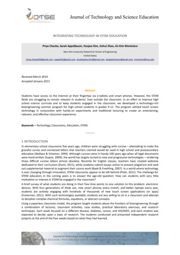
SYNOPSIS OF GLUCOSE METABOLISMA. Glucose is a chemical fuel which can be used to obtain energy, reducing power and carbonskeletons.1. Glucose is the major form in which carbohydrates absorbed through the intestinalepithelium are presented to cells. Common dietary disaccharides from which glucose is derived are:sucrose, a disaccharide of glucose and fructose,maltose, a disaccharide of glucose,lactose, a disaccharide of galactose and glucose.The intestinal epithelium splits disaccharides into monosaccharides andtransports them to the blood via the portal system.2. Different tissues take up glucose from blood at different concentrations depending ontheir glucose transporters (GLUTs) Blood glucose is maintained at about 5mM. Glucose transporters (GLUT) are integral membrane proteins that have 12membrane-spanning domains.Model of a mammalian glucose transporter. Thehydrophobicity profile of the protein points to thepresence of 12 transmembrane alpha helices.Chemical labeling and tryptic cleavage studies supportthis postulated topography. [From M. Muekler, C.Caruso S.A. Caldwin, M. Panico, M. Blench, H.R.Morris W.J. Allard G.E. Lienhard and H.F. Lodish,Science 229 (1985):941.]GLUT1 and GLUT3 - present on many cells, Km 1 mM. Since blood glucose isusually maintained at about 5mM, GLUT1 and GLUT3 transport glucose at aconstant rate independent of blood glucose concentration. GLUT 2 - present on liver and pancreatic beta cells, Km 15 - 20 mM The rateof entry of glucose into liver and pancreatic beta cells is proportional to theblood glucose levels. GLUT4 - present on muscle and fat cells, Km 5 mMInsulin causes an increase in the number of GLUT4 molecules in themembranes of muscle and fat cells and thus controls entry of glucose in thesecells. GLUT5 - actually a fructose transporter.3. Tissue-specific use of glucose as a major fuel Brain and muscle use glucose as a major fuel.Brain also uses ketone bodies as fuel during severe starvation Liver does not use glucose as its major fuel. Rather, liver exports glucose foruse by other tissues. -5-
B. Glycolysis, the TCA cycle and the pentose phosphate shunt transduce the chemical energy ofglucose into two forms:1. the high-energy phosphate bond of ATP2. the electron donors NADH, FADH2 and NADPH (referred to as reducing equivalents).C. Glycolysis all cells do glycolysis C6 2 C3 aerobic glycolysis produces a net gain of 2 ATP and 2 (NADH H ) anaerobic glycolysis produces a net gain of 2 ATP (e.g., in red blood cells andexercising muscle) Under anaerobic conditions NADH is used to reduce pyruvate tolactate to regenerate NAD so that glycolysis can continue.D. Loss of CO2 and gain of NADH in the conversion of pyruvate to acetyl CoAE. TCA cycle only to a limited extent in fat cells, which use acetyl CoA primarily to synthesize fat,NOT in red blood cells, which don't have mitochondria limiting in exercising muscle in which the TCA cycle and oxidative phosphorylationcan't keep up with the rate at which pyruvate is produced by glycolysis 2 CO2 released during the final oxidation steps of entering acetate derived from acetylCoA 1 ATP gained (as GTP) 4 reducing equivalents gained as 3 NADH and 1 FADH2 To yield energy indirectly, electrons from NADH and FADH2 are supplied to themitochondrial electron transport chain (to be discussed in later lectures), whichproduces ATP at the expense of the ultimate electron acceptor, O2. MUCH more ATP isobtained from these subsequent electron transport reactions than is directly obtainedvia glycolysis, the TCA cycle or the pentose phosphate shunt.F. Pentose Phosphate Shunt happens in all cells, to different extents the oxidative branch yields 2 reducing equivalents, as 2 NADPH, and one molecule ofpentose (C5) phosphate from each glucose-6-phosphate NADPH is important for reductive biosynthesis, particularly in fat cells, which produceand consume large amounts in the synthesis of fatty acids. Red blood cells useNADPH to maintain a reduced environment. the non-oxidative branch interconverts phosphate sugarsC5 C3, C4, C6, C7G. Glucose is stored as glycogen, a glucose polymer. in muscle, heart, liver, to a limited extent in fat cells - NOT in brain, NOT in red bloodcellsH. Intracellular generation of glucose1. Gluconeogenesis in liver, kidney requires a net utilization of energy C3 C6
2. Glycogenolysis - glycogen breakdown to glucose-1-phosphate and then conversionto glucose-6-phosphate in muscle, heart, liver, to a limited extent in fat cells, NOT in brain, NOT in redblood cellsI.Only the liver and kidney can generate glucose for export to the blood.J. Glucose provides cellular building material as well as fuel. Its breakdown providesprecursors for biosynthesis of amino acids, heme, fatty acids, cholesterol and otheressential molecules.K. The centrality of glucose in metabolism requires good homeostatic control of blood glucoseat about 5 mM to ensure adequate concentrations in all tissues, especially in the brain andmuscles. Coordination of glucose breakdown (glycolysis), glucose synthesis (gluconeogenesis)and glucose storage and release (glycogen metabolism) is achieved by hormonal(insulin, glucagon, epinephrine and norepinephrine) regulation (resulting in thecovalent modification of essential enzymes -primarily phosphorylation) and by tissuespecific differences in the distribution of key enzymes. Pancreatic alpha cells secrete glucagon in response to low blood glucose andglucagon causes the liver to release glucose to the blood. Pancreatic beta cells secrete insulin in response to high blood glucose. Insulinpromotes uptake of glucose by muscle and fat cells and promotes glucose storage inliver. The pancreatic insulin-secreting beta cells and the glucagon-secreting alpha cells arein a co-regulatory circuit. Secretion of insulin and associated molecules by the betacells inhibits the secretion of glucagon from the alpha cells. Thus, increased insulinrelease is coupled to reduced glucagon release (high blood glucose). Recent evidencesuggests that zinc, secreated with insulin from the beta cells may be the inhibitor ofglucagon secretion from the alpha cells. Glucagon, secreted by the alpha cells (lowblood glucose) causes the beta cells to secrete a low level of insulin. This relatively lowlevel of insulin secretion is not sufficient to counteract the higher levels of glucagonsecreted in response to low blood glucose. Various speculations exist concerning thephysiological significance of the low insulin secretion in response to glucagon.
METABOLIC ENERGYA.All living organisms require a continual input of energy to maintain the multitude of metabolicreactions in a state far from equilibrium (equilibrium death).Three major purposes for energy in biological organisms: Mechanical Work - e.g., muscle contraction Active Transport - to maintain chemical balance, e.g., cells need to continually pump outNa ions which are taken in as part of transport mechanisms for other molecules achieved by the Na /K ATPase, which uses energy to pump Na against a concentrationgradient. Active transport accounts for about 10% - 30% of the energy needs of anorganism. Synthesis of essential biological molecules.B.C.D.Adenosine Triphosphate [ATP] is the universal currency of energy in biological processes withincells. It is the link between energy-producing and energy-utilizing systems. Its hydrolysis yieldsuseful energy.Structural basis for the phosphoryl transfer potential of ATP electrostatic repulsion between proximal negative charges ADP and Pi have a larger number of resonance forms than ATPThe energy liberated in the hydrolysis of ATP can be harnessed to drive reactions that requirean energy input. Reactions that wouldn't proceed spontaneously, because they required an input ofenergy, can proceed if they are coupled to ATP hydrolysis. ATP hydrolysis shifts the equilibria of coupled reactions. The equilibrium of a reaction ischanged by 108 per ATP molecule hydrolyzed. Thus, if 3 molecules of ATP arehydrolysed in a coupled reaction, the equilibrium of the reaction would be changed by1024.
E.ATP is formed from ADP and Pi when fuel molecules are oxidized by chemotrophs (or when lightis trapped by phototrophs)This ATP-ADP cycle is the fundamental mode of energy exchange in biological systems. ATP turnover is high. The half-life of an ATP molecule in many tissues is approximately oneminute. A resting human uses approximately 40 kg of ATP in 24 hours approximately 1000tons in a lifetime. During strenuous exertion it can be as high as 0.5 kg / min.F.NADH and FADH2 are the major electron carriers in the oxidation (dehydrogenation) of fuelmolecules.In aerobic organisms the ultimate electron acceptor is O2. However, electrons are not transferreddirectly from fuel molecules and their breakdown products to O2. They are first transferred to theelectron carriers (substrate-level oxidations). The reduced forms of these carriers then transfertheir high-potential electrons to O2 by means of an electron transport chain located in the innermembrane of the mitochondria.1. Nicotinamide adenine dinucleotide [NAD ] structure - in NAD R H, in NADP R PO32 In the oxidation (dehydrogenation) of a substrate, the nicotinamide ring of NAD accepts a hydrogen ion and two electrons - the equivalent of a hydride ion. The reduced form of this electron carrier is called NADH. In the oxidized form, thepyridine ring nitrogen is tetravalent and carries a positive charge. In the reduced form,NADH, the nitrogen atom is trivalent.
Reaction type that uses NAD (typically, but not exclusively, oxidation of an alcohol to acarbonyl).2. Flavin adenine dinucleotide [FAD] The reactive part of FAD is its isoalloxazine ring. It accepts two electrons, and twoprotons. G.Reaction type that uses FAD (generally, but not exclusively, a dehydrogenation of asaturated compound with the formation of a double bond)NADPH is the major electron donor in reductive biosynthesis. In most biosynthetic reactions the precursors are more oxidized than the products soreducing power is required
e.g., in fatty acid biosynthesis a keto group is sequentially reduced via an alcohol and anunsaturated aliphatic chain. NADPH differs from NADH in that the 2'-OH of its adenosine moiety is esterified withphosphate. This extra phosphate is a tag that directs this reducing agent to discerningbiosynthetic enzymes.NADPH carries electrons in the same way as NADH.NADPH is used almost exclusively for reductive biosynthesis, whereas NADH is usedprimarily for the generation of ATP. H.Why have both NADP and NAD ? The cellular ratio of NADP /NADPH 0.014 The cellular ratio of NAD /NADH 700 The cell can thus maintain a high level of reducing equivalents for biosynthetic reactionswhile simultaneously maintaining high capacity for substrate-level oxidations so that energycan be generated.I.Coenzyme A (A for acetylation ) is a universal carrier and donor of acyl groups, J.The terminal sulfhydryl group is the reactive site, which forms thioesters with acyl (not onlyacetyl) groups.Acetyl CoA is a common intermediate in different catabolic pathways. glycolysis, beta-oxidation of fatty acids and amino acid degradation all supply the cellularpool of acetyl CoA.
THE PATHWAYSI. The major intracellular form of glucose is glucose-6-phosphate1. Phosphorylation of glucose to form glucose-6-phosphate by hexokinase: irreversible, regulated step ATP is expended feedback inhibited by glucose-6-phosphate, the product of the reaction relatively low Km for glucose (about 0.1 mM) Glucose-6-phosphate, unlike glucose, is negatively charged, which prevents its exit fromcells. The liver is a glucose buffer for the blood. Its activity assures constant glucoseconcentration (approx. 5mM) in the blood.o Glucokinase, a liver-specific enzyme whose biosynthesis is induced by insulin inresponse to high blood glucose levels, has a high Km (about 10 mM) and high Vmax forglucose, and is not product-inhibited. The liver does not use glucose as its preferredfuel. Rather, it stores glucose as glycogen when blood glucose levels are high (e.g.,after a meal), and recovers glucose from the stored glycogen and releases it into theblood when blood glucose is low (e.g., during fasting). The high Km and inducibility ofglucokinase allows the liver to store glucose as glycogen only when blood glucoselevels are high, conserving glucose for other tissues that use it as a primary fuel (e.g.,brain, heart, white muscle) when blood glucose levels are low. The non-productinhibition of glucokinase allows the liver to capture glucose for storage as glycogeneven when its intracellular levels of glucose-6-phosphate are high. Glucokinase genetranscription is repressed by glucagon, an hormone secreted by the pancreatic alphacells in response to low blood glucose levels. When glucose levels are limited (lowblood glucose) existing Glucokinase is bound by the Glucokinase Regulatory Protein[GKRP], which inactivates and sequesters it in the cell nucleus, thereby preventing theglucose produced by gluconeogenesis and glycogenolysis from being rephosphorylatedand used by the liver. Glucokinase gene transcription is stimulated by insulin, anhormone secreted by the pancreatic beta cells in response to high blood glucoselevels.o The pancreatic insulin-secreting beta cells and the glucagon-secreting alpha cells arein a co-regulatory circuit. Secretion of insulin and associated molecules by the betacells inhibits the secretion of glucagon from the alpha cells. Thus, increased insulinrelease is coupled to reduced glucagon release (high blood glucose). Recent evidencesuggests that zinc, secreated with insulin from the beta cells may be the inhibitor ofglucagon secretion from the alpha cells. Glucagon, secreted by the alpha cells (lowblood glucose) causes the beta cells to secrete a low level of insulin. This relatively lowlevel of insulin secretion is not sufficient to counteract the higher levels of glucagonsecreted in response to low blood glucose. Various speculations exist concerning thephysiological significance of the low insulin secretion in response to glucagon.o The major function of liver glycolysis is to obtain carbon skeletons and energy for thesynthesis of biomolecules that the liver exports for use or storage (e.g., fat) by othertissues.II. Glycolysis occurs in the cytosol, yielding 2 ATP, 2 pyruvate and 2 (NADH H ) from eachglucose moleculeThe first steps of glycolysis convert glucose-6-phosphate into a form that can be readily cleaved intophosphorylated three-carbon units. High energy phosphate (as ATP) is then generated from thethree-carbon units.
2.Isomerization of glucose-6-phosphate (aldose) to fructose-6-phosphate (ketose) byphosphoglucose isomerase: freely interconverted3.Phosphorylation of fructose-6-phosphate to form fructose-1,6-bisphosphate byphosphofructokinase 1 [PFK1]: THIS IS THE RATE-LIMITING STEP OF GLYCOLYSISAND THE MOST IMPORTANT SITE OF REGULATION. irreversible, regulated step ATP is expended responds to cell energy charge (ATP/AMP ratio), so thatallosteric inhibition by ATP is antagonized by AMP ATP lowers the affinity (increases the Km) of the enzyme for its substrate, fructose-6phosphate, thereby slowing glycolysis when the energy charge of the cell is high. Citrate, an early TCA cycle intermediate, also inhibits PFK1, by increasing itssensitivity to ATP. A high level of citrate indicates that alternate fuels such as fattyacids and ketone bodies (which will be discussed in later lectures) are available. inhibited by low pH, thereby guarding against excessive lactate formation fromglycolysis, which would cause a further drop in blood pH (lactic acidosis) activated allosterically by fructose-2,6-bisphosphate which increases the affinity forfructose-6-phosphate (lowers the Km) and decreases the sensitivity to ATP inhibition3a.Phosphorylation of fructose-6-phosphate to form fructose-2,6-bisphosphate byphosphofructose kinase 2 [PFK2], a remarkable protein with TWO, distinct active sites(bifunctional enzyme). One site has phosphofructose kinase (phosphofructose kinase 2[PFK2]) activity and the other site has fructose bisphosphatase (fructose bisphosphatase2 [FBPase 2] activity. Fructose-2,6-bisphosphate is the most important regulator of glycolysis in the liver. Itsabundance is a reflection of whether blood glucose levels are high or low (see below). "Feed-forward" allosteric activation by fructose-6-phosphate The kinase reaction is favored by the dephosphorylated form of the enzyme. The phosphatase reaction is favored by the phosphorylated form of the enzyme. Thisis NOT a reversal of the kinase reaction (don't re-create ATP). The liver enzyme is phosphorylated by cAMP-dependent protein kinase. cAMP levels rise in response to hormones such as glucagon and epinephrine andnorepinephrine, (receptors for which activate adenylate cyclase), or in response toevents that inhibit cAMP phosphodiesterase (e.g., caffine intake). cAMP-dependentprotein kinase consists of catalytic subunits and cAMP-binding regulatory subunits.The catalytic subunit is inactive when bound to the regulatory subunit, whichdissociates when it binds cAMP. Glucagon, secreted by pancreatic alpha cells in response to low blood glucose,activates cAMP-dependent protein kinase, switching phosphofructokinase 2 tophosphatase mode, and preventing build-up of fructose-2,6-bisphosphate, thusslowing glycolysis and conserving blood glucose at step #3 Conversely, when blood glucose is high, glucagon levels are low, insulin levels arehigh, the enzyme is dephosphorylated, synthesis of fructose-2,6-bisphosphate isfavored, and glycolysis is speeded up at step #3.In summary, the PFK2 kinase activity predominates in the "fed" state, the phosphataseactivity predominates in the "starved" state. The muscle isoform of PFK2/FBPase (bifunctional enzyme) is not phosphorylated(not regulated) in response to the starved state (muscle does not have glucagonreceptors). Furthermore, the skeletal muscle isoform does not have a site forphosphorylation by cyclic AMP-dependent protein kinase. In skeletal muscle,fructose 6-phosphate is the major regulator of PFK2/FBPase. However, the primaryregulator of muscle glycolysis is believed to be ATP (the supply of energy).The PFK2 activity of the cardiac muscle isoform of PFK2/FBPase is STIMULATED
by phosphorylation by cyclic AMP-dependent protein kinase (it has a differentphosphorylation site than the liver isoform), resulting in an increased rate of fructose2,6-bisphosphate synthesis and stimulation of phosphofructokinase 1 activity. The rateof heart muscle glycolysis therefore increases upon phosphorylation of PFK2/FBPase.Epinephrine causes an increase in heart rate and an increase of glycolysis (by causingan increase in fructose 2,6-bisphosphate concentration to stimulatephosphofructokinase 1) to supply extra energy for the increased heart rate. Skeletalmuscle PFK2/FBPase, because it does not have a phosphorylation site for cyclicAMP-dependent protein kinase, is not affected by epinephrine, but liver PFK2 activityis inhibited, thereby slowing liver glycolysis in response to epinephrine.4.Cleavage of fructose-1,6-bisphosphate to form dihydroxyacetone phosphate andglyceraldehyde-3-phosphate [G3P] by aldolase: reversible G3P is on the direct pathway of glycolysis5.Interconversion of dihydroxyacetone phosphate and glyceraldehyde-3-phosphate bytriose phosphate isomerase: reversible6.Oxidation of glyceraldehyde-3-phosphate to form 1,3-bisphosphoglycerate[1,3-BPG], a high-potential phosphorylated compound, by glyceraldehyde-3-phosphatedehydrogenase: reversible ATP NOT expended - part of the energy obtained in the oxidation of G3P is harvestedas an electron pair donated to NAD - the resulting NADH will be recycled back toNAD at reaction #11 under anaerobic conditions Carbon 1 of 1,3-BFG is at the oxidation level of a carboxylic acid. The phosphorylgroup of this mixed anhydride has high transfer potential. 1,3-bisphosphoglycerate is converted to form large amounts of 2,3bisphosphoglycerate in red blood cells by bisphosphoglycerate mutase. 2,3bisphosphoglycerate regulates the hemoglobin/O2 dissociation curve - deficiency ofglycolytic enzymes can cause hemolytic anemia. (Red blood cells, whose primaryfunction is to supply oxygen to tissues, are themselves restricted to anaerobicmetabolism, since they lack mitochondria.)7.Transfer of the high-energy phosphate bond from 1,3-bisphosphoglycerate to form ATPand 3-phosphoglycerate by phosphoglycerate kinase: reversible substrate-level phosphorylation; ATP is produced8.Interconversion of 3-phosphoglycerate and 2-phosphoglycerate by phosphoglyceratemutase: reversible9.Dehydration of 2-phosphoglycerate to form phosphoenolpyruvate [PEP] by enolase: reversible the dehydration elevates the transfer potential of the phosphoryl group10.Transfer of high-energy phosphate bond from phosphoenolpyruvate to form ATP andpyruvate by pyruvate kinase: essentially irreversible, highly regulated step substrate-level phosphorylation; ATP is produced "feed forward" activation by fructose-1,6-bisphosphate liver isozyme shows cooperative phosphoenolpyruvate binding, allosteric inhibition byATP and alanine (an amino acid synthesized from pyruvate in one metabolic step) inhibition by glucagon-triggered phosphorylation - This slows glycolysis in the liverwhen blood glucose is low, thereby conserving glucose for use by tissues that use itas a primary fuel. Recall that PFK2 (step 3) is also phosphorylated, and its kinase
activity is reduced, by the same mechanism, in response to low blood glucose levels.muscle isozyme is NOT regulated by phosphorylationA rare, hyperactive mutant pyruvate kinase is harmful to red blood cells because itdepletes the precursor pool, diverting substrate from the synthesis of 2,3bisphosphoglycerate, thereby impairing hemoglobin function.11. Regeneration of NAD through reduction of pyruvate (the electron acceptor) to form lactateby lactate dehydrogenase (fermentation): reversible Major option used by cells under anaerobic conditions to regenerate cytosolic NAD .Without O2, mitochondria can't resupply NAD - this step allows glycolysis to continueanaerobically by regenerating the NAD required in step #6. Anaerobic glycolysis isobviously necessary in red blood cells, which lack mitochondria. It is also importantwhen the oxygen supply is limiting, e.g., in exercising muscle, or anoxia due to impairedblood supply, as in stroke. The microbial fermentation repertoire is vast, and includes important commercialproducts such as ethanol, butanol and acetone. Coelocanths, "living fossil" fish, fromoxygen-poor deep sea strata, also employ alcoholic fermentation.Summary of aerobic glycolysis:Glucose 2 Pi 2 ADP 2 NAD 2 Pyruvate 2 ATP 2 NADH 2 H 2 H2OSummary of anaerobic glycolysis:Glucose 2 Pi 2 ADP 2 Lactate 2 ATP 2 H2ORemember that phosphofructokinase 1 is inhibited by low pH. The lactate that is formed underanaerobic conditions contributes to lower blood pH. Therefore, slowing glycolysis at thecommitted step slows lactate production and helps prevent acidosis due to excess lactate.Utilization of galactose:Common dietary disaccharides are sucrose, glucose(α 1 2)fructose, maltose, glucose(alpha1 4)glucose, and lactose, galactose(β 1 4)glucose. Lactase deficiency, impairing cleavageof lactose to monosaccharides, causes the digestive disorder lactose intolerance. Oxidation ofgalactose requires phosphorylation at carbon 1 by galactokinase followed byAttachment to UDP by the enzyme galactose-1-phosphate uridyl transferase.Galactose-1-phosphate UDP-glucose UDP-galactose glucose-1-phosphateUDP-galactose-4-epimerase, can act:UDP-galactose UDP-glucoseAfter exchange with galactose via transferase, glucose-1-phosphate is isomerized to glucose-6phosphate by phosphoglucomutase. Genetic deficiency of transferase (galactosemia) results ininappropriate metabolism of galactose to toxic forms such as galactitol.III. The Tricarboxylic Acid [TCA] Cycle occurs in mitochondria.Each turn of the cycle produces one high energy phosphate bond in the formation of GTP (highenergy phosphate transferable to ADP to form ATP) and 4 reducing equivalents (3 NADH and 1
FADH2). AFTER the O2-dependent processes of electron transport and oxidativephosphorylation, the total number of ATP produced per cycle is 10 (2.5 ATP from each NADHoxidized, 1.5 ATP from oxidation of FADH2 and 1 GTP ADP GDP ATP ) Two moleculesof pyruvate are generated from one molecules of glucose, fueling two turns of the TCA cycle.Gycolysis of 1 molecules of glucose (6 carons) yields a net of two molecules of ATP, 2molecules of NADH ( 5 ATP) and two molecules of pyruvate (3 carbons). Each of the twomolecules of pyruvate generated per glucose is converted to acetyl CoA (step # 12), producingone molecule of NADH ( 2 NADH, or 5 ATP per molecules of glucose). Acetyl CoA thenenters the TCA cycle.Thus one molecule of glucose completely oxidized yields 32 ATP.Anaerobic glycolysis to lactate nets only 2 ATP per glucose molecule.12.Formation of acetyl CoA from pyruvate by pyruvate dehydrogenase pyruvate moves between cytosolic and mitochondrial compartments by carriermediated transport. irreversible NADH produced, CO2 released deactivated by phosphorylation by a protein kinase activity stimulated by highNADH/NAD , acetyl CoA/CoA or ATP/ADP ratios pyruvate and ADP activate by inhibiting the kinase dephosphorylation (by a protein phosphatase), and consequently enzymatic activity, isstimulated by insulin. Ca2 activates the phosphatase in heart muscle to increase energy for rapidcontraction competitively inhibited by acetyl CoA, NADH nutrition buffs: 4 vitamins are required as co-factors: thiamin (as thiaminepyrophosphate, TPP), riboflavin (as FAD), niacin (as NAD ) and pantothenate (asCoenzyme A). Lipoate, non-essential in the diet, is also required. Alcoholism,combined with poor nutrition, can deplete thiamin, thereby blocking the TCA cycle. Forsuch malnourished individuals, glucose without simultaneous thiamin supplementationis harmful, because its aerobic metabolism is compromised.13.Condensation of oxaloacetate and acetyl CoA by citrate synthase to form citrate Irreversible feed-back inhibited by citrate (the product of the enzymatic reaction) ordered binding of substrates: oxaloacetate binding changes enzyme conformation toaccept acetyl CoA Remember, citrate is an allosteric inhibitor of reaction #3, the rate-limiting step ofglycolysis catalyzed by PFKI. (Citrate can exit the mitochomdria to the cytosol, whereit is converted back to acetyl CoA and oxaloacetate. The acetyl CoA provides carbonskeletons for fatty acid biosynthesis, and the oxaloacetate provides electrons for thereductive biosynthesis of fatty acids. Fatty acid biosynthesis will be discussed in thecourse segment on lipid metabolism.)14.Interconversion of citrate, cis-aconitate and isocitrate by aconitase reversible15.Conversion of isocitrate to alpha-keto-glutarate by isocitrate dehydrogenase irreversible oxidative decarboxylation NADH produced, C02 released major regulation step of the cycle responsive to the energy charge of the cell the cooperative binding of isocitrate and NAD is enhanced by ADP (reduced energy) competitively inhibited by NADH (increased reducing power for ATP generation) Ca2 , released from sarcoplasmic reticulum in contracting muscle lowers the Km andspeeds up the velocity of the enzyme to increase energy for muscle contraction16.Conversion of alpha-keto-glutarate to succinyl CoA by alpha-keto-glutaratedehydrogenase irreversible oxidative decarboxylation NADH produced, CO2 released
product inhibited by succinyl CoA and NADHCa2 , released from sarcoplasmic reticulum in contracting muscle lowers the Km andspeeds up the velocity of the enzyme to increase energy for muscle contraction17.Formation of succinate from succinyl CoA by succinyl CoA synthetase (succinatethiokinase) substrate-level phosphorylation; GTP produced18.Conversion of succinate to fumarate by succinate dehydrogenase FADH2 produced, a C C double bond is formed Succinate dehydrogenase is a membrane-bound protein located on the innermitochondrial membrane, unlike the other enzymes of the TCA cycle, which arelocated in the mitochondrial matrix.19.Conversion of fumarate to malate by fumarase20.Oxidation of malate to form oxaloacetate by malate dehydrogenase NADH producedSummary of the TCA cycle:Acetyl CoA 3 NAD FAD GDP Pi 2 H2O 2 CO2 3 NADH FADH2 GTP 2H CoAAlthough O2 does not participate in the TCA cycle, the cycle
E. ATP is formed from ADP and P i when fuel molecules are oxidized by chemotrophs (or when light is trapped by phototrophs) This ATP-ADP cycle is the fundamental mode of energy exchange in biological systems. ATP turnover is high. The half-lif