Transcription
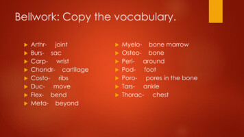
Bellwork: Copy the vocabulary.Arthr- joint Burs- sac Carpwrist Chondr- cartilage Costoribs Ducmove Flex- bend Meta- beyond Myelo- bone marrowOsteo- bonePeri- aroundPod- footPoro- pores in the boneTars- ankleThorac- chest
TheSkeletalSystem
Standards 8)Outline basic concepts of normalstructure and function of all body systems,and explain how homeostasis ismaintained. (Ourfocus is on the skeletal system)
Objectives Studentswill describe the function ofthe skeletal system. Students will name and locate thebones of the skeleton. Students will analyze bones usedwhen engaging in different actions.
Functions of the Skeletal System Support Structuralsupport Framework for attachmentDiscuss: Based on other body systemswe have studied, what does the skeletonsupport?Name specific examples. Storage Calcium reserve Energy reserves (lipids in yellow marrow)
Functions of the Skeletal System Blood Cell Production AKAhemopoiesis (formation of blood’scellular components RBC and WBC produced in red marrowProtection Surrounds soft tissues and organsLeverage for movement Changemagnitude and direction of forcesgenerated by skeletal muscles
Structures of the Skeletal System:Types of Bones Long Bones Longer than they are wide Ex: humerus, femurShort Bones Roughly equal length and width Ex: carpals, tarsalsFlat Bones Thin and broad Ex: ribsIrregular Bones Complex shapes Ex: pelvic girdle, vertebrae
The Axial Skeleton The axial skeleton is blueand includes the:skull vertebral column sternum ribs hyoid bone (or laryngeal)
The SkullFrontal View
The SkullLateralView
TheVertebralColumn(TheSpinalColumn)Draw andlabel
Discussion With a partner discuss Applying your knowledge of thenervous system, what organ does thespinal column protect?
Sternum and Rib Cage12 sets ofRibsTrue,False,Floating
DiscussionApplying what your know aboutmedical terminology, what cavity dothe ribs protect?
The Appendicular Skeleton The appendicular skeleton is beigeand includes shoulder girdles arms wrists hands pelvic girdle legs ankles feet.
The Shoulder Girdle
The ArmUpper ArmLower Arm
The Hand
The Pelvic GirdleWhy do you think the shape of the pelvisIs different between males and females?
Leg bonesUpper LegLower Leg------Patella
AnkleandFoot
ActivityMake sure your worksheets are labeled accurately. Use your book, page 310.In your small group, use your skeleton to evaluate which bones are used in dailyactivities such as: Brushing your teeth Driving a car Throwing a football Running a raceEnrichment: Go back to your diagram and give the everyday “layman’s” terms to as manybones as possible. (For example: jaw for mandible.) Explain why it is important to know the layman’s terms as well as the correctmedical terminology. When would you use each?
BELLWORK: Day Two COMPAREAND CONTRAST THE THREETYPES OF CONNECTIVE TISSUE: CARTILAGE(p. 309) LIGAMENT (p. 312) TENDON (p. 320)
Connective Bone Tissue Cartilage: Acts as cushion between bones; articular cartilagelocated on ends of bones and acts as shock absorber,preventing ends from grinding together when you move.Ligaments: tough, whitish bands that connect from bone to boneand can withstand heavy stress. Tendons:cord-like structures that attach muscle to bone.
The knee joint:-cartilage-ligaments
Standards 8)Outline basic concepts of normalstructure and function of all body systems,and explain how homeostasis ismaintained. (Ourfocus is the skeletal system)
Objectives Explainhomeostasis of the body relating tothe skeletal system Defineossification Identifyboneand label the parts of the long Compare/contrastjointsthe different bone
Homeostasis and Mineral Storagein the Skeletal System 99% of bodily calcium is deposited in the skeleton Nervous System use Ca ions Ca concentration is heavily regulated Parathyroidhormone (PTH) – elevates Ca levels inbody fluids Bones become weaker Calcitonin - depresses Ca levels in body fluids Bones become stronger These hormones work together to maintainhomeostasis!
Growth and Formation of Bone Begins6 weeks after fertilization,stops around age 25Is it possible for some people to growtaller even after high school? Ossification– process of tissuereplacement by bone-http://www.youtube.com/watch?v 6f90506PvS8&feature related
Growth and Formation of Bone Requirements: Prenatal – minerals absorbedfrom mother (loses bonemass) Consume Ca and P from diet Vitamin D3 allows absorptionof Ca and P Vitamins A and C needed forosteoblast activityDiscuss: How can a womanretain or gain the necessaryminerals her body needs duringpregnancy?
Structure of the Long Bones Diaphysis Central shaftBone marrow Yellow vs. red (lipids or fat vs. RBCs)Epiphysis Enlarged ends Covered with articular cartilageCompact bone Dense/solid Found in diaphysisSpongy bone Network of bony rods w/ spaces Found in epiphysisPeriosteum Covers outer surface of boneEndosteum Lines the marrow cavityLabel in your notes.
MoveableBone Joints-Saddle-Ball and Socket-Pivot-Hinge-Ellipsoidal/Condyloid-Gliding
Moveable Bone Joints Pivot joint: turnstile movement in neck and forearm Ball and socket joint: hip and shoulder; all forms of movement,including rotation Hinge joint: allow opening and closing movement in knees andelbows Gliding joint: wrists and ankles; provides sliding back and forthmovement Saddle joint: shaped like saddle, found in thumb; can rock upand down or side to side Condyloid joint: oval shaped bone end fitting into elliptical cavityin other bone so there is movement from one plane to anotherbut no rotation as found in fingers and toes Ellipsoidal joint: provide two axes of movement through samebone like joint formed at wrist with radius and ulna
Classification of Joint MovementGet up and let’s move!!! Flexion: bending a joint anddecreasing angle between involvedbones Extension: straightening a joint Abduction: moving away frombody’s midline Adduction: moving toward midlineof body Circumduction: circular armmovement of a pitcher Rotation: bone “spins "on its axis Supination: turning hand palm up Pronation: turning hand palm down
Classification of Joint Movement Eversion: The movement of the soleof the foot away from the medianplane. Inversion: The movement of the soletowards the median plane. Dorsiflexion: Flexion of the foot in anupward direction — compareplantar flexion, which is flexing thefoot down. Retraction: The movement of abody part in the posterior direction,or being drawn back. Protraction: The movement of abody part in the anterior direction, orbeing drawn forwards.
Immoveable Joints Afixed jointbetween bonesconnected byfibrous tissue (forexample, thesutures of the skull). At what time did these bonesneed to be able to move?
Cartilaginous Joint:the joint space is covered in dense connective tissue Inmales this mayshift slightly attimes. Infemales this jointis vital to provideroom duringvaginal childbirth.
Activity Completethe “Meeting at the Joint”worksheet. Complete cutting out your skeleton. Identify and label the bones of yourskeleton. We will place our skeletons on thebulletin board and then label thejoints.
“Simon Says” Review game!!Which one doesn’t belong! (Quiz)
Extended Learning!?!? Research Theythe carpals and tarsals.each have specific names. Whatare they? Labeleach on the hand and foot.
Bellwork: Page 316 Drawand describe the followingconditions related to the spine: Kyphosis Lordosis Scoliosis
Directed Reading Activity In your group of three choose one of the followingdirected reading from the website: Care Considerations with Patients with Spinal CordInjuries Total Knee Replacement and Imaging Computed Tomography of Facial Fractures Each person in your group will choose a differentdirected reading. You may not do the same one. After you answer the questions, then go to the ExtendedLearning Assignments tab on the class website. Complete the task for the corresponding professionaljournal.
Myelo- bone marrow . medical terminology. When would you use each? BELLWORK: Day Two COMPARE AND CONTRAST THE THREE TYPES OF CONNECTIVE TISSUE: CARTILAGE (p. 309) LIGAMENT (p. 312) TENDON (p. 320) Connective Bone Tissue Cartilage: Acts as cushion between bones; articular cartilage located on ends of bones and acts as shock absorber, preventing ends from