Transcription
Forensic Science, Medicine and Pathology (2021) 92-yCASE REPORTFox sign in a case of terminal stage pancreatic cancer and suggestionsfor diagnosisJulian Prangenberg1· Elke Doberentz1 · Burkhard Madea1Accepted: 19 May 2021 / Published online: 30 June 2021 The Author(s) 2021AbstractSkin signs in acute pancreatitis are well-known and frequently discussed manifestations accompanied by unfavorable prognoses although they may rarely appear in clinical and forensic medicine. In 2018, the district attorney’s office ordered a forensicautopsy for a 74-year-old man with terminal stage pancreatic cancer. The autopsy was ordered based on accusations of thedeceased’s widow regarding alleged medical malpractice and poor hospital care. The widow filed a grievance about multipleunsuccessful attempts to draw blood from her husband in addition to a diaper dermatitis at the right groin. An autopsy andadditional histological examinations were performed. After considering all findings, the diaper dermatitis was eventuallyassumed to be a Fox sign caused by acute pancreatitis, and the allegations of medical malpractice were refuted. This caseled us to identify another case with suspected cutaneous manifestations in pancreatic disease. We performed immunohistochemical staining on those two cases and six control cases to examine whether there was detectable presence of pancreaticlipase and trypsin in the skin discolorations and whether it could be used as a feasible method to verify skin signs associatedwith pancreatitis. Based on our findings, a minor disseminated lipase and trypsin staining should be considered regular andis therefore not conclusive of a skin sign associated with pancreatitis. Moreover, trypsin does not seem to be as suitable aslipase for this suggested immunohistochemical method. Nevertheless, this method might be a useful addition for determiningthe origin of skin discoloration and verifying skin signs associated with pancreatitis.Keywords Skin sign associated with pancreatitis · Pancreatitis · Fox’s sign · Grey Turner’s sign · ImmunohistochemistryIntroductionDermatologic findings, which are frequently observed during autopsies, can provide valuable differential diagnosticclues regarding their underlying etiology. Such findingssometimes even indicate cause of death [1, 2]. These findings include various skin signs in acute pancreatitis that aredefined based on their particular expansion and location ofemergence. A commonly accepted view on pancreatitis isthat the premature activation of digestive enzymes withinthe pancreas leads to autodigestion of the organ [3–5]. Theenzymes include proteases such as trypsin, elastase, and pancreatic lipase [6]. Skin signs associated with pancreatitisare presumably caused by the expansion of intra- and retroperitoneal hemorrhages along anatomic structures, which* Julian Prangenbergjulian.prangenberg@ukbonn.de1Institute of Legal Medicine, University Hospital Bonn,Stiftsplatz 12, 53111 Bonn, Germanymanifest in the subcutaneous tissue at specific locations,depending on the path of expansion [7]. However, thesesigns do not solely occur in acute pancreatitis, and manyother causes have been identified, including necrotizingpancreatitis, intraperitoneal hemorrhage, portal hypertension, liver cirrhosis anticoagulation with warfarin, or deepvein thrombosis [8–13]. Furthermore, they indicate a poorprognosis [14]; therefore, it seems reasonable to raise awareness regarding the potential clinical impact they entail. GreyTurner’s sign [15] and Cullen’s sign [16] represent the bestknown signs and are frequently mentioned in textbooks andjournals. A lesser-known skin sign is the Fox sign, whichemerges from the extraperitoneal expansion of pancreaticfluid along the fascia of the psoas and iliacus until its subcutaneous manifestation below the inguinal ligament [17].By contrast, the Fox sign is rather rare in clinical practice,and some physicians may have never encountered such acase themselves.In 2018, we encountered an autopsy that was orderedby the district attorney’s office owing to alleged medical
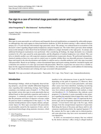
Forensic Science, Medicine and Pathology (2021) 17:486–492malpractice and poor hospital care in which the Fox sign wasassumed to be present and later used to refute the raised allegations. Furthermore, we identified another case with skinsigns associated with suspected pancreatitis and examinedhow to identify skin signs in suspected pancreatitis usingimmunohistochemistry.MethodsTwo forensic autopsy cases, in which the deceased exhibited skin discolorations at typical locations for skin signsin pancreatitis at the external examination, were identifiedbetween 2018 and 2019. The presence of those skin signsand an additional pancreatic disease was the inclusion criterion. Skin samples of the discolorations were obtained, andhistologic examinations and immunohistochemical stainingwere performed. Six autopsy cases served as controls. Fivecontrols had no skin discolorations or relevant organ pathologies; one case showed a reddish-livid discoloration at theright flank but no pancreatic pathologies. The cause of deathwas hanging in three cases and gunshot to the head, severehead injury, and myocardial infarction in one case each.The immunohistochemical staining used the following antibodies: rabbit monoclonal anti-pancreatic lipaseantibody (EPR6276) ab124915 and rabbit monoclonalanti-trypsin antibody (EPR19498-15) ab211491 (Abcam,Cambridge, United Kingdom). In accordance with the manufacturer’s instructions, the immune complexes were visualized using the Dako Envision Dual Link System (DakoDenmark, Glostrup, Denmark). After staining and preparation, the slides were microscopically examined using theOlympus BX 53 photomicroscope with the Olympus SC 180camera (Olympus Corporation, Tokyo, Japan) with a sensorFig. 1 Suspected Fox sign at theright groin (case 1)487size of 4.8 2.7 mm at an eyepiece magnification of 10 andan objective magnification between 4 and 20 .Case 1The district attorney’s office ordered a forensic autopsy ofa 74-year-old male owing to allegations regarding medicalmalpractice. The patient had a known pancreatic carcinomaand was taken to the hospital for angina and dyspnea. Duringhospitalization, a pulmonary embolism and hepatic metastases were diagnosed. The patient was also treated with anunspecified anticoagulating agent owing to cardiac arrhythmia.Despite curative treatment, his medical condition deteriorated,and he died a few days after admission. The clinical causeof death was stated as a metastasized pancreatic carcinomawith hepatic failure. The widow filed a grievance about thetreatment, including multiple unsuccessful attempts to drawblood from her husband during his in-patient stay and diaperdermatitis at the right groin due to alleged poor hospital care.External examination of the 75-year-old male (96 kg, 175 cm)showed distinctive anemia and pale skin and mucosa. Both armsand the right groin displayed extensive reddish-livid discoloration(Fig. 1). Internal examination revealed a progressed pancreaticcarcinoma with multiple metastases in the liver, lungs, lymphnodes, and abdominal wall. Furthermore, the autopsy identifieda thyroid tumor, a space-consuming lesion in the mediastinum,an exceeding of the critical heart weight (660 g), renal infarctionsand shock-kidneys, old cerebral infarctions, and cystitis. No signsof medical malpractice were found, and the nursing care wasevaluated as unobjectionable. The assumed diaper dermatitis wassuspected to be a Fox sign due to acute or necrotizing pancreatitis. The cause of death was eventually defined as a metastasizedterminal stage pancreatic carcinoma.
488Forensic Science, Medicine and Pathology (2021) 17:486–492Fig. 2 Livid discoloration at theright flank (case 3)Additional histological investigations were performedto confirm the presumption of the Fox sign. Histologicalinvestigations identified necrotizing pancreatitis due to apancreatic adenocarcinoma. The skin sample of the rightgroin revealed an edema of the subcutaneous tissue flushedwith intact erythrocytes, fatty tissue necrosis, and slightperivascular infiltration of lymphocytes. An acanthosis orspongiosis as an indicator of underlying dermatitis wasnot detected. Immunohistochemical staining of trypsinand pancreatic lipase in the skin sample was performedto substantiate the assumption of the Fox sign. Immunohistochemical analysis did not show any reddish stainingfor trypsin and little disseminated staining for pancreaticlipase in the dermis.Fig. 3 Extensive reddish staining of lipase in the epidermis and dermis (case 3, anti-pancreatic lipase antibody, 200 )
Forensic Science, Medicine and Pathology (2021) 17:486–492489Fig. 4 Extensive reddish staining of lipase in the epidermis and subcutaneous tissue (case 3, anti-pancreatic lipase antibody, 200 )Case 2A 46-year-old male (55 kg, 167 cm) complained of abdominal pain and blood in his vomit the past few days. He initiallyrefused admission to the hospital. The paramedics were thencalled to his apartment, but he died after unsuccessful resuscitation in his apartment.External examination showed a pale, bluish discoloration with central yellowish staining over his right costal arch(Fig. 2). Forensic autopsy revealed a cachectic constitution,a fatty liver with beginning cirrhosis, a necrotic and inflamedpancreatic head with surrounding hemorrhage, and hemorrhage in the right-sided mesocolon and right renal pelvis.The cause of death was determined to be necrotizing pancreatitis with internal bleeding.Immunohistochemistry did not show any signs of reddishstaining for trypsin; however, there was extensive stainingfor pancreatic lipase in the epidermis and dermis and widespread staining throughout the pancreas owing to necrotizingpancreatitis (Figs. 3, 4, 5).Control casesMacroscopically uninjured abdominal skin of five malesaged between 43 and 66 years (mean, 52.4 years) and theskin discoloration of an 89-year-old female with an initiallysuspected Grey Turner’s sign were examined. No relevantorgan pathologies or other factors described in the currentliterature that potentially cause skin signs were present. Thecause of death was hanging in three cases, gunshot to thehead in one case, and severe head injury in one case.Immunohistochemical analysis occasionally showed verylittle disseminated staining for pancreatic lipase in the dermis, but there was no extensive staining as shown in case 2.One case displayed moderate circumscribed staining in theepidermis for trypsin (Figs. 6 and 7).DiscussionIn forensic analyses, determining the origin and etiology ofskin discoloration to exclude or confirm bodily harm can becrucial. This, among other factors, requires knowledge ofthe described skin signs in pancreatitis and other conditionsthat may cause these specific discolorations. The literaturedescribes certain conditions that potentially cause the Foxsign, such as necrotizing pancreatitis, deep vein thrombosis,and rupture of an abdominal aortic aneurysm [8, 13, 17].In case 1, a pulmonary embolism was diagnosed in thehospital before the man’s death. Because pancreatic carcinoma has the highest risk for the development of vein thrombosis among other carcinomas [18] and approximately 93%of thromboses originate in the inferior vena cava and pelvicand leg veins [19], a deep vein thrombosis could also have
490Forensic Science, Medicine and Pathology (2021) 17:486–492Fig. 5 Extensive widespread staining of lipase in the pancreas due to necrotizing pancreatitis (case 3, anti-pancreatic lipase antibody, 40 )caused the Fox sign. There were no signs of inflammationindicating a diaper dermatitis in the skin. Immunohistochemical analysis showed a low level of staining for pancreatic lipase in the subcutaneous skin and no staining fortrypsin. However, the pancreatic lipase staining was slightlymore distinct than in the control cases, and after considering all factors, the discoloration was assumed to be a Foxsign. To the best of our knowledge, identifying a presumeddiaper dermatitis as a Fox sign and thus refuting allegationsof medical malpractice and poor hospital care appear torepresent a novelty. Furthermore, this case emphasizes theimportance and explanatory power of forensic autopsies andcomplementary histological examinations regarding allegedmedical malpractice.The male in case 2 had a fatty liver with the beginningof cirrhosis, a necrotic and inflamed pancreatic head withsurrounding hemorrhage, and hemorrhage in the right-sidedmesocolon and right renal pelvis. The cause of death wasdetermined as necrotizing pancreatitis with internal bleeding. There were no signs of reddish staining for trypsin;however, there was extensive staining for pancreatic lipase inthe epidermis and dermis that was much more intense thanin any of the included cases. Considering the immunohistochemical results and because the person had two conditionsthat are known to cause such discoloration, Grey Turner’ssign was assumed to be present in this case.Overall, the differential diagnostics of skin discolorationsshould include the distinction from hematoma or ecchymosisdue to blunt force or needle puncture sites because this couldfeign the origin of skin signs associated with pancreatitis[20]. In both cases, there was no clear evidence of bluntforce or needle punctures although small injections are notalways visible and therefore cannot be ruled out with absolute certainty.Regarding the control group, one case showed circumscribed staining of trypsin in the epidermis and very slightdisseminated staining of lipase in the dermis. This mightbe owing to cross-reactions with the trypsin antibody andother serine proteases present in human skin [21]. Talaset al. [22] examined the expression of human elastase 1 inthe skin that showed results similar to those found in bothour study and control cases. Lipase can be detected in thesebaceous glands and in the external root sheath of the hairfollicle [23]. Therefore, lipase enzymes were believed to bedissolved during staining, which resulted in their detectionin normal skin. Skin discoloration at the right flank observedin the 89-year-old female may be explained by the use ofanticoagulants. The use of an anticoagulating agent withwarfarin is described in the literature to form skin discoloration at this location [12]. Unfortunately, no informationon anticoagulating agents or other medication was available.Thus, the presence of Grey Turner s sign was considered
Forensic Science, Medicine and Pathology (2021) 17:486–492491Fig. 6 Circumscribed reddish staining of trypsin in the epidermis (control case, anti-trypsin antibody, 200 )unlikely and the discoloration presumably had a differentcause.Based on our findings, minor disseminated staining oflipase and trypsin should be considered regular and aloneshould not lead to the conclusion of a skin sign associatedwith pancreatitis. Considering all cases, trypsin does notseem as suitable as lipase for this suggested immunohistochemical method. Furthermore, in the three cases withFig. 7 Disseminated reddish staining of trypsin in the epidermis (control case, anti-pancreatic lipase antibody, 200 )
492skin signs associated with suspected pancreatitis, it remainedunclear when the discoloration first occurred; therefore, boththe antemortem and postmortem degradation of the enzymesmust be considered, which might have led to the indistinctstaining pattern in two cases.Conclusively, additional immunohistochemical investigations regarding the presence of pancreatic lipase may beuseful in cases with unclear skin discoloration. However,considering the relative rarity of this phenomenon, furtherand ideally multicenter studies with a sufficient number ofcases are warranted to establish and verify reliable stainingpatterns for the examined enzymes.Key Points:1. Dermatologic findings are frequently observed duringautopsies and can provide valuable information.2. A variety of conditions can cause skin discoloration inspecific locations.3. We performed immunohistochemical investigations toprove the presence of pancreatic enzymes in the skin.4. Trypsin does not appear to be a suitable marker for verifying skin signs associated with pancreatitis.5. Pancreatic lipase may be a useful marker for determiningthe etiology of skin discoloration.Funding Open Access funding enabled and organized by ProjektDEAL. This research received no specific grant from any fundingagency in the public, commercial, or not-for-profit sectors.DeclarationsEthical approval This article does not contain any studies with humanparticipants or animals performed by any of the authors.Conflict of interest The authors declare that they have no conflict ofinterest.Open Access This article is licensed under a Creative Commons Attribution 4.0 International License, which permits use, sharing, adaptation, distribution and reproduction in any medium or format, as longas you give appropriate credit to the original author(s) and the source,provide a link to the Creative Commons licence, and indicate if changeswere made. The images or other third party material in this article areincluded in the article’s Creative Commons licence, unless indicatedotherwise in a credit line to the material. If material is not included inthe article’s Creative Commons licence and your intended use is notpermitted by statutory regulation or exceeds the permitted use, you willneed to obtain permission directly from the copyright holder. To view acopy of this licence, visit http:// creat iveco mmons. org/ licen ses/ by/4. 0/.Forensic Science, Medicine and Pathology (2021) 17:486–492References:1. Birngruber CG, Riße M, Kettner M, Schnabel A, Ramsthaler F,Verhoff MA. Über die so genannten Hautzeichen bei akuter Pankreatitis. Arch kriminol. 2016;238:42–56.2. Cocks M, Sander I, Crain B. Frequency of dermatologic findingsat autopsy. J Forensic Sci. 2018;63:1867–9.3. Bhatia M, Brady M, Shokuhi S, Christmas S, Neoptolemos JP,Slavin J. Inflammatory mediators in acute pancreatitis. J Pathol.2000;190:117–25.4. Bhatia M, Neoptolemos JP, Slavin J. Inflammatory mediators astherapeutic targets in acute pancreatitis. Curr Opin Investig Drugs.2001;2:496–501.5. Bhatia M. Novel therapeutic targets for acute pancreatitis andassociated multiple organ dysfunction syndrome. Curr Drug Targets Inflamm Allergy. 2002;1:343–51.6. Bhatia M, Wong FL, Cao Y, Lau HY, Huang J, Puneet P,Chevali L. Pathophysiology of acute pancreatitis. Pancreatology.2005;5:132–44.7. Meyers MA, Feldberg MA, Oliphant M. Grey Turner’s signand Cullen’s sign in acute pancreatitis. Gastrointest Radiol.1989;14:31–7.8. Bem J, Bradley EL. Subcutaneous manifestations of severe acutepancreatitis. Pancreas. 1998;16:551–5.9. Dhawan SS, Alsafwah S, Shirwany A. Intraperitoneal hematomamanifest as Cullen’s sign after femoral cardiac catheterization.Clin Cardiol. 2007;30:361.10. Bonani M, Franzen D, Anabitarte P. Images in emergency medicine. Cullen’s sign and Grey-Turner’s sign. Ann Emerg Med.2008;51:448–58.11. Di Bisceglie AM, Richart JM. Spontaneous retroperitoneal andrectus muscle hemorrhage as a potentially lethal complication ofcirrhosis. Liver Int. 2006;26:1291–3.12. Masha L, Bernard S. Grey Turner’s sign suggesting retroperitonealhaemorrhage. Lancet. 2014;383:1920.13. David M, Pelberg J, Kuntz C. Grey Turner’s sign. Q J Med.2013;106:481–2.14. Lankisch PG, Weber-Dany B, Maisonneuve P, Lowenfels AB.Skin signs in acute pancreatitis: Frequency and implications forprognosis. J Intern Med. 2009;265:299–301.15. Grey-Turner G. Local discoloration of the abdominal wall as asign of acute pancreatitis. Br J Surg. 1919:394–5.16. Cullen TS. A new sign in ruptured extrauterine pregnancy. Am JObstet Gynecol. 1918;78:457–60.17. Fox JA. A diagnostic sign of extraperitoneal haemorrhage. Br JSurg. 1966;53:193–5.18. Kessler CM. The link between cancer and venous thromboembolism: A review. Am J Clin Oncol. 2009;32(Supplement):S3-7.19. Dt Ärztebl. 1997;94:A-301–311 (Heft 6).20. Schädler J, Birngruber CG, Püschel K, Schröder AS, Sperhake J-P.Verfärbungen der Bauchdecke bei Pankreatitis. Rechtsmedizin.2018;28:191–201.21. de Veer SJ, Furio L, Harris JM, Hovnanian A. Proteases:Common culprits in human skin disorders. Trends Mol Med.2014;20:166–78.22. Talas U, Dunlop J, Khalaf S, Leigh IM, Kelsell DP. Humanelastase 1: Evidence for expression in the skin and the identification of a frequent frameshift polymorphism. J Invest Dermatol.2000;114:165–70.23. Jimenez-Acosta F, Planas L, Penneys NS. Lipase expression inhuman skin. J Dermatol Sci. 1990;1:195–200.Publisher’s Note Springer Nature remains neutral with regard tojurisdictional claims in published maps and institutional affiliations.
journals. A lesser-known skin sign is the Fox sign, which emerges from the extraperitoneal expansion of pancreatic uid along the fascia of the psoas and iliacus until its sub-cutaneous manifestation below the inguinal ligament [17]. By contrast, the Fox sign is rather rare in clinical practi