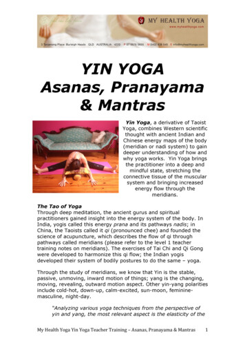
Transcription
PRANAYAMAthe eigAppropriateprovided tiHAVPRANAYAMAAKLONAVALAKAIVALYADHAMAKaivalyadhama S M Yoga Mandir SamitiSwami Kuvalayananda Marg,Lonavla, District Pune, Indiawwwkdham.com Swami Kuvalayananda
POPULAR YOGAPRANAYAMASWAMI KUVALAYANANDAConantKAIVALYADHAMA,Swami Kuvalayananda Marg,Lonavla (410103), INDIA
Swami Kuvalyananda, 1966PREFACE"pb garee we esu fm ARP THe GA ASATFirst Published : January, 1931 Fifth Edition : January, 1972Sixth Edition : 1977(Reverence to Madhava, the Supreme Bliss !Seventh Edition : 1983Blessed by Him the dum shall grow eloquentEighth Edition : 1999——Do—Ninth Edition : 2000and the lame shall stride across a Mountain)It is after fifteen years that we are presenting to the public ourhandbook of Pranayama. No doubt, in the meanwhile we publishedPranayama Part Two, in 1958. But the Pranayama Part Twoalthough containing six varieties of Pranayama that had not beenincluded in Part One and also giving very valuable articles on thesubject of Pranayama, could not present the subject of Pranayamain its proper perspective. Thus the public remained starved ofPrāņāyāmic food for a very long time. During all these years thedemand for a tolerably complete handbook on this subjectcontinued.Tenth Edition : April, 2005Eleventh Edition : April, 2010Printed in IndiaISBN : 81 - 902803-6-8All rights reserved.Price : Rs. 200/-Now we have great pleasure in presenting this verycomprehensive handbook on the subject of Pranayama and weare sure it will receive the same appreciation and patronage fromthe public that our publications have been receiving for the lastfour decades. In the present handbook we have included all theeight varieties of Pranayama that are advocated by the authors ofHatha-Yoga, with a somewhat cursory reference to the fourvarieties of that subject as taught by Pataiijali. The lengthy articlePublished byPrinted aton ‘Respiration’ and also an article on the ‘Physiological andShri O. P. Tiwari, Secretary,Ace Enterprises,Kaivalyadhama, S. M. Y. M. Samiti,Madhu-Raj Nagar,Swami Kuvalayananda Marg,137/A Paud Road,Lonavla - 410 403Pune - 410 038Spiritual Values of Pranayama’ which form an important part ofthe subject as they provided an essential background for clearlyunderstanding it, but which did not appear in Part, have beenretained. Thus we have tried our best to make the present handbook
ivPrāņāyāmaof Prāņāyāma as comprehensive as possible. We must, however,make it clear to our readers that though this handbook is completein itself, it is no substitute for our guarterly journal YogaMimamsa, because that journal contains vast scientificinformation in the form of actual Laboratory experiments and alsomany articles on Pranayama. This information could not beincluded in this handbook because of its limited scope and alsobecause of the type of readers for whom it is intended. Some ofthe articles which had appeared in Pranayama Part Two and whichwere borrowed from Yoga-Mimāņmsā have also been omitted here.These omissions, however, will not come in the way of our readers’understanding the subject in its basic form.The great importance of the practice of Prāņāyāma is clearlybrought out in the last Chapter of this handbook but the treatmentof the subject is from the modern point of view. Hence it wasthought desirable to note here something that our ancient authorshave said about Prāņāyāmic values, both physiological andspiritual.Manu, one of the greatest ancient law-givers, observes thatPrāņāyāma is a specific remedy against the evil tendency of themind and the sense organs.'? Kulluka commenting on theseObservations explains the evil tendencies of the mind as 'Loveand Hatred' ;and the evil tendencies of the sense organs as theattractions of these organs towards the external objects? Thesenatural tendencies ofthe mind and the sense organs are condemnedas evil because success in Yoga depends upon the introversion ofthe mind and the sense organs. A similar view is expressed by the1. gaoi werd mr mer Pema, M.S. VI712. wma)M.S.VI723. sbagfawawavesm: KulltikaPreface*author of Hatha Pradipika* saying that some Yoga experts wereof the same opinion, that is they considered Prāņāyāmic alonewould be sufficient to overcome the evil tendencies of the mindand sense organs.Pataiijali, the greatest exonent of Yoga Sastra gives in his twoSūtras,* namely II. 52 and 53, a clear exposition of the spiritualvalues of Pranayama. The spiritual development of a student ofYoga depends upon two things. First, he must eliminate the factorthat obscures the spiritual light and secondly, he must be able tofix his wandering mind. According to Patatījali a student of Yogais able to make a beginning in both these directions by practisingPranayama.Thus far we have taken notice of the importance of Pranayamafrom the point of view of the ancient authors of the subject ofYoga. We had to touch this point here as it was not discussedeither in Part One or Part Two of Pranayama. previously published.Now we shall indicate some additional features about thepresent handbook.As usual we have very rightly illustrated the subject that isunder discussion in this handbook. Every effort is made tomake the handbook thoroughly instructive as well as interesting.Cautions have been pronounced at every step, making the textbook areliable and safe guide for a practical student of Pranayama.Inspite of this fact we would strongly advise everyone first to gethimself trained under an expert, if possible, and then take up thishandbook for guidance.4. STIPRAwd SIETI ven sft HPI 38.5. qq: sf were: Pat. Y. S. IL 52.RETa Aa Ara: Pat.Y.S. I53.
viPranayamaThree important appendices have been attached to thishandbook. The general hints contained therein have been basedupon our clinical experience at the Arama. The insertion of theseappendices in this handbook, is intended to make it throughlyuseful to the practical student of Yoga.The glossary very tersely gives anatomical and physiologicalinformation which has been detailed in Yoga-Mimamsa. It will,however, serve as a valuable guide to a practical student of Yogaand will make him independent of any text-book of anatomy andphysiology.From what has been said upto now in this Preface, thishandbook can be called to be the fourth impression of Pranayamabecause the largest part of it is taken from Part One although itcontains some chapters borrowed from Part Two.The present handbook of Pranayama owes its publication toMessrs Popular Prakashan of Bombay. They have spared no painsto make it as attractive, instructive and helpful as possible. Thisfirm of publishers has already put in the market our handbook ofAsanas. Our best thanks are due to them for having undertaken topublish our writings simply for the love of our ancient cultureinterpreted in the light of modern science and thought.We have no words to express adequately our gratitude to ourreaders. They have taken very sympathetically to whatever wehave written on the science and practice of Yoga inspite of manya shortcomings for which these writings suffer.We cannot close this short Preface without mentioning a wordabout our service to Yoga. Our personal share in this service isvery small. The overwhelmingly largest share belongs to ourĀšramite Brothers. They are highly qualified university men mostdisinterestedly devoted to the cause of Yoga. Even those brotherswho have no university qualification are men of sublime culturePrefaceviiand have given decades of their precious life to the cause of Yoga.May God bless them all !May the cause of Yoga prosper and may it bring peace to thewar-weary world !KUVALAYANANDAKaivalyadhama,Lonavala, (C.Rly.)15th March, 1966
CONTENTSPREFACEKaivalyadhama is happy to present the 11th edition ofPRANAYAMA to the readers and lovers of Yoga. As you knowSwami Kuvalayananda was the first person to initiate and promotethe scientific investigation into the traditional aspect of Yoga.Based on scientific findings he had critically edited widelyacclaimed books of Asana and Pranayama. The work we initiatedwill always be remembered in the World of Yoga. He was not aperson seeking fame as he always saidrerit Arete sitfrerecit Ferre aisya TEM fai fan 1(Yogarahasya of Dattatreya 3.3)Meaning that an honour to a Yogi is poison while disrespecthelps him to grow and act as an ambrosia. The book Pranayamais still regarded as the best, though he left us in the year 1966. Wecould not improve upon his work. Needless to say, it containsmost systematic, scientific and techniques explained is simple andterse manner. I am sure readers will continue to practice and takeadvantage of his valuable contribution as he said “Yoga has a complete message for humanity. It has a messagefor the human body. It has a message for the human mind. And ithas also a message for the human spirit. Will rich men with theirmoney and young intelligent and healthy men with their selflessservice, come forth to help the Kaivalyadhama to take thismessage, to the farthest corner of the World ?"7)- O. P. sanas Appropriate to Prāņāyāma31-46IILPranayama in . 33XI.Mūrcchā134-136XII.Plāvinī137-139XIII. Physiological andSpiritual Values of Prāņāyāma140-152Appendix - I153-161Appendix - IT162-163Appendix - III164Glossary165-182
xiLIST OF ILLUSTRATIONSFIG. 24, Correct Standing Position for Ujjayai.FIG. 25. Jiana-Mudra or the Symbol of Knowledge.FIG. 1. Median Section through the Head Indicating theRespiratory Tract etc.FIG. 26, Preparation for Closing the Nostrils.FIG. 2. Face with the Mobile Nose Cut off and the Nasal Bonesand Cavities, with the Upper jaw-Bones Exposed.FIG. 28. The right Nostril Closed.FIG.The pharynx Exposed.FIG.The Vocal Cords.FIG.The Trachea and the Bronchial Tubes.FIG.The Thorax Exposed.FIG,The Ribs.FIG.Nāsāgra Drsti or The Nasal Gaze.FIG. Kūsttādēdat.SsBhriimadhya-Drsti or the Frontal Gaze.FIG. 10. Uddiyana in Sitting.FIG. 11. Uddiyana in Sitting (Side View).FIG. 27. Ugly Contortions of the Face.FIG. 29. The Left Nostril Closed.FIG. 30. Both The Nostrils Closed.FIG. 31. Both The Nostrils Open.FIG. 32. Starting Inhalation with Controlled Abdominal Muscles(Front View).FIG. 33. Starting Inhalation with Controlled Abdominal Muscles(Side View).FIG. 34, Full Inhalation with Controlled Abdominal Muscles.(Front View).FIG. 12. Uddiyana in Standing.FIG. 35. Full Inhalation with Controlled Abdominal Muscles.(Side View).FIG. 13. The Carotids Exposed.FIG. 36. Full Inhalation with Protracted Abdomen. (Front View).FIG. 14. Jālandhara-Bandha or the Chin-Lock. (Front View).FIG. 15. Jālandhara-Bandha or the Chin- Lock (Side View).FIG. 37. Full Inhalation with Protracted Abdomen (Side View).FIG. 16. Preparation for Padmāsana.FIG. 39. Full Exhalation (Side View).FIG. 17. Padmāsana or the Lotus Pose.FIG. 40. AbdomenFIG. 18. Preparation for Siddāsana.FIG. 19. Siddāsana or the Accomplished Pose.FIG. 20. Preparation for Svastikāsana.FIG. 38. Full Exhalation (Front View)and Thoraxat the end of Pūrakainand Thoraxat the end of RecakainKapalabhati.FIG. 41. AbdomenKapālabhāti.FIG. 21. Svastikāsana or the Auspicious Pose.FIG. 42. The Tongue Arranged as a Bird's Beak.FIG. 22. Preparation for Samāsana.FIG. 43. The Mouth Widely Opened.FIG. 23. Samasana or the Symmetrical Pose.
CHAPTER IABBREVIATIONS-Āšv.Sr.Ghe. Agera-sufum.d.y. unn o -Ji. Mu. ViPāt.Y.S.Brhadyogi Ya. Sm.therefore desirable that a student of Pranayama is acquainted withaga-arr.Baud. Dh."eraatanatomy and physiology of . Ņya.vowdtfrer-8s. 3 BP.— -PRANAYAMA is a Yogic exercise in respiration. It issome important details of the respiratory system. Hence, wepropose to describe in this chapter a few broad features of theRespiration consists of the alternate expansion and contractionof the thorax, by means of which air is drawn into or expelledfrom the lungs. In this chapter we shall first consider the variousorgans that are directly concerned with this passage of air to andfrom the lungs; and then we shall see how these organs act in thedifferent stages of respiration.SCHEME OF TRANSLITERATIONa-aw-ā;g-i -i;s-uaWRwonw-l5w-e-o;sū; -aiaft - au;The organs of respiration may be enumerated as the nose, thepharynx, the larynx, the trachea, the bronchi and the lungs. Nervesand blood-vessels connected with these parts may also be lookedupon as organs of respiration. Generally, however, the nose is notincluded in such an enumeration. But when we want to write onATM - m; fart - h;respiration from the Yogic point of view, the nose has not only toak o uw-knAGBed-gw-5uzhmw.g-i;sūbe included but also to be studied in detail. In dealing with theseorgans we shall proceed along the passage of the air from outsidezba-tix-dv-dhw-ninto the lungs and as we meet with the nose first, we shall starta-tag - th;q-d;q - dh;A-n;Abi,Ropthis chapter with a description of that organ."-yWÉow-bnt-n«ak-g o w-saloq&-hsg-ksa-t;a-vw-jiTur Nose - - We shall be able to study the nose both externallyand internally with the help of a mirror and of the charts given inthis chapter. In using this mirror the reader should allow light tocome from his back and fall upon the mirror held in his front. The
2Pranayamamirror should be so adjusted as would easily throw reflected lightinto the nostrils when the head is thrown backward, but wouldnot dazzle the eye.We always see our nose in the mirror, But what we generallysee is only the external part of the nostrils. Let our reader have apeep into the internal portions of the nostrils also. He will findthat by the side of the dividing wall, up in the cavity there is ahole in each of the two nostrils. He might think that these holesare a sort of communication between the nose and some otherorgan situated inside the head. But he would be seriously wrongin thinking that way. In fact the external nose is only a small partof the organ anatomically known as the nose. The most importantpart of it is situated inside the head, behind the two holes ourreader has seen, and above the hard palate that forms a part oftheroof of his mouth. We shall presently examine this internalpartof the nose. But before we do so let us finish with the externalpart of that organ.If we feel our nose with our fingers, we find that a very largepart of it is mobile. This is because this portion of the nose,including the dividing septum, is made of different cartilagesattached to one another by a tough. Fibrous membrane and coveredover by the skin. If our reader again looks into his mirrorandexamines the inside of his nostrils, he will find that theskin whichcovers the external surface of the nose, continues inward andalsocovers the lower chamber of the internal nose. He will find eventhe lower portion of the septum clothed similarly in skin.Thisportion of the nasal cavity is called the vestibule. From thewallsof this vestibule, our reader will find, a number of stoutand stiffRespiration3hair projecting. Out of these hair, those that grow from the frontpart project backward, whereas those that rise from the back partproject forward. Thus a sievelike arrangement is provided just atthe entrance of the nose. The external nose has two ends. Theupper end which is connected with the forehead is called the rootand the lower end which is free, is called the apex of the nose.If we again feel the flexible part of our nose and examine theupper as well as posterior borders of it, we find them hard. This isbecause the elastic part of the nose is attached to bones at the topbehind. In Fig.2 the mobile nose has been removed and bones towhich it is attached are exposed to our view. Our reader willimmediately see that the bony borders which carried the flexibleattachments of the nose, represent a picture exactly similar to thatof the heart drawn in the playing cards. Let us observe a few factsabout this bony aperture that directly concern us. Below theforehead and between the eyes there are two bones which we canfeel externally. They are called the nasal bones (1) and form whatis known as the bridge of the nose. A vertical line (3) is seendividing the aperture into two exact halves. This represents thenasal septum. The circular curves at the bottom belong to the twoupper jaw-bones. (5). These curves mark the lower borders of thetwo holes observed by our reader in the back part of his externalnose.We have already stated that the external nose is only a smallpart of the real organ. The most important part of it is situatedinside the head. We have now removed the mobile nose and cometo the entrance of the inner cavities. Let us now note what is thereinside the aperture that lies before us in Fig. 2.
4PranayamaThe aperture which is divided into two halves by the nasalseptum leads to two cavities that tare roughly speaking ovalshaped.These cavities continue backward and open into the throat withRespiration*lie just above the roof of the mouth. The lateral walls of the nasalcavities are somewhat complicated. They are also made of bonesThey rise from below at some distance from the septum, but asholes similar to those we observe on the bony surface before us.they rise they incline towards the septum to meet it at the roof.Each of these cavities has a floor, a roof, and a medical and aSome idea of this inclination of the lateral walls can be had fromlateral wall.Fig. 2. Here the bony aperture represents the external borders ofThe septum which divides these cavities stands for the medialwall of both. It is made of bones all along except in front where athese nasal walls. So these cavities are broad below, but narrowdown at the top. From the lateral walls arise inside the cavitieslarge cartilage fills up the gap. The floor is also bony. A referenceto Fig. la will show this floor in a median section. It is made oftwo bones. The front part is made of the upper jawbones and thescrolllike bones. One ofthem has been marked in Fig. 2 (4). Theseback part, of the hard palate bones. There are two upper jawbones and two hard palate bones. The upper jaw-bones are arrangedside by side and form our upper jaw in which the teeth are set.Behind the upper jaw, the hard palate bones stand side by sideNow we consider the roof. This is also a bony structure. Theand form our hard palate. The lower surface of the two upper jawbones and the hard palate bones go to form the roof of our mouth.If we try to feel the roof of our mouth with our finger from theupper teeth backward, we will meet with two hard surface. One isrough and the other is smooth, The rough surface which we feeljust above our teeth is presented by the upper jaw-bones, and thehard but smooth surface that we cross when the finger is movedbackward belongs to the hard palate bones. Behind this the softpalate can easily be felt. Fig. 1 shows that the upper surface of theupper jaw-bones and the hard palate bones are horizontal and thatis why the floor of the nasal cavities is also horizontal. So theupper jaw-bones and the hard palate bones at once form the roofof the mouth and the floor of the nose. That means the nasal cavitiesscroll-like bones extend all long the length of the lateral walls andopen backward into the throat.floor of the nasal cavities is horizontal; but the roof is arched. Anidea of this arch can be had from Fig. 1b, where the septum isshown arching at the top. Now this septum rises direct to meet theroof, and therefore the upper curve of the septum fairly representsthe arch of the roof. We may note especially two bones that formthe roof. The nasal bones that have already been noticed form thefront slope of the arching roof. Behind these nasal bones the roofis formed by a big bone called ethmoid. The ethmoid is at oncethe roof of the nose and a part of the floor of the brain. That meansit separates the nasal cavities from the brain. Thus the nasal cavitiesstand between the mouth and the brain; and open backwards intothe throat just above the soft palate.Upto now we have studied the bony structure of external noseas well as of the nasal cavities. Now we have to note that thesehard surfaces recovered over with mucous membrane. This mucousmembrane is a continuous piece that lines out the inside of the
6Pranayamaexternal nose except the vestibule, but it also clothes the floor, theroof and the walls of the nasal cavities and continue into thepharynx. When we examine this mucous membrane covering thenasal cavities, we find that functionally two areas are to be mappedout. The upper area covers one third of the total surface of thenasal cavities and the lower area the remaining two-thirds. Havingthe sense of smell situated in it, the upper district is called theolfactory region, the lower district being called the respiratoryregion, as it constitutes the passage for air breathed in and outduring respiration, That means the cavities of the nose have gottwo separate tracts for the two different functions they perform.If we divide the height of the nostrils into three equal parts, theuppermost part is used for smelling and two lower pats are usedfor breathing. None should suppose, however, that the uppermostpart is not at all available as a passage for respiration. Although inthe normal and quiet breathing only the lower two parts are used,Respiration7tract. In fact it is marked by the presence of a rich venous plexus.These anatomical facts have a physiological significance. As notedabove in ordinary quiet breathing, the air moves through the lowertwo-thirds of the nasal area. The air in the upper one-third isscarcely disturbed. Now that portion of the mucous membranewhich lines the passage of the air must be stouter than the portionwhere the air is stationary the hours of normal breathing. Thegreater vascularity of the respiratory tract has also a purpose toserve. When external air is to be breathed into the lungs, it mustbe warm and moist, otherwise it may have an injurious effectupon the delicate structure of the lungs. Now the large supply ofvenous blood which is present in the plexus situated in therespiratory region, raises the temperature of the air duringinspiration and also moistens it.It has been stated above that the sense of smell is situated inin forced breathing the upper third is also utilized. The sense ofsmell, however, is confined to the upper third only, having nothingto do with the two lower regions. This is because the sense ofsmell depends upon the presence of the olfactory nerve-endingsthe upper one-third of the nasal cavities. This sense consists ofwhich are distributed only over the upper third area. (Vide Fig.as we have seen above, separates the nasal cavities from the brain.1(4)).Two features which distinguish the mucous membrane coveringthe olfactory region from that covering the respiratory tract,deserve our attention here. The mucous membrane which clothesthe respiratory tract is thick and spongy whereas that which linesthe olfactory region is softer and more delicate. The otherdistinguishing feature is the very great vascularity ofthe respiratoryvery fine nerve filaments from twelve to twenty in number. Theyare distributed like a thick brush both on the septum and the lateralwalls. Here these nerves descend through the ethmoid bone which,Through the ethmoid the olfactory nerves reconnected with theolfactory bulb which in its turn is joined to the base of the brainthrough the olfactory tract. Fig. 1 illustrates the olfactory nerves,the bulb, the tract as also the base of the brain. When the fineendings of the olfactory nerves are stimulated by particles carryingodour with them, sensation of smell is experienced.Following facts may be remembered with advantage in regardto the sense of smell.
8PranayamaRespirationThe delicacy of the sense of smell is very remarkable. It has3STue PHarynx--It has been stated above that the nasal chambersbeen calculated thāt even 1000000000 ofa grain of musk can beposteriorly open into the throat. These openings are situated abovedistinctly smelt.the soft palate and below the base of the cranium. This portionBut when the particles issuing from an odoriferous substanceare very few, their presence in the air may not be appreciated innormal breathing, because they pass through the respiratorypassage only and are not presented to the nerve-endings in theolfactoty region. If under such circumstances a sudden sniff ismade, air is forced even into the olfactory tract and the faint odourmarks the beginning of the pharynx. Now if our reader again lookscan be detected.Even a liberal proportion of odoriferous particles in the air failmaking itself felt. For the sense of smell to be excited, the mucousmembrane must be neither too dry nor too moist. So When onecatches cold his sense of smell is dulled owing to the presence ofexcessive moisture. There is another reason also why one doesnot smell properly during cold. The swelling of the mucousmembrane covers the nerve filaments rendering them inaccessibleto the odoriferous particles.into the mirror with his mouth widely opened, he will observethat there is something like a wall of flesh covered over with themucous membrane, stretching behind the tongue and the softpalate. This wall arches above the soft palate in something like adome. It is exactly under this dome that the posterior openings ofthe nose are located. Below the tongue the wall descends in theform of a sack that ends in two openings, one of which leads tothe aesophagus and the other to the larynx (Vide Fig. 3). From thebackward openings of the nasal chambers to the lower openingsleading to the aesophagus and the larynx, the same canal stretchescontinuously and is known as the pharynx. Our reader need notbe told that the mouth is only an opening in the anterior wall ofthe pharynx. So upto now we have noticed five openings of thepharynx, two nasal, one oral, one aesophageal and one laryngeal.There are two more orifices which pierce the pharynx. They areIt is a matter or common experience that perfumes make theStrongest impression to start with. Afterwards they grow fainterand even their presence is lost upon us, if we continue long to bein their presence. This circumstance can be accounted for by theof the ears. The portion of the pharynx that is situated above thefatigue of the sense of smell. The olfactory apparatus is soonsoft palate is called the nasal part of the pharynx, that situatedexhausted and fails us on that account. If, however, we take abehind the mouth and the tongue is called the oral part, whereasround in the fresh air, the apparatus is refreshed and we can againappreciate the odour.the remaining portion is called the Jaryngeal part.situated in the side walls of the pharynx, one on each side, abovethe soft palate. They are called Eustachian orifices, because theymark the openings of the Eustachian tubes which run to the cavitiesThe pharynx is used for the passage of the air as it is breathedin and breathed out. In inhalation the air drawn through the nasal
10Pranayamacavities passes across the nasal and oral parts of the pharynx andthen getting down the larynx goes into the trachea and the lungs.In exhalation the air expelled from the lungs follows the reversepath. At the time of breathing the aesophagus as well as theEustachian tubes remain closed and the possibility of the air goinga wrong way is avoided. Again the soft palate leaves, between itand the back wall of the pharynx, an opening sufficient for freemovement of the flowing air. Hence, there is no obstruction in theway of respiration.At the time of speaking, however, the soft palate completelycovers the upper part of the pharynx, so that no air can find itsway upward into the nasal part. But in some person, the soft palateis defective, there being a small cleft in it. When these peopleattempt speaking some of the air from the lungs escapes throughthis cleft above the palate, and finds its way through the nasalpassages that are ever open, adds nasalized element to their voice.Our reader knows that he uses a part of the pharynx also forswallowing. Food travels through the oral part into the laryngealpharynx and then gets into the aesophagus. The question is whyfood going down the pharynx does not run into the larynx and isalways pushed down the aesophagus. For this purpose we have torefer to a small organ named epiglottis.The epiglottis is situated at the root of the tongue (Vide Fig. 3)and serves as cover for the larynx in times of need. In the act ofswallowing, the larynx is raised, and the descending morsel lowersthe epiglottis which meeting the raised larynx completely coversits mouth. Thus the larynx being closed, food finds its way to theaesophagus or gullet. The rising of the larynx can be felt byRespiration1!anybody by lacing his fingers on the middle of his throat andimitating the act of swallowing. If, however, through mistake evena small particle of food gets the wrong way, we mean gets intothe larynx, violent coughing ensues, the system forcibly tries toexpel the intruder. Food does not get into the nasal part, becausethe soft palate completely shuts out that portion during the act ofswallowing.The mucous membrane covering the nose is continuous withthe pharynx. It is also to be noted that it continues to cover all thepassages leading from the pharynx. It is this circumstance whichmakes a trouble starting with the throat very often spreads to thenose, the ear and the larynx. That is why running of the nose,deafness of the ear and coughing are on many occasions seengoing together.Tur Lanvxx---We have noted above that the pharynx has twopassages opening from its lower end. They are the aesophagusand the Jarynx. Both the aesophagus and the larynx pass throughthe nēck, the former is situated in the posterior part of it, whereasthe latter is situated in the anterior portion. The larynx begins i
PRANAYAMA the eig Appropriate provided ti LONAVALA KAIVALYADHAMA Kaivalyadhama S M Yoga Mandir Samiti Swami Kuvalayananda Marg, Lonavla, District Pune, India wwwkdham.com PRANAYAMA Swami Kuvalayananda VALYAD HAMA K A . POPULAR Y