Transcription
Diagnostic MedicalMicrobiologyLaboratory ManualDr. Abdelraouf A. ElmanamaPh.D. MicrobiologyMedical Technology DepartmentIslamic University-Gaza2007
Table of ContentsIntroductionGeneral View on the Parameters Used in the Process of MicroorganismIdentificationGeneral InformationUrine CultureBlood CultureCerebrospinal Fluid (CSF)Body Fluid Culture, SterileConjunctival DischargeEar DischargeGenital Culture and sensitivityPus(wound, Abscesses, Burns and sinuses) culture & sensitivitySputum Culture and SensitivityStool Culture: Routine, Salmonella & ShigellaStool Culture: E. coli O157:H7Stool Culture: Vibrio spp.Throat Swab for Beta-Haemolytic Streptococcus Culture, Group A OnlyIsolation and Identification of Enterobacteriaceae and Pseudomonas: Part 1Isolation and Identification of Enterobacteriaceae and Pseudomonas: Part 2Isolation and Identification of Streptococci and EnterococciIsolation and Identification of StaphylococciIsolation and Identification of Neisseriae, Mycobacteria, and AnaerobesSerology, Part 1: Direct Serologic TestingSerology. Part 2: Indirect Serologic TestingUsing Antimicrobial Chemotherapy to Control MicroorganismsAppendix: Common Antibacterial AntibioticsENUMERATION OF MICROORGANISMSKOH SMEARAbdelraouf A. Elmanama[2]Ph.D Microbiology
INTRODUCTIONThis diagnostic microbiology manual is designed to be used in conjunctionwith lecture textbook and other resources. Microbiological techniques aredifferent in many ways when compared with other laboratory disciplines.Although results are not obtained in a short time, the time required to performthe test is very short. Most of the techniques are simple, yet requires a greatdeal of theoretical background to be correctly interpreted. For this reason,each laboratory exercise was supplemented with the theory behind it.As a guide for the student, each experiment was started by the titleOBJECTIVE, which means what is required from the student to learn uponcompletion of the exercise. It is in the form of questions (name, define,discuss, etc.). Students should try answering all the questions when readingand working each exercise.Before performing any of the exercises in this manual, one should read thesafety precaution and measures as well as the exercise (the materialsneeded, the procedures, and the expected results). This will ensure the safetyof the student and also will ensure good results.General View on the Parameters Used in the Process of MicroorganismIdentificationBefore one can proceed to identify a microorganism, the characteristics of thatorganism have to be determined in details. The major characteristics whichare observed include the following:A. Cultural CharacteristicsIn clinical terms, it is the shape, size, color, elevation and other characteristicsof the colony formed on the culture plate. In taxonomy, it includes the nutrientrequirements for the growth of the organism and the physical factors such astemperature, pH and the incubation period. These factors are used to identifycertain pathogenic species but less commonly used in routine procedures.The cultural characteristics of a microorganism usually vary depending on themedia used and many other factors. Some experienced microbiologists couldhave a good guess about the identity of a microorganism just by its culturalcharacteristics, but this was proven to be a bad technique. Students as well asmicrobiologists are advised to follow strict procedures for the identification ofisolates from clinical specimens.B. Morphology and StainingThis includes the microscopic appearance of a stained preparation of theorganism. Useful information to be taken into account, are the size of theindividual cells, cell shape and arrangement and staining reaction if differentialstaining procedures is used.Abdelraouf A. Elmanama[3]Ph.D Microbiology
EXAMPLE: A gram stained film prepared from a pure culture of certainmicroorganism shows the following:-Small spherical cells "Cocci"-Arranged in clusters-Gram-positive violet in colorSome laboratories which have a little facility could give the report of amicrobiological examination of a clinical specimen just by stating theirmorphological characteristics and the sensitivity testing results.C. BIOCHEMICAL CHARACTERISTICSFrequently, the identity of a species requires detailed knowledge of itsbiochemical activities, since other characteristics are not sufficiently distinctiveor differential. For example, the bacterium Escherichia coli, a normalinhabitant of our intestinal tract, is indistinguishable microscopically fromSalmonella typhi, the bacterium that causes typhoid fever. However, if thesetwo bacteria are examined for their metabolic (or biochemical) characteristics,they are found to be very different and distinguishable on this basis.Numerous laboratory techniques are available for the characterization ofmicroorganisms. In general, the microorganism is grown in the presence of aspecific substrate, after which the culture is examined to determine whatchemical changes have taken place. This subject will be discussed in detailsin other parts of the handout.D. SEROLOGICAL CHRACTERISTICSSometimes, to identify a species as E. coli is insufficient, for the reason thatsome strains of this organism are non-pathogenic and others are highlyassociated with diseases. Serological testing in such case will identify theexact strain number based on testing against prepared specific antisera.In-Vivo serological tests (skin tests) are of great value in the diagnosis ofmany bacterial, fungal and viral infections.E. OTHER CHARACTERISTICSTo identify some strains of bacteria, one may need to look for othercharacteristics than those mentioned above. Phage typing and animalinoculation are examples of uncommon techniques used in the identificationprocess.Abdelraouf A. Elmanama[4]Ph.D Microbiology
General InformationThe microbiology laboratory is considered to be vital and the take the greatamount of the general work load of the laboratory. Receiving and recordingspecimens, culturing, staining, isolation and identification of pathogens anddoing sensitivity tests for the isolated pathogens are the major tasks.Who Can Request Laboratory Services1. All licensed physicians, dentists and optometrists.2. All public health nurses and physicians assistants.3. Local Health Departments.4. Communicable Disease Specialists.Reports shall be given only to the submitter. Private individuals will not receivereports.Information For Microbiology Laboratory StaffGeneral Requirements for Collecting and Submitting SpecimensProper collection and adequate amounts of specimen are required. Thefollowing criteria should be used as guidelines:Medical Group employees who handle laboratory specimens have relativelyhigh rates of work-related hepatitis and other transmittable diseases. Looselycapped containers and soiled requisitions sent to the laboratory are asignificant risk to all who come in direct contact with these contaminatedmaterials or areas contaminated by such materials. Therefore, laboratory staffwill not accept soiled laboratory requisitions/leaking specimen containers.When needed, a written test request must include the following information: Patient detailsHospital No.Name: First name and family nameSexDate of birth/AgeAddressSocial security no. (insurance)For females: whether pregnant or lactatingDetails of illnessPresenting signs/symptomsDuration/date of onsetRecent travel historyImmunizationsAbdelraouf A. Elmanama[5]Ph.D Microbiology
Identification of SpecimensType of specimen (Exact source and nature of specimen)Collection date and timeLaboratory numberLaboratory findingsTests requestedOrdering physicianAlso, ALL specimens must be properly labeled with the patient's full name,date and time of collection, and specimen source.Swab Specimens: Separate specimens must be submitted for each specificrequest, i.e., one for bacterial culture, one for fungal culture, etc.Special Culture / Specimen Requirements:Anaerobic Specimens: Submit in anaerobic transport containers or in asealed syringe with no bubbles.SPECIMEN COLLECTIONProper specimen collection, container labeling, and culture requests are theresponsibility of the ordering physician. Technologists in the ClinicalMicrobiology Laboratory will be familiar with specimens of choice and propercollection techniques.The technologist in the laboratory will directly handle specimens of clinical andenvironmental source which are received from the Postal Service or handcarried to the laboratory.The technologists will handle the clinical specimens completely by thefollowing procedure.SPECIMEN HANDLING AND STORAGESpecimen containers and requisitions will be delivered to the ClinicalMicrobiology Central Processing Area (CPA) within the specified period(depending on the specimen source and type) of collection. Upon receipt, theCPA staff will check requisitions for completeness. Specimens will be storedproperly until they are picked-up by the microbiology staff. The CPA staff willassign numbers for the specimens and indicate them on the originalrequisitions. When STAT requests are received, the CPA staff willimmediately notify the microbiology supervisor and arrange for the immediatedelivery of the specimen.Abdelraouf A. Elmanama[6]Ph.D Microbiology
SPECIMEN DELIVERYThe Clinical microbiology Laboratory recommends that a specimen should betransported to the laboratory as soon as possible (a maximum delay isindicated for each type of specimen; see next sections for individualinstructions). If more than one specimen is received for one type of analysis,the Processing Area staff will note on the requisition "Duplicate Specimen" inred ink.Actively growing cultures of organisms for identification should be submittedon tubed media appropriate for the organism being submitted. Seal the tubewith water proof tape. All specimen containers should be closed tightly orsealed in order to prevent leakage and contamination. Media in Petri dishesor liquid cultures are not an acceptable transport media.LABELING, LOGBOOKUpon receiving the specimen and requisition with complete data., record it inthe microbiology log book in numeral order. The number assigned to thespecimen is written on the specimen container and the requisition form,culture media containers, and culture media plates. In addition, date and timeof processing and the name of the patient should be written clearly on allAbdelraouf A. Elmanama[7]Ph.D Microbiology
culture plates, tubes, slides or whatever used in the processing of thespecimen.SPECIMEN REJECTION CRITERIA.General IssuesIn general, specimens for the microbiology laboratory are unacceptable if anyof the following conditions apply:1. The information on the label doesn’t match the information on therequest form.2. The specimen was transported in an improper container or at wrongtemperature .3. The quantity of the specimen is insufficient to carry out all the requiredexamination.4. Leaking specimenBACTERIOLOGY Blood received in blood culture bottles is unsuitable for fungalisolation.Saliva is unacceptable for culture. Submit "deep cough orinduced sputum."Multiple urine, stool, sputum, or routine throat specimens senton the same day from the same source from the same patient.Other specimens unsatisfactory for cultures are: Specimens in fixative (Formalin).Dried out swabs.Foley Catheter tips.24 hour urine/sputum for routine bacteria, or fungi.Urine held two hours or more at room temperature.Fluids received in culturette tubes.Swab material for anaerobic culture not in the proper anaerobictransport.Gram stains for Neisseria gonorrhea on vaginal or anal cryptspecimens are not diagnostic and will not be performed.Stool specimens for culture from a patient who has been aninpatient greater than 5 days will not be performed.Anaerobic cultures on vaginal, cervical, urine (unless suprapubictap), sputum or fecal specimen.Every effort should be made to contact the physician or unit if aspecimen is rejected. The physician will be informed about the reason/s forspecimen rejection.Abdelraouf A. Elmanama[8]Ph.D Microbiology
REPORTING OF MICROBIOLOGY RESULTS(Pathogenic Organisms versus Normal Flora)Reporting of Bacteriology Results.CSF, Body Fluids, Blood, and Wounds: Positive gram stains, uponpreliminary examination, will be reported to the physician. Identification of anorganism isolated will be performed on all aerobic organisms as appropriate.Ear: Potential pathogens, i.e., S. aureus, gram-negative organisms will beidentified and antimicrobial sensitivities performed.Eye: Report identification on any organisms isolated.appropriateSensitivities asGastrointestinal: Routine screen for Salmonella, Shigella, and specialcultures for Campylobacter, Vibrio, and E. coli 0157:H7. The lab will reportPseudomonas and Staphylococcus aureus. Proteus will be reported on thepediatric patients. Negative cultures will be reported as “No EntericPathogens Isolated.”Lower Respiratory (Aspirates): Report any pathogenic organisms isolatedNasal/Nasopharyngeal: Report any gram negative rod, S. aureus, S.pneumoniae, H. Influenzae, N. meningitidis, Group A Streptococcus.Skin: Predominant organism will be identified.performed on coagulase negative Staph.No sensitivity will beSputum: Specimens evaluated as adequate for culture are screened forpotential pathogens. Legionella must be specifically requested.Throat Cultures: Routinely screened for Group A Strep. Reported asPositive for Beta-hemolytic Strep. Group A, or Negative for Group A Strep.Physician must specify if a culture is to be screened for other than group AStrep. A positive Strep list is provided daily to Primary Care and EmergencyRoom for immediate follow-up.Mouth Cultures: Must specify organism of interest to be screened, i.e., C.albicans, C. diptheriae, etc., or Cystic Fibrosis patient for potentialpathogens. Tooth sockets from dental clinic will be screened for anypredominating organism.Urines: Report identification and antimicrobial sensitivities on colony countsgreater than 10,000. Female Urine will be screened for S. saprophyticus.Susceptibility testing will not be performed on Streptococci (other thanEnterococci), Corynebacterium species, or Lactobacillus. Plates withthree or more organisms will be reported as "three or more organisms, pleaserepeat." Mixed flora of less than 10,000 colonies each will be reported as"Normal Skin Flora."Abdelraouf A. Elmanama[9]Ph.D Microbiology
Vaginal/Cervical:Report predominant organism.Mixed cultures ofLactobacillus, diphtheroids, staphylococcus, alpha streptococcus,Acinetobacter, members of Enterobacteriaceae and yeast will beconsidered Normal Vaginal Flora.Reporting of Susceptibility Testing Results.After initial identification and susceptibility testing, susceptibility testing will beperformed every 4-5 days on biotypically identical organisms isolated from thesame patient from the same site. Cultures will be held 48 hours should theclinician feel that repeat susceptibility testing is indicated.Reporting of TB/MYCOLOGY Results.All acid-fast and fungal organisms isolated will be reported.Abdelraouf A. Elmanama[10]Ph.D Microbiology
Urine CultureAim of the testAn etiological diagnosis of bacterial urinary tract infection by quantitativecultivation of the urine with identification and susceptibility test of the isolatedbacteria(s).Types of specimenUrine (Midstream urine), suprapubic aspiration, catheterized urine.Criteria of specimen rejectionUn-refrigerated specimen older than 2 hours may be subject to overgrowthand may not yield valid results; unlabeled specimen; mislabeled specimen;specimen in expired transport container; 24 hours urine specimens.Pathogens and commensalsUrine specimenCommon pathogenscommensal floraNeisseria gonorrhoeaethe urine is sterile except for theurethral mucosa which support thegrowth of microflora as:Diphtheroid bacilliLactobacillus sppCoagulase negative Staphylococciα Haemolytic StreptococciBacillus sppNon pathogenic Neisseria spp.Anaerobic cocciCommensal MycobacteriumCommensal Mycoplasma spp.E. coli and other EnterobacteriaceaeEnterococcus sppStaphylococcus aureusStaph saprophyticusCorynebacterium jeikeiumAcinetobacter sppPseudomonas sppGardnerella vaginalisβ-haemolytic streptococciSalmonella spp (early stage of infection)ParasitesSchistosoma haemetobiumTrichomonas vaginalisPre specimen processingPatient preparingInstruct the procedures for the patientSpecimen collection Collection of midstream urine for bacterial investigation:Patient not needing assistance:Give the patient a suitable container.Instruct the patient before the collection, preferably withillustration.Tell the patient not to touch the inside or rim of the container.Abdelraouf A. Elmanama[11]Ph.D Microbiology
Male:1. If not circumcised, draw back the foreskin.2. Begin to urinate, but pass the first portion into the toilet.3. Collect the mid-portion of urine into the container, and pass the excess intothe toilet.Female:1. Squat over the toilet and separate the labia with one hand.2. Void the first portion of urine into the toilet.3. Collect the mid-portion of urine into the container and pass the excess intothe toilet.Infants:- Have ready: Clean, preferably sterile container of appropriate size or aplastic bag, cotton wool or gauze pads, handwarm soapy water.1. Clean the external genitals.2. Give the child as much liquid as possible just prior to the collection.3. Seat the child on the lap of the mother, nurse or ward attendant.4. Collect as much urine as possible in the container or plastic bag when thechild urinates.Note: First morning specimens yield highest bacterial counts from overnightincubation in the bladder, and are the best specimens. Colony countinterpretation standards are based on controlled studies from first earlymorning collections. Forced fluids or random specimens dilute the urine andmay cause reduced colony counts. Hair from perineum will contaminate thespecimen. The stream from a male may be contaminated by bacteria frombeneath the prepuce. Bacteria from vaginal secretions, vulva or distal urethramay contaminate transport. Organisms from hands or clothing mightcontaminate. Receptacle must be sterile. Read Patient Preparation.Who will collect the specimenMidstream urine is collected by the patient. If disabled, nursing staff will assistin collection. For catheterized specimen, nursing staff will collect thespecimen. Suprapubic aspiration is performed by the physician.Quantity of specimenTo fill line on transport tube ( 20 mL)Abdelraouf A. Elmanama[12]Ph.D Microbiology
Time relapse before processing the sampleThe maximum time allowed for processing a urine sample is 2 hours from thetime of collectionStorageAt room temperature unless delay is inevitable; it must be refrigeratedAbdelraouf A. Elmanama[13]Ph.D Microbiology
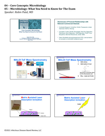
Specimen processingMedia1. Blood Agar,2. MacConkey Agar3. Nutrient AgarCulturing procedure Mix the urine sample to re-suspend microorganism present. Dip a 1 µl calibrated loop in vertical position in the urine andremove the loop and use the collected fluid to inoculate anutrient agar plate that will be used for urine plate count. Take another loop to streak Blood agar and another loop tostreak MacConkey agar plates. Streak the Nutrient agar plate to cover all surface area.A plate count of 10,000 CFU/ml of pure culture should be considered positiveand isolated organism should be identified and sensitivity test will beperformed.UrineWet mountBlood AgarMacConkey AgarNutrient AgarPost specimen processingInterfering factors:Patient on antibiotic therapy.Improper sample collection.Result reporting:Report wet mount as an initial report.Report the isolated pathogen and its sensitivity pattern as a final report.Turn around time:Wet mount results should be available 1 hour after specimen receipt.Isolation of a possible pathogen can be expected after 2-3 days. Negativeculture will be reported out 1-2 days after the receipt of the specimen.Additional informationA single culture is about 80% accurate in the female; two containing the sameorganism with count of 105 or more represents 95% chance of true bacteriuria;three such specimens mean virtual certainty of true bacteriuria. Urinary tractinfection is significantly higher in women who use diaphragm-spermicideAbdelraouf A. Elmanama[14]Ph.D Microbiology
contraception, perhaps secondary to increased vaginal pH and a higherfrequency of vaginal colonization with E. coli. A single clean voided specimenfrom an adult male may be considered diagnostic with proper preparation andcare in specimen collection. If the patient is receiving antimicrobial therapy atthe time the specimen is collected, any level of bacteriuria may be significant.When more than two organisms are recovered, the likelihood of contaminationis high; thus, the significance of definitive identification of the organisms andsusceptibility testing in this situation is severely limited. A repeat culture withproper specimen collection including patient preparation is often indicated.Periodic evaluation of diabetics and pregnant women for asymptomaticbacteriuria has been recommended.2 Institutionalized patients, especiallyelderly individuals, are prone to urinary tract infections, which can be severe.Cultures of specimens from Foley catheters yielding multiple organisms withhigh colony counts usually represents colonization of the catheter and not truesignificant bacteriuria. Most laboratories limit the number of organisms whichwill be identified when recovered from urine to two. Similarly, most do notroutinely perform susceptibility tests on isolates from presumablycontaminated specimens. Failure to recover aerobic organisms from patientswith pyuria or positive Gram's stains of urinary sediment may indicate thepresence of mycobacteria or anaerobes. As the number of patients who arechronically catheterized increases, so does the controversy on whatconstitutes a diagnostic specimen. Few clinical studies have been performedto support the identification of more then two organisms or implicate usual siteflora (eg, diptheroids, alpha or gamma streptococci, and coagulase-negativestaphylococci other than S. saprophyticus).Abdelraouf A. Elmanama[15]Ph.D Microbiology
Blood CultureAim of the testAn etiological diagnosis of bacteremia by aerobic and anaerobic cultivation ofthe blood, with identification and susceptibility test of the isolated organism(s).Blood culture should be made for cases with suspected septicemia,endocarditis, and bacteremia secondary to localized infections (pneumonia,intraabdominal abscesses, pyelonephritis, epiglottitis, meningitis). In this casethe blood culture may provide an etiological diagnosis of the localizedinfection.Types of specimenWhole bloodCriteria of specimen rejectionBlood collected in tubes or bottles other than aerobic and anaerobic bloodculture bottles. If the information on the label does not match that of therequest form. Specimens for anaerobic blood culture received in aerobicbottles or vice versa.PathogensBlood is a sterile body fluid and normally contains commensalsCommon pathogensStreptococcus sppStaphylococcus aureusListeria monocytogenesCorynebacterium jeikeiumHaemophilus influenzaSalmonella typhiPseudomonas aeruginosaFungiCandida albicansOther candida sppHistoplasma capsulatumBacteroides fragilis and other anaerobic bacteriaCoagulase negative staphylococciEnteric gram negative bacilliNeisseria meningitidesNon fermenter gram negative bacilliCryptococcus neoformansCoccidoides immitisPre specimen processingPatient preparingThe major difficulty in interpretation of blood cultures is potentialcontamination by skin flora. This difficulty can be markedly reduced by carefulattention to the details of skin preparation and antisepsis prior to collection ofthe specimen.Abdelraouf A. Elmanama[16]Ph.D Microbiology
Skin preparation: First cleanse the vein puncture site with isopropanol. Thenuse tincture of iodine or povidone iodine to disinfect the site usingprogressively larger concentric circles. Iodine should remain in contact withskin for about 1 minute or until dry to ensure disinfection. The vein puncturesite must not be palpated after preparation. Blood is then drawn. Followingvein puncture, alcohol is used to remove the iodine from the site.Specimen collectionBlood cultures should be drawn prior to initiation of antimicrobial therapy. Ifmore than one culture is ordered, the specimens should be drawn separatelyat no less than 30 minutes apart to rule out the possibility of transientbacteremia by self-manipulation by the patient of mucous membranes in themouth caused by brushing teeth, etc or by local irritations caused byscratching of the skin.The time of collection must be indicated. Strict aseptic technique is essential.If present remove the plastic cap from the blood culture bottles, swab thestoppers with tincture of iodine or povidone iodine and allow to dry. Collect 20mL blood in a sterile plastic syringe and inoculate at least 10 mL blood (asindicated on bottle) into each bottle or use Vacutainer and butterfly collectionset and monitor the fill using the graduations on the side of the bottle. Formore information about the amount of blood, please refer to the blood bottlesmanufacturer’s user guide.Quantity of specimenVolume inoculated in sets of culture bottles for aerobic and anaerobiccultivationChildren below 2 yearsI mL of venous blood in 2 bottlesChildren 2-5 years2 mL of venous blood in 4 bottlesChildren 6-10 years3 mL of venous blood in 4 bottlesChildren 11-15 years5 mL of venous blood in 4 bottlesChildren above 15 years and adults 5 mL venous blood in three sets of bottles (6 bottles).StoragePre-incubate or maintain specimen at room temperature. Do not refrigerateContainerOne aerobic and one anaerobic blood culture bottle. Do not vent.Specimen processingMediaAerobic Blood culture bottleAnaerobic Blood culture bottleMacConkey AgarBlood AgarChocolate AgarAbdelraouf A. Elmanama[17]Ph.D Microbiology
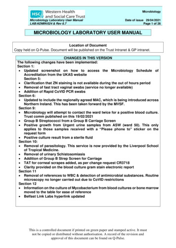
MethodBlood is injected to both aerobic and anaerobic bottles and incubated for up to10 days at 37 oC. Discard as negative after the 10 days incubation period isexpired. During the incubation period, a gram stain and subculture ontoappropriate media should be done. See diagram below:B lo odA ero b icA naero b icG ram S tainB lo o d A garG ram stainC ho co late A garM acC o nkeyA garB lo o d A garPost specimen processingInterfering factorsPatient on antibiotic therapyResult reporting:Any isolated organism will be reported. Antibiotic sensitivity will also beincluded with the report.Turn around timeInitial blood culture results will be reported as soon as it shows growth. Finalresults with sensitivity will be issued after 24-48 hours of the initial report.Negative results will be issued after 10 days of culture submission.Interpretation of Positive Blood Cultures Virtually any organism, including normal flora, can cause bacteremiaA negative culture result does not necessarily rule out bacteremia;false-negative results occur when pathogens fail to growA positive culture result does not necessarily indicate bacteremia;false-positive results occur when contaminants grow.Gram-negative bacilli, anaerobes, and fungi should be consideredpathogens until proven otherwise.The most difficult interpretation problem is to determine whether anorganism that is usually considered normal skin flora is a truepathogen.Abdelraouf A. Elmanama[18]Ph.D Microbiology
LimitationsThree negative sets of blood cultures in the absence of antimicrobial therapyare usually sufficient to exclude the presence of bacteremia. One set isseldom ever sufficient.1 Prior antibiotic therapy may cause negative bloodcultures or delayed growth. Blood cultures from patients suspected of havingBrucella or Leptospira must be requested as special cultures. Consultationwith the laboratory for special culture procedures for the recovery of theseorganisms prior to collecting the specimen is recommended. Yeast often areisolated from routine blood cultures. However, if yeast or other fungi arespecifically suspected, a separate fungal blood culture should be drawn alongwith each of the routine blood culture specimens. See separate listing forproper collection of Blood Fungus Culture. Mycobacterium avium complex(MAC) is frequently recovered from blood of immunocompromised patients,particularly those with acquired immunodeficiency syndrome, AIDS. Specialprocedures are required for the recovery of these organisms; Contact lab.Abdelraouf A. Elmanama[19]Ph.D Microbiology
Cerebrospinal fluid (CSF)Aim of the testDiagnosis of the bacteria or fungal meningitis by microscopic examination andculture with identification and susceptibility test of the isolated organism.Types of specimenCSFCriteria of specimen rejectionNon sterile container and the general causes for rejection stated in theintroductionPathogen and commensalsInfection of C.S.FCSF is a sterile fluid and does not contain any commensals, however, careshould be taken not to contaminate the specimen with skin normal flora duringcollection.Common bacterial pathogenHaemophilus influenzaeNeisseria meningitisStreptococcus pneumoniaeGroup A & B streptococciGram negative bacilliListeria monocytogenesTreponema pallidum (rare)Brucella (rare)Salmonella (rare)Toxoplsma (rare)Microbes that cause chronic meningitisM. tuberculosisCryptococcus neoformansCoccidoides immitisHistoplasma capsulatumBlastomyces dermatitidesCandida spp.NocardiaActinomycesPre specimen processingSpecimen collectionWho will collect the specimenOnly physiciansAbdelraouf A. Elmanama[20]Ph.D Microbiology
Quantity of specimen3 ml of CSF is sufficient for cultureTime relapse before processing the sampleCSF is an emergency specimen and should be processed immediatelyStorageRoom TemperatureSpecimen processingMedia 2 Blood AgarChocolate AgarMacConkey AgarFluid ThioglycollateCult
Abdelraouf A. Elmanama Ph.D Microbiology [3] INTRODUCTION This diagnostic microbiology manual is designed to be used in conjunction with lecture textbook and other resources. Mic