Transcription
Respiratory SystemKristine Krafts, M.D.
Respiratory System Objectives Describe the components and functions of theconducting and respiratory portions of therespiratory system. Describe the embryologic steps in the developmentof the respiratory system. Describe and compare the characteristicmicroscopic features and functions of the differentregions and airways of the respiratory system,including lining epithelium, glands, cartilage, smoothmuscle and elastic fibers.
Respiratory System Lecture Outline Introduction Conducting portion Respiratory portion Pleura
Respiratory System Lecture Outline Introduction Anatomy Embryology
Respiratory System Lecture Outline Introduction Anatomy
Anatomy of therespiratory system
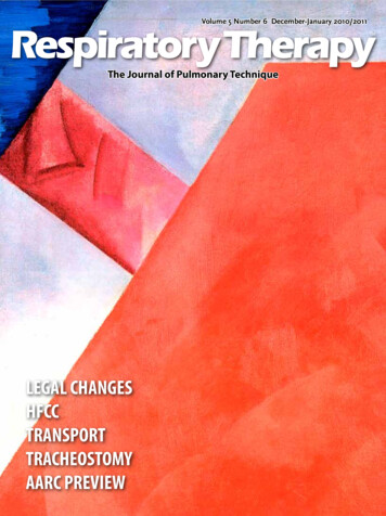
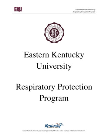
Two Functional Parts of the Respiratory Tract Conducting portion Conduct air to and from alveoli Condition the air (warm, humidify, filter) Respiratory portion Gas exchange
TracheaPrimary bronchusSecondary bronchiRight lungConductingportionLeft lungBronchiole (1 mm or less in diameter)Terminal bronchioleRespiratory bronchioleAlveolar ductRespiratoryportionAlveolar sac
Respiratory System Lecture Outline Introduction Anatomy Embryology
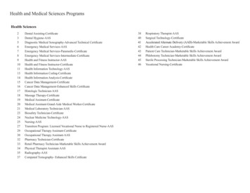
PharynxEsophagusLungsStomachLiverPancreasDuodenum (½)GallbladderStuff that develops from the foregut
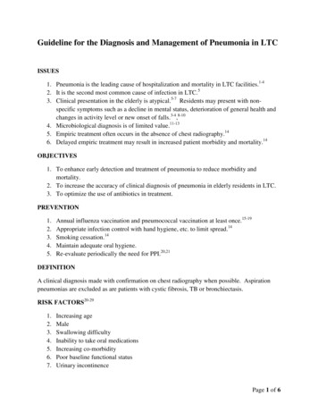
Endoderm gives riseto the epithelium andglands of the larynx,trachea, bronchi andthe pulmonaryepithelium.Mesoderm gives riseto the connectivetissue, cartilage andsmooth muscle ofthe respiratory tract.Development of respiratory diverticulum (bud)
Development of bronchiand bronchioles The lung bud divides intotwo bronchial buds, whichdivide and form secondaryand tertiary bronchi. As bronchi develop, thesurrounding mesenchymeforms cartilage, smoothmuscle, connective tissueand capillaries.
When can the baby breathe? By week 24, respiratory bronchioles are presentand lungs are vascularized. Respiration is possible– but chances of survival outside placenta areslim. Weeks 24-26: terminal alveolar sacs develop andtype II pneumocytes secrete surfactant (decreasessurface tension) By week 26-28: there are enough alveolar sacs andsurfactant to allow a prematurely born infant tosurvive without medical intervention.
Strange but true 95% of mature alveoli develop after birth! The newborn infant has only 1/6 – 1/8 thenumber of alveoli as adults. Most alveolar growth is done by age 8.
Lungs at Birth At birth, the lungs are half-filled withamniotic fluid. This fluid is cleared through the mouth andnose by pressure on thorax during delivery.It also is reabsorbed into pulmonary bloodvessels and lymphatics.
Respiratory System Lecture Outline Introduction Conducting portion Nasal cavity Larynx Trachea Bronchi Bronchioles
Walls of the Conducting System The walls of the conducting system change inthickness and composition from region to region. Components include: Epithelium (“respiratory epithelium”) Lamina propria Mucous and serous glands Cartilage Smooth muscle Adventitia
Pseudostratified columnarepitheliumGoblet cellsCiliaRespiratory epithelium
Respiratory Epithelium Cell Types Ciliated columnar cells: most abundant cell type.Cilia beat in unison and move mucus and trappedparticles to oropharynx, where it is swallowed orexpectorated. Goblet cells: produce mucus. Basal cells: stem cells that replenish epithelium.Hard to see. Brush cells: Columnar cells. No cilia but haveapical microvilli. Hard to see. Neuroendocrine cells: epithelial cells containinghormones. Hard to see.
Respiratory System Lecture Outline Introduction Conducting portion Nasal cavity
Nasal Cavity Respiratory epithelium everywhere except atthe top (which has specialized olfactoryepithelium). Serous and mucous glands and numerousblood vessels in lamina propria. Nasal septum: midline structure consisting ofbone and hyaline cartilage. Nasal fossa: chambers on each side of septum.
Respiratory System Lecture Outline Introduction Conducting portion Nasal cavity Larynx
Epiglottis coverslaryngeal opening duringswallowing.Laryngeal cartilagessupport the wall of thelarynx and serve asattachments for vocalismuscles.Core of elastic cartilage.Superior surface:nonkeratinized stratifiedsquamous epithelium.Inferior surface:respiratory epitheliumFalse vocal folds arecovered by respiratoryepitheliumVocal folds are coveredby nonkeratinizedstratified squamousepitheliumLarynx and epiglottis
False vocal foldsTrue vocal foldsVestibuleVocalis muscleLarynx, coronal section
Epiglottis: super low-power view
Epiglottis: elastic cartilage
Epiglottis: elastic cartilage
Larynx: true and false vocal cords
Respiratory System Lecture Outline Introduction Conducting portion Nasal cavity Larynx Trachea
Trachea Extends from larynx and divides into twoprimary bronchi. Contains 16-20 C-shaped hyaline cartilage ringswith the dorsal opening bridged by smoothmuscle (trachealis muscle). Lined by respiratory epithelium. Seromucous glands in lamina propria andsubmucosa.
Trachea dividesinto 2 primarybronchi.Trachea
Trachea: super low-power view
Trachea: respiratory epithelium
Trachea: hyaline cartilage
Respiratory System Lecture Outline Introduction Conducting portion Nasal cavity Larynx Trachea Bronchi
Bronchi Trachea divides into two primary bronchi, whichdivide into secondary bronchi. Secondary bronchi divide into tertiary bronchi,which supply bronchopulmonary segments. Tertiary bronchi divide into smaller bronchi,which divide into bronchioles.
Morphologic Changes as Bronchi Branch Bronchi undergo 9-12 branchings. As branching progresses: Connective tissue decreases in thickness Relative amount of smooth muscle andelastic tissue increases Cartilage disappears (gone by bronchioles)
Lung super low-power view
Bronchus
Bronchus
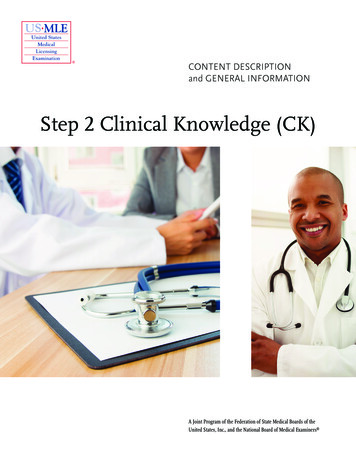
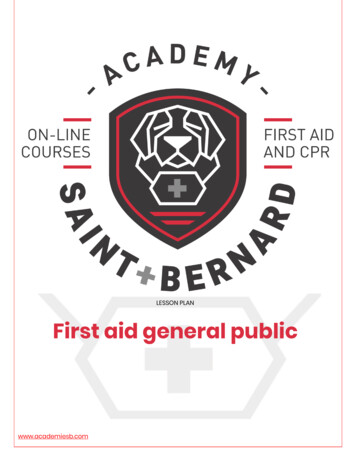
Wall of Bronchus Respiratory epithelium (E)Lamina propria (LP)Smooth muscle (SM)Hyaline cartilage (C)Nerves (N) and blood vessels (V)Note the order of structures:E à LP à SM à C
PseudostratifiedcolumnarepitheliumGoblet cellsCiliaBronchus: respiratory epithelium
Respiratory System Lecture Outline Introduction Conducting portion Nasal cavity Larynx Trachea Bronchi Bronchioles
Bronchioles NO glands or cartilage. Larger bronchioles have respiratory epithelium.Smaller bronchioles have low columnarepithelium. In asthma, the smooth muscle in thebronchioles constricts, causing difficultybreathing.
Fibrousconnectivetissue ispresent butno cartilageor glands!Respiratoryepithelium isfoldedSmoothmuscleBronchiole
Smaller bronchioles have simple columnar epithelium
Terminal Bronchioles Simple cuboidal epithelium with cilia. Also: Clara cells (non-ciliated epithelial cellswith secretory granules). No goblet cells. As you go down the respiratory tract, gobletcells are lost before cilia.
Terminal bronchiole
Clara cellsIs there anything they don’t do?Make surfactant components, break down mucus,detoxify harmful substances, transfer IgA, fight bacteria
Respiratory System Lecture Outline Introduction Conducting portion Respiratory portion Respiratory bronchioles Alveoli
Respiratory System Lecture Outline Introduction Conducting portion Respiratory portion Respiratory bronchioles
Airways Preceding AlveoliRespiratory bronchioles As you go distally along the respiratorybronchioles, alveoli increase in number. Cilia are gone by the end of the respiratorybronchiole.Alveolar ducts Back-to-back alveolar openings along wall Smooth muscle between alveolar openingslooks like knobs
Respiratory bronchiole
Alveolar duct
Alveolar duct (AD), alveolar sac (AS) and alveolus (A)
Respiratory System Lecture Outline Introduction Conducting portion Respiratory portion Respiratory bronchioles Alveoli
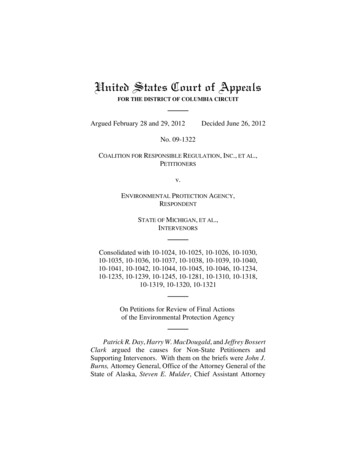
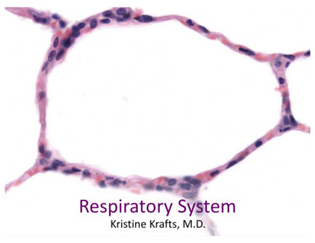
Alveoli Sac-like structures with super-thin walls so O2 andCO2 can diffuse between air and blood. Separated by interalveolar septae, which containcapillaries. Cells lining interalveolar septae: Type I cells (thin, flat squamous cells) Type II cells (pneumocytes): produce surfactant Alveolar macrophages (dust cells)
Alveolus
Type I Cells Cover 95% of alveolar surface Simple squamous cells with thin cytoplasm Blood-air barrier includes (from air to blood): Type I cells Fused basal laminae of type I cells andcapillary endothelial cells Capillary endothelial cells
Type II Cells (Pneumocytes) Cover 5% of alveolar surface. Large cuboidal cells with round nuclei. Typical secretory cell structure. Lamellar bodies incytoplasm make and store surfactant. Surfactant decreases surface tension in alveoli andprevents collapse of alveoli during expiration.
Alveoli lined by type I and II cells
Alveolar Macrophages (Dust Cells) Found on surface of alveoli, within alveoli and ininterstitial connective tissue. Remove debris and particles that escape mucusand cilia in conducting portion of respiratory tract
Macrophages (dust cells)
Respiratory System Lecture Outline Introduction Conducting portion Respiratory portion Pleura
Parietal and Visceral Pleura The outer surface of the lung and the inner surfaceof the thoracic cavity are covered by the pleura,which is a serous membrane (serosa). Parietal pleura lines the thoracic cavity; visceralpleura covers the lungs. Serous membranes consist of simple squamousepithelial cells called mesothelium plus a thin layerof connective tissue. The pleural cavity contains serous fluid made by thepleura.
Respiratory System Lecture Outline Introduction Conducting portion Respiratory portion Pleura
Sac-like structures with super-thin walls so O 2 and CO 2 can diffuse between air and blood. Separated by interalveolar septae, which contain capillaries. Cells lining interalveolar septae: Type I cells (thin, flat squamous cells) Type II cells (pneumocytes): produ