Transcription
Current Atherosclerosis Reports (2022) 23-9GENOMICS AND GENOMICS (R.A. HEGELE AND L. BRUNHAM, SECTION EDITORS)More than Just a Monolayer: the Multifaceted Role of Endothelial Cellsin the Pathophysiology of AtherosclerosisMarion Mussbacher1 · Klaudia Schossleitner2Astrid Schrammel1 · Johannes A. Schmid5· Julia B. Kral‑Pointner3,4· Manuel Salzmann3,4 ·Accepted: 7 March 2022 / Published online: 11 April 2022 The Author(s) 2022AbstractPurpose of the Review In this review, we summarize current insights into the versatile roles of endothelial cells inatherogenesis.Recent Findings The vascular endothelium represents the first barrier that prevents the entry of lipoproteins and leukocytesinto the vessel wall, thereby controlling two key events in the pathogenesis of atherosclerosis. Disturbance of endothelialhomeostasis increases vascular permeability, inflammation, and cellular trans-differentiation, which not only promotes thebuild-up of atherosclerotic plaques but is also involved in life-threatening thromboembolic complications such as plaquerupture and erosion. In this review, we focus on recent findings on endothelial lipoprotein transport, inflammation, cellulartransitions, and barrier function.Summary By using cutting-edge technologies such as single-cell sequencing, epigenetics, and cell fate mapping, novelregulatory mechanisms and endothelial cell phenotypes have been discovered, which have not only challenged establishedconcepts of endothelial activation, but have also led to a different view of the disease.Keywords Endothelial cells · Atherosclerosis · Endothelial dysfunctionIntroductionEndothelial cells (ECs) comprise the innermost layer of allblood vessels and act as gatekeepers of vascular homeostasis by regulating vascular permeability, leukocyte adhesion/extravasation, vascular tone, and hemostasis. Depending* Marion Mussbachermarion.mussbacher@uni-graz.at* Johannes A. Schmidjohannes.schmid@meduniwien.ac.at1Department of Pharmacology and Toxicology, Universityof Graz, Graz, Austria2Department of Dermatology, Skin and Endothelium ResearchDivision, Medical University of Vienna, Vienna, Austria3Ludwig Boltzmann Institute for Cardiovascular Research,Vienna, Austria4Department of Internal Medicine II/Cardiology, MedicalUniversity of Vienna, Vienna, Austria5Institute of Vascular Biology and Thrombosis Research,Medical University Vienna, Schwarzspanierstr. 17,1090 Vienna, Austriaon the location and the vascular bed, ECs are exposed tovarious degrees and types of shear stress, which not onlyinfluence their shape, but also affect intracellular signalingand gene expression. Thus, endothelial cells from veins,which are exposed to low blood pressure and flow, differfrom those of arteries that are subjected to high pressureand require a robust layer of smooth muscle cells (SMCs)to maintain mechanical stability. By regulating contractility of this smooth muscle layer, arterial ECs adjust localblood flow and contribute to the supply of the underlyingtissue with oxygen, nutrients, and transmitters. Apart fromvenous and arterial vascular beds, ECs are found in capillaries and lymphatic vessels, where they contribute to exchangeof fluid and proteins. With respect to the barrier functionof the endothelial layer, we distinguish between continuous, fenestrated, and discontinuous endothelium. The firstis found in most vascular beds and takes on its extreme inthe brain, where the tight junctions of the monolayer formthe blood–brain-barrier. Fenestrated endothelium with poresup to 100 nm is found at anatomical sites such as kidney andintestine, where transcellular transport processes or filtrationtasks must be fulfilled. Finally, discontinuous endothelium13Vol.:(0123456789)
484occurs in certain organs like liver or spleen with pores up to200 nm and additional gaps up to 1 µm, which sometimeslack the basal membrane. Thus, ECs can vary significantlyin form and function, while they are also characterized bya high degree of plasticity allowing them to change theirphenotype according to local requirements.The endothelial layer is covered by a dense glycocalyx—a gel-like layer of proteoglycans and extracellular matrixcomponents, which is involved in transendothelial transport,e.g., of lipoproteins, and which can be lost or reduced ininflammation [1].Under homeostatic conditions, ECs prevent platelet activation, blood clotting, and leukocyte adherence/infiltration by secreting/expressing mediators such as nitric oxide(NO), prostacyclin, tissue plasminogen activator (t-PA), andantithrombin III [2]. However, once activated by inflammatory stimuli, ECs upregulate cellular adhesion moleculessuch as E-selectin, intercellular adhesion molecule (ICAM),and vascular cell adhesion molecule (VCAM), which trigger leukocyte rolling on the endothelial surface followed byleukocyte diapedesis. Local immune cell activation fuels tissue inflammation, which increases endothelial permeabilityenabling the entry of plasma proteins to the interstitial space.Inflammatory activation of endothelial cells and endothelial dysfunction are key events in the initiation of atherosclerosis. This was elegantly demonstrated in a long-termfollow-up study demonstrating that patients with endothelialdysfunction have a significant risk for the development ofcardiovascular events [3, 4]. In atherosclerosis, disturbedendothelial homeostasis facilitates the permeation and trapping of lipoprotein particles in the subendothelial space,which become oxidatively modified and are sensed as danger-associated molecular pattern (DAMP) by both innate andadaptive immune cells. In addition, some ECs may transdifferentiate into mesenchymal cells (endothelial to mesenchymal transition, EndMT) and promote atherogenesis by lossof cell–cell contacts and deposition of extracellular matrix[5]. Besides initiating immune responses and transdifferentiation processes, modified lipoprotein particles are taken upby macrophages and smooth muscle cells leading to the formation of foam cells and fatty streak build-up. Progressiveintracellular lipid overload and curtailed efferocytosis causecell death and necrotic core formation leading to the thickening of the vascular wall and limiting local blood flow andoxygen supply. Consequently, arterial constrictions causeeven larger areas of turbulent or oscillatory blood flow, further contributing to EC activation. Lipid-rich plaques with athin fibrous cap are prone to plaque rupture, which exposesthrombogenic material to the circulation and initiates lifethreatening atherothrombotic occlusion. This detrimentalprocess is accelerated by expression of pro-coagulatorytissue factor (TF) and anti-fibrinolytic plasminogen activator inhibitor (PAI-1) by the luminal endothelium [6].13Current Atherosclerosis Reports (2022) 24:483–492Alternatively, local endothelial denudation due to endothelial apoptosis may trigger thrombus formation by plaqueerosion [7].Given the multiple important roles of ECs throughout theinitiation and progression of atherosclerosis, understandingmolecular mechanisms that regulate EC fate and functions ispivotal for the development of future therapies. This reviewsummarizes recent findings and provides insights into further perspectives (Fig. 1).Genetics and Genomics of ECs with an Emphasison InflammationAtherosclerosis is a multifactorial disease associated withvarious risk factors such as dyslipidemia, diabetes mellitus,smoking, and hypertension. Although heritable factors areassumed to account for over 50% of disease risk [8], knowledge on underlying genetics is limited. Genome-wide association studies (GWAS) provided the first unbiased evidenceof hundreds of loci associated with cardiovascular diseaseand underlying atherosclerosis. Besides well-known lipidmetabolism regulating genes such as low-density lipoproteinreceptor (LDLR) and proprotein convertase subtilisin/kexintype 9 (PCSK9), genetic variants for inflammatory geneshave been identified in GWAS. These include C-X-C motifligand 12 (CXCL12), SH2B adaptor protein 3 (SH2B3),AB0, human leukocyte antigen (HLA), interleukin 6 receptor(IL-6R), interleukin 5 (IL-5), platelet endothelial cell adhesion molecule 1 (PECAM1), protein C receptor (PROCR),and antisense non-coding RNA in the INK4 locus (ANRIL),whereby SH2B3, PECAM1, and ANRIL are expressed bybut not limited to endothelial cells (summarized in [9]).These GWAS studies have recently been extended by singlecell epigenomics of human atherosclerotic lesions, which ledto the identification of cell-type specific alterations that predispose for coronary artery disease and myocardial infarction [10]. For ECs, the most prominent changes were seenin chromatin regions that are responsible for cell adhesion,wound healing, and angiogenesis [9]. For a direct comparison of dysregulated EC signatures, another group used bloodoutgrowth endothelial cells isolated from patients with andwithout coronary endothelial dysfunction for microarrayanalysis [11]. Thereby, they identified high mobility groupbox1 (HMGB1) and laminin gamma1 (LAMC1) to be dysregulated in ECs at an early step of atherogenesis. HMGB1is sensed as damage-associated molecular pattern (DAMP)and enhances inflammatory responses such as binding ofnuclear factor “κ-light chain enhancer” of activated B cells(NF-κB) molecules to DNA [11]. NF-κB is a family oftranscription factors closely involved in inflammation andatherogenesis [12]. EC-specific activation of NF-κB via itskey kinase IκB kinase 2 (IKK2) significantly acceleratesthe development of atherosclerosis by increasing leukocyte
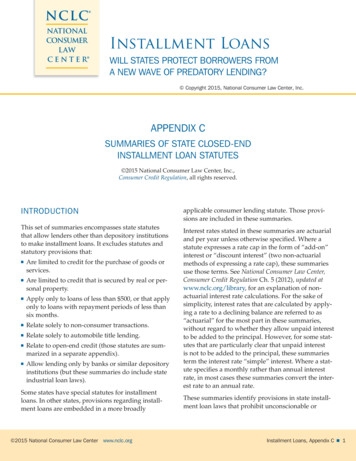
Current Atherosclerosis Reports (2022) 24:483–492485Fig. 1 Multifaceted role of endothelial cells in atherogenesis. Underhomeostatic conditions (upper part of the scheme), endothelialcells (EC) are covered by a dense glycocalyx and constantly releasenitric oxide (NO), which is synthesized by endothelial NO synthase (eNOS). Tissue plasminogen activator (t-PA), antithrombin III(ATIII), and prostacyclin, which is produced by cyclooxygenases(COX), prevent platelet activation and blood clotting. Bone marrowderived endothelial progenitor cells (EPC) allow EC renewal. Smoothmuscle cells (SMCs) are in a “contractile” state, enabling the adjustment of the vascular tone. Under atherosclerotic conditions (lowerpart of the scheme), the glycocalyx is degraded by plasminogen activator inhibitor (Serpine1) and EC permeability is increased due todownregulation of junctional proteins (e.g., occludin and claudin-5).Mechanosensitive cation channels such as Piezo1 detect turbulentflow and cause the downregulation of Krüppel-like factor 2 (Klf2)and forkhead box O1 (Foxo-1). Endothelial dysfunction is characterized by reduced eNOS expression and decreased NO bioavailabilitydue to elevated levels of reactive oxygen species (ROS). Transcytosisof low-density lipoproteins (LDLs) is mediated by scavenger receptor class B (SR-B1) and potentially activin-like kinase (ALK1). ECinflammation facilitates leukocyte recruitment by increasing the surface expression of E-selectin, vascular cellular adhesion molecule 1(VCAM), and intercellular adhesion molecule 1 (ICAM). Activationof NF-κB is further enhanced by high mobility group box1 (HMGB1)and initiates the assembly of the EC inflammasome, resulting in therelease of interleukin 1β (IL-1β). Endothelial-to-mesenchymal-transition (EndMT) is characterized by decreased expression of CD31 andeNOS/NOS3 as well as increased levels of fibroblast activation protein (FAP) and alpha-actin 2 (ACTA). Whereas transforming growthfactor β (TGFβ) promotes EndMT and SMC transition towards amacrophage-like state, fibroblast-like growth factor (FGF) inhibits EndMT. The (athero)thrombotic risk is increased by endothelialexpression of tissue factor (TF) and release of von Willebrand factor (vWF). Endothelial apoptosis increases the number of circulatingCD146 ECs in the blood streaminflux and promoting a phenotypic transition of smooth muscle cells towards a macrophage-like phenotype [13]. Anotherimportant mediator of inflammation is the inflammasome,a multiprotein complex composed of a sensor protein andinflammatory caspases, which are in some cases linkedby an adapter protein [14]. NF-κB can upregulate varioussensor proteins, which bind the adaptor protein apoptosisassociated speck-like protein containing a CARD (ASC),thereby recruiting and activating caspase 1, which convertspro-IL-1β to active IL-1β [15]. The latter is then secretedvia gasdermin D-containing pores, activating NF-κB onnearby cells and amplifying the inflammatory circuit. Acrucial type of inflammasomes contains NLR family pyrindomain containing 3 (NLRP3), which was first discoveredin macrophages as a mechanism for caspase-1 activation andIL-1β processing [16]. However, later it became clear thatthis fundamental driver of inflammation is also operativein other cells of the vasculature [17]. In endothelial cells,13
486Forkhead box P (Foxp1) has been identified as an importanttranscription factor repressing the expression of inflammasome components such as NLRP3, caspase 1, and IL-1β,which significantly contribute to the development of atherosclerosis [18]. Using transgenic mice, Zhuang et al. showedthat EC-specific overexpression of Foxp1 decreases plaqueprogression and EC-specific Foxp1 knockout acceleratesdisease development [18]. Interestingly, FoxP1 was identified to be regulated by Krüppel-like factor 2 (Klf2) and bothproteins are decreased in atheroprone vessel regions, whichare exposed to disturbed blood flow [18].Moreover, recent studies identified transforming growthfactor-β (TGF-β) as an additional inflammatory driver in atherosclerosis [19 ]. In strong contrast to its anti-inflammatoryrole in smooth muscle cells, endothelial TGF-β signalingpromoted vascular permeability and vessel inflammation.Both EC-specific deletion of TGF-β receptors and nanoparticle-targeted suppression of endothelial TGF-β signalingdelayed the onset of atheroma formation and reduced leukocyte recruitment and as well as EndMT [19 ], underliningthe need for additional research into the cell type-specificprerequisites for drug development.Cellular TransitionsNovel technologies such as single-cell RNA sequencing(scRNA-seq) revealed the tremendous heterogeneity ofendothelial cells as well as other cell types across and withinvascular beds with more than 78 EC clusters being identified in 11 murine tissues [20 ]. In the murine aorta, threemajor EC populations have been discovered: (i) C D34highECs (approx. 56%), which strongly express CD34 and lymphocyte antigen 6a (LY6A); (ii) T HY1high ECs that areenriched for Thy-1 cell surface antigen (THY1), transcription box-1 (TBX1), and fms-related tyrosin kinase 4 (FLT4),and “activated ECs,” which expressed genes such as nitricoxide synthetase 3 (NOS3), endothelin 1 (EDN1), angiotensin converting enzyme (ACE), and VCAM1 [21]. Whereas CD34high and THY1high ECs were both associated withendothelial proliferation and vascular development, activated ECs were enriched in pathways associated with vesseldilation and blood pressure regulation [21]. These data weresupported by a study from Kalluri and colleagues, whichrevealed specialization towards lipoprotein handling, angiogenesis, and extracellular matrix production in two of the ECsubsets [22]. The heterogeneity of endothelial cells, smoothmuscle cells, and immune cells in single-cell datasets fromatherosclerosis samples has been summarized in detail [23].In the murine aorta of ApoE / animals on high fat diet(HFD), endothelial cells upregulated classical markers, suchas adhesion molecules VCAM-1, ICAM-1, E-selectin, andinflammatory markers such as IL-6, transcription factorhypoxia-inducible factor-1α (HIF1α), but also a plethora of13Current Atherosclerosis Reports (2022) 24:483–492G-protein coupled receptors [24]. Under high-salt, high-fat,or high-plasma glucose diet, an additional EC populationwith occludin (OCLN)low and plasminogen activator inhibitor 1 (SERPINE1)high characteristics emerged, which pointedto increased EC permeability under conditions of disturbedEC homeostasis as OCLN is associated with tight junctionand SERPINE-1 mediates extracellular matrix degradation[21]. In parallel, the “activated EC” cluster was reducedunder the same conditions, which coincided with decreasedendothelium-induced vasodilation [21].During plaque growth ECs are exposed to a changingmicroenvironment which can involve hypoxia, oxidativestress, varying shear stress, and sustained inflammation (e.g.,TGFβ). This triggers EC transformation towards states, inwhich endothelial properties are suppressed and mesenchymal cell behavior is increased. The phenotypic modulation iscalled EndMT and characterized by decreased expression ofendothelial markers such as CD31 and NOS3 and increasedlevels of mesenchymal markers such as fibroblast activationprotein (FAP) and alpha-actin 2 (ACTA) [25]. Moreover,EC-specific lineage-tracing experiments using yellow fluorescent protein (YFP) expressed in an EC-specific mannerafter tamoxifen-injection revealed that EndMT is commonin atherosclerotic lesions of A poE / mice that are fed aHFD and is linked to plaque instability in humans. Expression of FAP fibroblast-like cells increased in these miceto about 25% of plaque cells and counting of YFP-positivecells suggested that upon 30 weeks of HFD approximately45% of FAP cells were derived from endothelial cells [25].This process seemed to destabilize the plaque via enhancedexpression of collagen-degrading matrix metalloproteinasesand lower synthesis of collagen [25]. Among eight identifiedEC clusters obtained from scRNA-seq of aortic and cardiacECs derived from LDLR / mice fed a diabetogenic HFD,three EC clusters expressed mesenchymal marker genes[26]. Further metabolic, transcriptomic analysis of theseEndMT clusters revealed diminished fatty acid oxidation,increased inflammation, and extracellular matrix organization [26]. TGFβ and fibroblast growth factor (FGF) signaling cascades are both activated by inflammatory cytokinesand oscillatory shear stress. However, they display opposing roles in the induction EndMT: Whereas TGFβ promotesEndMT [19 ], endothelial FGF receptor (FGFR) signaling isatheroprotective and knockout of FGFR substrate 2a dramatically increases atherosclerotic plaque burden and neointimaformation by extensive EndMT and subsequent fibronectindeposition [27].Besides ECs, also other cell types are subjected to phenotypic transitions, with vascular SMCs constituting probablythe most prominent fraction. It has been shown that inflammation drives SMCs from a contractile to a so-called synthetic state, with higher expression of extracellular matrixcomponents and reduction of contractile elements (for
Current Atherosclerosis Reports (2022) 24:483–492review, see 28). Genetic cell-lineage tracing elucidated thatSMCs can further change their identity towards macrophagelike cells in the course of atherosclerosis and it has beenpostulated that more than 80% of SMC-derived cells lackclassical SMC-markers in advanced lesions, while adopting different cellular phenotypes such as macrophage-like,mesenchymal-stem cell-like, or myofibroblastic cells [29].Recent single-cell genomic analyses revealed the importanceof fibromyocytes [30] and also noted that SMCs can adoptan intermediate state between stem cells, ECs, and monocytes (31). Moreover, the inflammatory environment in theplaques also affects the polarization of macrophages derivedfrom infiltrating monocytes, which show a broad spectrumof activation states [32].Altogether, these findings underline that atherosclerosisleads to a drastically altered environment, which results insignificant de-differentiation and trans-differentiation processes of all the different cells of the vasculature.Shear StressLocated at the blood/vessel interface, ECs are constantlyexposed to the hemodynamic forces of blood flow, whichaffect ECs on a transcriptional, epigenetic, and protein level.Whereas unidirectional, laminar shear stress is consideredatheroprotective, pulsatile disturbed shear stress such asoscillatory flow that is typically found at arterial branchesand vessel wall irregularities induces chronic inflammation [33] and thus enhances atherogenesis. Shear stress isrelayed by various mechanosensors such as integrins (e.g.,integrinα5—annexin A2 interaction [33]), CD31/PECAM-1,VE-cadherin, and vascular endothelial growth factor receptor 2 (VEGFR2). The interplay between the mechanosensitive cation channel Piezo1, the purinergic P2Y2 receptor, and Gq/G11-mediated signaling has been identified to mediateinflammatory signaling and atherosclerosis under disturbed,but not under laminar shear stress [34]. Moreover, plexin D1(PLXND1) is required for the response of endothelial cellsto shear stress and regulates the site-specific distribution ofatherosclerosis [35]. However, disturbed flow also inducedanti-inflammatory feedback pathways, such as the endothelialadrenomedullin-calcitonin-like receptor (CALCRL), whichsignals through cAMP and reduces endothelial inflammationand lesion formation [36].Endothelial DysfunctionEndothelial dysfunction (ED) represents an early hallmarkof atherosclerosis. It is characterized by a shift of physiological endothelial function towards impaired vasodilation,chronic inflammation, and thrombosis. A key scenario of EDcomprises reduced formation and/or bioavailability of thegaseous transmitter NO, which is enzymatically produced487by NOS3/eNOS [37]. As a consequence of reduced endothelial NO concentrations, the soluble guanylyl cyclase (sGC)/cGMP/protein kinase G (PKG) cascade in adjacent SMCsis impaired. From the mechanistic viewpoint, differentprocesses have been identified to contribute to ED. Theyinclude uncoupling of eNOS (i.e., formation of superoxide at the expense of NO production due to submaximalcofactor concentrations), aberrant eNOS phosphorylationand dephosphorylation, inhibition of eNOS by endogenousmethylarginines, and accelerated breakdown of NO due tosupraphysiological production of ROS by different enzymatic sources [38]. eNOS function is complexly regulatedat levels of transcription, translation, protein maturation,and cofactor assembly. Posttranslational control of eNOSactivity by multisite phosphorylation/dephosphorylationis well characterized; however, other protein modificationsincluding S-nitrosylation, acetylation, and glutathionylationhave been reported to modulate enzymatic activity (37). In arecent report, Ser 615 has been identified as site of hydroxyllinked N-acetyl-glucosamine (O-GlcNAc) modification thatentails reduced eNOS activity under conditions of glucosedysregulation observed in type-2 diabetes [39].Moreover, posttranscriptional modulators of gene expression and altered micro RNAs (miRNA) expression patternshave been described in settings of ED, atherosclerosis,hyperglycemia, obesity, and senescence. With respect toendothelial cells, miR-92a has been reported to regulateeNOS mRNA levels via targeting Klf2 [40]. Moreover,vascular expression of miR-221 and miR-222 has beendescribed to be upregulated in initial atherogenic stages andtheir expression levels have been negatively correlated witheNOS signaling [41]. In addition, miR-195 and miR-532that have been suggested as biomarkers of thrombosis werefound inversely correlated to eNOS expression [42].In this context, the synergistic interaction between eNOSand silent information regulator 1 (SIRT1) as well as itsupstream modulation by miRNAs has gained scientific interest [43]. Belonging to the group of N AD -dependent proteindeacetylases, SIRT1 activates eNOS via lysine deacetylation [44]. Moreover, SIRT1 upregulates protein expressionof eNOS in a forkhead box O1- (Foxo-1) or KLF2-dependent manner [45]. Conversely, via a positive feedback loop,eNOS-derived NO positively regulates protein expression ofSIRT-1. At the transcriptional level, SIRT1 is regulated by anumber of miRNAs including miR-34a [46].Besides miRNAs, two long noncoding RNAs (lncRNAs),termed spliced-transcript endothelial-enriched lncRNA(STEEL) and lncRNA that enhances eNOS expression(LEENE), have been recently described to increase eNOSmRNA expression via KLF2 [47]. Targeting and/or modulation of endothelial miRNAs and lncRNAs that directly orindirectly affect eNOS expression might represent a novel13
488therapeutic approach to counteract ED and associatedpathologies.Lipoprotein TransportFor decades, passive movement of LDL particles across acompromised endothelial barrier was believed to induceatherogenesis; however, only recently it became clear thatLDL can be transported via endothelial transcytosis, whichrepresents an active process that is mediated by severalendothelial receptors such as scavenger receptors class Btype 1 (SR-B1) [48 ] and activin-like kinase 1 (ALK1).Transcytosis of LDL by SR-B1 requires dynamic interaction with dedicator of cytokinesis 4 (DOCK4), which acts asguanine nucleotide exchange factor for Ras-related C3 botulinum toxin substrate 1 (RAC1) and is essential for LDL-SRB1 internalization. EC-specific knock-out of SR-B1 showedreduced uptake of LDL by artery wall macrophages andseverely decreased atherogenesis in the absence of changesin plasma lipid levels or aortic inflammation [48 ]. Interestingly, endothelial deletion of the alarmin HMGB1 wasreported to downregulate SR-B1 levels via its upstream transcription factor sterol regulatory element-binding protein 2(SREBP2) [49], which likewise decreased lesion size in ECspecific HMGB1 / LDLR / mice [49], linking inflammation and LDL transcytosis.By combining a genome-wide RNAi screen of endothelialcells with GWAS data sets, ALK1—a member of the TGF-βreceptor superfamily—was identified as additional receptor to mediate endothelial/aortic LDL uptake in vitro andin vivo [50]. However, data on the role of ALK1 in atherosclerosis are still missing and limited by its role in angiogenesis, as loss-of-function mutations in ALK1 cause vascularmalformations in humans [51]. Interestingly, both SR-B1and ALK1 are localized in caveolae, which are specializedmembrane microdomains involved in endocytic processes[52]. Absence of caveolin 1—the main protein constituent of caveolae—in LDLR / mice reduces LDL transportacross the endothelium and decreases vascular inflammationin early-stage atherosclerosis independent of eNOS activity [53, 54]. Although binding of LDL and oxidized LDL(oxLDL) to CD36 and oxLDL receptor 1 (LOX-1) have beendemonstrated, none of these receptors affects endothelial LDLtranscytosis [49].In addition to endothelial transcytosis, endothelialbreaches and hemorrhage were found in the intima of carotidarteries of A poE / mice at sites of local flow perturbation,which attracts leukocytes and promotes fatty streak formation [55]. Thus, recent findings indicate that fatty streaks canbe the result of active transport and retention of oxLDL aswell as deposition at sites of injury [55].13Current Atherosclerosis Reports (2022) 24:483–492Barrier FunctionActivation of endothelial cells results in a reduction of vascular barrier function, increased paracellular and transcellular transport, and, enhanced deposition of fatty streaks inareas of disturbed flow. Pathways that increase endothelialpermeability include targets of HIF1-α and NF-κB, such asvascular endothelial growth factors, IL-1β, IL-6, and tumornecrosis factor α (TNFα), but also TGFβ or ROS. In recentyears, it has become known that metabolic programs andatherosclerosis are linked by eNOS uncoupling, excess ROS,and the production of advanced glycation products [56].Additionally, adenosine monophosphate–activated proteinkinase (AMPK) can phosphorylate eNOS to decrease NOformation [57] and it can control tight junction protein cingulin to regulate barrier function [58]. Disturbing eNOS candirectly and indirectly lead to the disruption of endothelialbarriers [59].Yet, the vascular system harbors mechanisms to counteract these disruptive processes for example by the activationof protective Tie2 signaling through platelets and von Willebrand factor (vWF) [60]. Transcriptional processes downstream of sphingosine-1-phosphate receptor 1 (S1PR1) inaortic endothelial cells showed suppression of the NF-κBpathway and allowed the identification of an endothelial cellsubset at aortic branch points [61] that is protected fromactivation by inflammatory stimuli. Furthermore, recentfindings in the field have shown that the barrier function ofendothelial cells to small molecules and the transmigrationof immune cells are controlled by independent processes.Barrier function towards leak is controlled by tight andadherens junction complexes [62], whereas transmigrationis controlled by specific receptor-ligand interactions in combination with substrate stiffness and internal endothelial cellstiffness [63, 64]. Endothelial ICAM-1 and VCAM-1 clusters form cellular protrusions to prepare the endothelial cellfor transmigration of leukocytes. The amount of ICAM-1and VCAM-1 on the cell surface is a response to TNFα orIL-1β signals that determine whether a paracellular or transcellular route is taken for diapedesis [65]. During diapedesis,the paracellular space is sealed by RhoA activation and acontractile actin ring [66]. The combined effects of disruptive and protective programs and the uncoupling of barrierfunction for small molecules and immune cells will help usunderstand the formation of atherosclerotic plaques.Revascularization and Plaque StabilityIt is widely recognized that revascularization of plaques andplaque stability are governed by inflammation and mechanical forces [67, 68]. Meanwhile, many studies have shownthat high plasticity of endothelial cells, smooth musclecells, and fibroblasts plays a major role in the emergence
Current Atherosclerosis Reports (2022) 24:483–492of highly proliferative subsets of cells th
thase (eNOS). Tissue plasminogen activator (t-PA), antithrombin III (ATIII), and prostacyclin, which is produced by cyclooxygenases (COX), prevent platelet activation and blood clotting. Bone marrow-derived endothelial progenitor cells (EPC) allow EC renewal. Smooth muscle cells (SMCs) are in a "contractile" state, enabling the adjust-