Transcription
Vector Hazard Report:Southern U.S./ Northern MexicoArthropod-borne disease threats and vector species profiles includingbionomics, distribution and identification resourcesCompiled by David Pecor, 2018
PrefaceThis product was produced by the Walter Reed Army Institute of Research,Walter Reed Biosystematics Unit (WRAIR-WRBU)This document provides summarized information on major vectors and vector-borne diseasesreported from the Southwest United States and Northern Mexico. Information related to the identification, distribution, medical importance, control and surveillance of vector species are included. Forupdated information on the current hazards known from this region, please use the near-real timehazard assessment links on page 3. Each page of this document is also hyperlinked via the table ofcontents to allow easy navigation and access to information most relevant to the reader. View theVector Hazard Report Quick Guide pages for updated information about current outbreaks and regional climate as well as vector identification resources. Detailed bionomics data for each vectorspecies is available on the vector species ecology profile pages for mosquitoes and ticks.The target audience for this document are commanders, medical planners, preventive medicinepersonnel, and particularly medical entomologists.You Can Request a Vector Hazard Report by contacting theWRBU: NMNH-WRBU@si.edu2
Vector Hazard Report Quick GuideReal-Time Threat Assessment ResourcesVisit these websites for regularly updated information aboutcurrent vector-borne disease threats and regional climate.U.S. Dept. of State Travel AlertsHealth.mil ReportsCDC Current Outbreaks ListWHO Outbreak NewsHealthMap OutbreaksVectorMap Current ClimateAccuWeather Current RadarAdditional ResourcesWHO Country Profile: MexicoCDC Travelers Guide: MexicoGlobal Mosquito Vectors of Arboviruses of the World3
Vector Hazard Report Identification GuideVector Identification ResourcesVectorSourceMosquitoes of the U.S. (Morphology)Ward, R. D. (2005). Identification and Geographical Distribution of theMosquitoes of North America, North of Mexico, By RF Darsie Jr. and RAWard, pp. 416. University Press of Florida, USA, 2005. ISBN0 8130 27845. US 75.00.-. Parasitology, 131(4), 580-580.Mosquito, Aedes albopictus (Molecular)Ruiling, Z., Tongkai, L., Dezhen, M., & Zhong, Z. (2018). Genetic characters of the globally spread tiger mosquito, Aedes albopictus (Diptera,Culicidae): implications from mitochondrial gene COI. Journal of VectorEcology, 43(1), 89-97.Mosquitoes (Molecular)Beebe, N. W. (2018). DNA barcoding mosquitoes: advice for potentialprospectors. Parasitology, 1-12.Mosquito, Aedes (Morphology)Rueda, L. M. (2004). Pictorial keys for the identification of mosquitoes(Diptera: Culicidae) associated with dengue virus transmission. WalterReed Army Inst Of Research Washington Dc Department Of Entomology.Mosquitoes (Morphology)Varnado, W. C., Goddard, J., & Harrison, B. (2012). Identification guideto adult mosquitoes in Mississippi. Mississippi State University ExtensionService, Starkville, MS.Mosquitoes, Anopheles of Central America(Morphology)Note: Key also available as mobile appWilkerson, R. C., Strickman, D., & Litwak, T. R. (1990). Illustrated keyto the female anopheline mosquitoes of Central America and Mexico. Journal of the American Mosquito Control Association, 6(1), 7-34.Ticks of the U.S. (Morphology)Bischof, Michael. Interactive Identification Key for the Hard Ticks(Ixodidae) of the Eastern U.S.Ticks of the U.S. (Morphology)Keirans, J. E., & Litwak, T. R. (1989). Pictorial key to the adults of hardticks, family Ixodidae (Ixodida: Ixodoidea), east of the Mississippi River. Journal of Medical Entomology, 26(5), 435-448.Ticks of the U.S. (Morphology)Kleinjan, J. E., & Lane, R. S. (2008). Larval keys to the genera of Ixodidae (Acari) and species of Ixodes (Latreille) ticks established in California. The Pan-Pacific entomologist, 84(2), 121.Fleas (Morphology)Ewing, H. E., & Fox, I. (1943). The fleas of North America: classification,identification, and geographic distribution of these injurious and diseasespreading insects (No. 500). US Department of Agriculture.4
Table of ContentsVector-borne Disease ThreatsVector Species ProfilesMosquito:Mosquito:Aedes aegyptiAedes albopictusAedes atropalpusAedes dorsalisAedes melanimonAedes triseriatusAedes vexansAnopheles freeborniAnopheles pseudopunctipennisAnopheles punctipennisAnopheles quadrimaculatusCoquillettidia perturbansCulex nigripalpusCulex quinquefasciatusCulex restuansCulex tarsalisCuliseta inornataMalariaDengue VirusChikungunya VirusZika VirusWest Nile VirusTick:Lyme DiseaseTickborne Relapsing FeverRocky Mountain Spotted FeverRickettsiosisAnaplasmosisEhrlichiosisColorado Tick Fever VirusFlea:PlagueTriatominae:Chagas DiseaseTick:Amblyomma americanumAmblyomma cajennenseAmblyomma maculatumDermacentor andersoniDermacentor variabilisHaemaphysalis longicornnisIxodes pacificusIxodes scapularisRhipicephalussanguineusU.S. Civilian Vector Control ContactsFlea:Xenopsylla cheopisCtenocephalides felisTriatominae:Triatoma gerstaeckeriTriatoma incrassataTriatoma indictivaTriatoma lecticulariaTriatoma neotomaaTriatoma protractaTriatoma recurvaTriatoma rubidaTriatoma sanguisuga5
Monthly Climate MapsClick here to view the maps described below(Updated monthly, NASA Earth Observations, WorldClim)RainfallThis map displays accumulated rainfall for the past month.Consistent Above and Below Average RainfallThis map displays areas with consistently above or below averagemonthly rainfall based on the previous three months. Above averagerainfall may mean increased mosquito breeding habitat in areas withpoor drainage. Below average rainfall can lead to increased domesticwater storage providing increased mosquito breeding habitat.Drought-Breaking RainThis map displays areas receiving above average rainfall for theprevious month with below average rainfall for the previous 12months. Drought-breaking rain may indicate suitable conditions forvectors and diseases in a stressed environment or population.Temperature AnomalyThis map displays areas where the earth’s temperature was warmer orcooler at the surface during the daytime over the expected averagemonthly temperatures (averaged over 2001-2010).Land Surface TemperatureThis map displays the temperature of the earths surface during the daytime.Back to table of contents6
MalariaBackgroundDisease Background:Human malaria is caused by protozoan species in the genus Plasmodium that aretransmitted by the bite of an infective female Anopheles mosquito. Clinical symptoms of malaria vary with thespecies. The most serious malaria infection, Plasmodium falciparum malaria, can produce life-threateningcomplications, including renal and hepatic failure, cerebral damage, and coma. Case fatality rates among childrenand nonimmune adults exceed 10% when not treated. The other human malarias, vivax, malariae and ovale, are notlife-threatening except in the very young, the very old, or persons in poor health. Illness is characterized by malaise,fever, shaking chills, headache, and nausea. The frequency of fever, occurring daily, every other day, or every thirdday, is characteristic of the species. Nonfatal cases of malaria are extremely debilitating. Relapses of improperlytreated malaria can occur years after the initial infection in all but falciparum malaria. Plasmodium malariae infections may persist for as long as 50 years, with recurrent febrile episodes. Although local transmission of Malaria isquite rare in the United States since the 1950’s, a number of imported cases are reported at hospitals annually. Sincevectors of Plasmodium sp. are still present in the U.S., there remains a risk of vector species feeding on infected humans and spreading the disease.Transmission Cycle: Female mosquitoes of the genus Anopheles are the exclusive vectors of humanmalaria. Plasmodium species undergo a complicated development in the mosquito. When a female Anopheles ingests blood containing the sexual stages (gametocytes) of the parasite, male and female gametes uniteto form a motile ookinete that penetrates the mosquito’s stomach wall and encysts on the outer surface ofthe midgut. Thousands of sporozoites are eventually released, and some of these migrate to the salivaryglands. Infective sporozoites are subsequently injected into a human host when the mosquito takes a bloodmeal. The time between ingestion of gametocytes and liberation of sporozoites, ranging from 8 to 35 days,is dependent on the temperature and the species of Plasmodium. Malaria parasites develop in the mosquitovector most efficiently when ambient air temperatures are between 25 o and 30o C. Parasite development isprolonged during cool seasons and at high altitudes, and may exceed the life expectancy of the vector.Once infected, mosquitoes remain infective for life. Vector competence is frequently higher with indigenous strains of malaria. This decreases the likelihood that imported strains from migrants will become established.Additional Resources: CDC Malaria Travel Alert Malaria Atlas Project Newman, R. D., Parise, M. E., Barber, A. M., & Steketee, R. W. (2004). Malaria-related deaths among US travelers, 1963–2001. Annals of internal medicine, 141(7), 547-555. Bannister, B. (2000). Malaria in Mexico. Weekly releases (1997–2007), 4(10), 1649. Maldonado, Y. A., Nahlen, B. L., Roberto, R. R., Ginsberg, M., Orellana, E., Mizrahi, M., . & Campbell, C. C.(1990). Transmission of Plasmodium vivax malaria in San Diego County, California, 1986. The American journal of tropical medicine and hygiene, 42(1), 3-9.Back to table of contents7
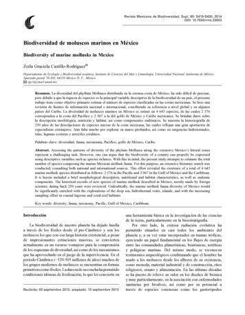
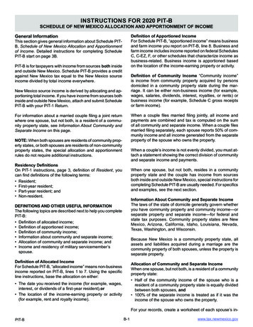
MalariaDisease Distribution1.U.S.: Number of imported cases of malaria reported to CDC, 20152.Mexico: Incidence rate associated with malaria (per 100 thousand inhabitants).Back to table of contents8
Dengue, Chikungunya and Zika VirusesDisease Background:Dengue fever (Breakbone fever, Dandy fever) is an acute febrile disease characterized bysudden onset, fever for 3 to 5 days, intense headache, and muscle and joint pain. It is commonly called break bone fever because of the severity of pain. There is virtually no mortality in classical dengue. Recovery is complete, but weakness and depression may last several weeks. Dengue is caused by a Flavivirus and includes four distinct serotypes(dengue 1, 2, 3 and 4). Recovery from infection with one serotype provides lifelong immunity from the same serotypebut does not protect against other serotypes. Dengue hemorrhagic fever (DHF) and associated dengue shock syndrome(DSS) were first recognized during a 1954 dengue epidemic in Bangkok, Thailand. DHF/DSS have spread throughoutSoutheast Asia, Indonesia and the southwest Pacific, Latin America and the Caribbean. DHF requires exposure to twoserotypes, either sequentially or during a single epidemic involving more than one serotype. DHF is a severe diseasethat produces high mortality in children. Chikungunya refers to an infection by the Chikungunya virus (CHIKV). Thename means “that which bends up” in the native language of southeastern Tanzania, and refers to the symptoms ofChikungunya fever. Chikungunya fever (CHIK) symptoms typically include a sudden high fever and severe joint pain.Headache, back pain, muscle pain, nausea, vomiting, arthritis, rash, and conjunctivitis may also occur. Unlike Dengue,CHIK is currently thought to be nonfatal. Outbreaks of CHIKV historically have occurred in Africa and Asia. In 2007,the virus was found to be spreading in northern Italy and in December 2013 was found in the Caribbean. Zika virus, anarbovirus related to CHIKV and DENV, has recently been reported from Latin America and Southern U.S. Originallydescribed from the Zika forest in Uganda, the virus has since spread to Southeast Asia, the Caribbean, South, Centraland North America. Symptoms are mild and include fever, rash, headache, joint pain and conjunctivitis. Zika virus poses a significant threat to women who are pregnant as Zika virus infection is associated with a birth defect called microcephaly.Transmission Cycle:Dengue, chikungunya and Zika viruses are exclusively associated with Aedes mosquitoesin the subgenus Stegomyia. Aedes aegypti appears to be the primary vector of these viruses although Ae. albopictus alsoplays a significant role in the transmission cycle.Additional Resources: CDC Dengue Fever Background CDC Chikungunya Background CDC Zika Virus Backgroind AFPMB Technical Guide No. 47. Aedes Mosquito Vector Control. Office of the Assistant Secretary of Defense(Energy, Installations and Environment). 2016.Back to table of contents9
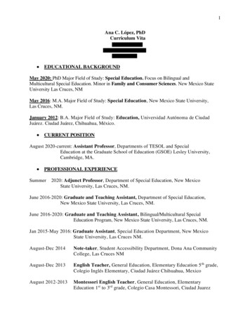
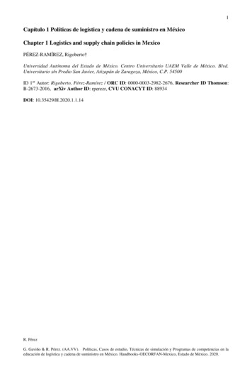
Dengue VirusNumber of imported cases ofDengue virus by U.S. county,CDC, 2016-2017.Bhatt, S. et al. 2013. TheGlobal Distribution andBurden of Dengue.Nature, 496: 504-507.Back to table of contents10
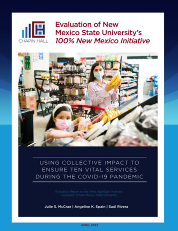
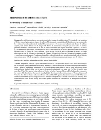
Chikungunya VirusChikungunya under the baseline and RCP 8.5 climate change scenarios in North- and Central America. Left: Climaticsuitability, right: hazard index. Climate change scenarios represent the mean model output obtained through the 5GCMs. Climatic suitability output is scaled to the over-all global minimum (0) and maximum (0.623) values observed in any model. Source: Tjaden, N. B., Suk, J. E., Fischer, D., Thomas, S. M., Beierkuhnlein, C., & Semenza, J.C. (2017). Modelling the effects of global climate change on Chikungunya transmission in the 21 st century. Scientific reports, 7(1), 3813.Back to table of contents11
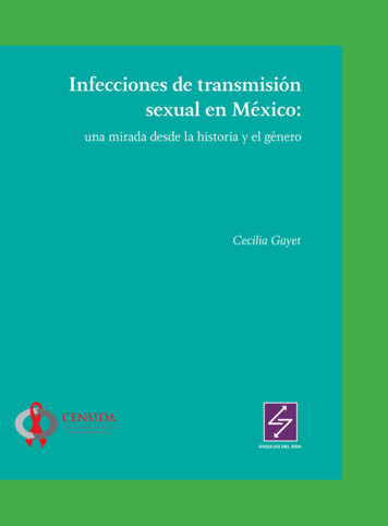
West Nile VirusDisease Background: West Nile fever is a mosquito-borne illness characterized by fever, headache, muscular pain, and rash. Occasionally, serious complications involve the liver and nervous system. The etiological agent, West Nile virus (WNV), is namedafter the district of Uganda where the virus was first isolated. It is a Flavivirus closely related to viruses causing Japaneseencephalitis and St. Louis encephalitis. Infection with WNV is most often asymptomatic. The incubation period ranges from 1 to 6days and clinically resembles a mild dengue-like illness.Transmission Cycle: WNV has been isolated from numerous wild birds and mammals. Serological surveys have demonstratedWNV antibodies in wild and domestic bird species, wild mammals such as lemurs, rodents and bats, and domestic animals such ascamels, horses, mules, donkeys, goats, cattle, water buffalo, sheep, pigs and dogs. However, birds are considered to be the primaryreservoir for WNV and may reintroduce the virus during seasonal migrations. Infections in most mammals fail to produce viremiashigh enough to infect potential vectors. WNV has been isolated from several species of mosquitoes in nature, and they are recognized as the major vectors, especially Culex spp. WNV has also been recovered from bird-feeding ticks and mites. A natural birdtick zoonotic cycle has been suggested, but the role of ticks in the natural transmission of WNV has not been well defined. Mosquitoes are clearly implicated in the transmission of WNV to humans. WNV replicates quickly in mosquitoes when temperatures exceed 25C. Infected mosquitoes can transmit WNV for life.Additional Resources: CDC West Nile Virus Background Lanciotti, R. S., Roehrig, J. T., Deubel, V., Smith, J., Parker, M., Steele, K., . & Hall, R. A. (1999). Origin of the West Nilevirus responsible for an outbreak of encephalitis in the northeastern United States. Science, 286(5448), 2333-2337.Number of reported cases of West Nile Virus, CDC 2016-2017Mexican states sampled for antibodies to West Nile virusand Venezuelan equine encephalitis virus in equines. Unshaded states were not sampled. The location of the WestNile virus isolation from a dead Common Raven is shownby a star. Source: Estrada-Franco, J. G., Navarro-Lopez, R.,Beasley, D. W., Coffey, L., Carrara, A. S., Da Rosa, A. T.,. & Ramirez, P. P. (2003). West Nile virus in Mexico: Evidence of widespread circulation since July 2002. EmergingInfectious Diseases, 9(12), 1604.Back to table of contents12
Borrelia DiseasesLyme DiseaseLyme disease is caused by the bacteria Borrelia burgdorferi. Although this disease is rarely fatal, it can bequite debilitating with significant long-term health effects. The primary vector of Lyme disease is Ixodes scapularis or more commonly known as the blacklegged or deer tick. Along the West coast the common vector ofLyme disease are Ixodes pacificus or Western black-legged tick. The bacteria is transferred to humans via ticksaliva as it feeds on blood. These ticks are quite small and are easily overlooked. The CDC reports around30,000 new cases of Lyme disease per year but estimates that as many as 300,000 cases may be diagnoses annually. Most cases are reported from the upper Mid-West and New England areas, however cases have beenreported from California as well as all the U.S. Southern border states. Since the early 2000’s there has been asteady increase in the number of Lyme cases in the U.S. Lyme disease in not thought to be widespread in Mexico. For more information see the United States Army Public Health Command FAQ sheet on Lyme disease.Source: CDC Tickborne Diseases: Lyme DiseaseTickborne Relapsing FeverTickborne Relapsing Fever (TBRF) caused by the bacteria Borrelia hermsii and B. turicatae. TBRFis most common in the arid South West U.S. and most cases occur in the summer when ticks areactive. Symptoms are flu-like and initially occur within 7 days of exposure, then repeated febrileillness every 7 days until treated. For more information about RMSF visit the CDC.Number of reported cases of tickborne relapsing fever —United States, 1990–2011Source: Centers for Disease Control and Prevention.Tickborne relapsing fever—United States, 1990–2011.Back to table of contents13
Rickettsia DiseasesRocky Mountain Spotted FeverRocky Mountain Spotted Fever (RMSF) RMSF is caused by the bacteria Rickettsia rickettsia and is primarily spreadthrough the bite of an infected Dermacentor variabilis (brown dog tick) and Dermacentor andersoni (Rocky Mountain wood tick). If left untreated, RMSF can be fatal with a mortality rate of 20-80%. For more information visit theCDC RMSF webpage. See also: Army Public Health Center Fact Sheet: Lyme Disease.RickettsiosisRickettsia parkeri Rickettsiosis is another Rickettsia bacterial infection. It is transmitted to humans via Amblyommamaculatum (Gulf Coast tick) in the southeastern and mid-Atlantic states, as well as parts of southern Arizona. Formore information visit the CDC Rickettsia parkeri Rickettsiosis webpage.Reported incidence rate* of spotted fever rickettsiosis,† by county — United States, 2000–2013. Source: Biggs, H. M. (2016).Diagnosis and management of tickborne rickettsial diseases: Rocky Mountain spotted fever and other spotted fever group rickettsioses, ehrlichioses, and anaplasmosis—United States. MMWR. Recommendations and Reports, 65.Back to table of contents14
Other Tick-borne DiseasesAnaplasmosis, also known as, human granulocytic ehrlichiosis (HGE), is caused by the bacteria Anaplasma phagocytophilum . Vectors of anaplasmosis Include Ixodes scapularis and Ixodes pacificus ticks. The CDC has receivedmost reports of anaplasmosis infection from the Northern mid-West and Northeast. See also: Bakken, J. S., & Dumler,S. (2008). Human granulocytic anaplasmosis. Infectious disease clinics of North America, 22(3), 433-448.Human Ehrlichiosis is primarily caused by three species of bacteria within the genus Ehrlichia, Ehrlichia chaffeensis, Ehrlichia ewingii and Ehrlichia muris eauclairensis. The primary vector of this disease are Amblyomma americanum or lone star ticks. Cases of ehrlichiosis are most concentrated in the Southeastern and South-central UnitedStates. See also: Dumler, J. S., Madigan, J. E., Pusterla, N., & Bakken, J. S. (2007). Ehrlichioses in humans: epidemiology, clinical presentation, diagnosis, and treatment. Clinical infectious diseases, 45(Supplement 1), S45-S51.Colorado Tick Fever is caused by Colorado tick fever virus (CTFV) and is transmitted to humans via an infected bitefrom a Dermacentor andersoni (Rocky Mountain Wood tick). CTFV typically occurs at higher elevations. Above5,000 feet. See also: Brackney MM, Marfin AA, Staples JE, et al. Epidemiology of Colorado tick fever in Montana,Utah, and Wyoming, 1995–2003. Vector Borne Zoonotic Diseases 2010;10:381–385.Reported incidence rate* of rate* of anaplasmosis, by county2000–2013. Source: Source: Biggs, H. M. (2016). Diagnosis andmanagement of tickborne rickettsial diseases: Rocky Mountainspotted fever and other spotted fever group rickettsioses, ehrlichioses, and anaplasmosis—United States. MMWR. Recommendations and Reports, 65.Reported incidence rate* of Ehrlichia chaffeensis ehrlichiosis,by county — United States, 2000–2013. Source: Source: Biggs,H. M. (2016). Diagnosis and management of tickborne rickettsial diseases: Rocky Mountain spotted fever and other spottedfever group rickettsioses, ehrlichioses, and anaplasmosis—United States. MMWR. Recommendations and Reports, 65.Source: Army Public Health Command Fact SheetNumber 18-058-0317. Colorado Tick Fever15
Plague (Pestis, Black Death)Disease Background: Plague is a zoonotic bacterial disease involving rodents and their fleas, some species of which occa-sionally transmit the infection to man and other animals. The infectious agent, Yersinia pestis, causes fever, chills, myalgia,nausea, sore throat and headache. Bacteria accumulate and swelling develops in the lymph nodes closest to the infected bite.Since most flea bites occur on the lower extremities, the nodes in the inguinal region are involved in 90 percent of cases. Theterm bubonic plague is derived from the swollen and tender buboes that develop. Plague is most easily treated with antibioticsin the early stages of the disease. However, untreated bubonic plague has a fatality rate of 50%. Infection may progress to septicemic plague with bloodstream dissemination of the bacteria to diverse parts of the body. Secondary involvement of thelungs results in pneumonia. Pneumonic plague is of special medical significance since respiratory aerosols may serve as asource of person-to-person transmission. This can result in devastating epidemics in densely populated areas. Untreated pneumonic or septicemic plague is invariably fatal but responds to early antibiotic therapy. To ensure proper diagnosis, medicalpersonnel should be aware of areas where the disease is enzootic. Plague is often misdiagnosed, especially when travelers ormilitary personnel develop symptoms after returning from an enzootic area.Transmission Cycle: Plague is a disease of rodents. It is maintained in nature among wild rodents and their fleas. This zo-onotic cycle is termed sylvatic, campestral, rural, or wild. Plague and can be very complex, involving many rodent and fleaspecies. Some rodents are highly susceptible to infection, resulting in high mortality. Although large numbers of dead anddying rodents are a good indication of an epizootic of plague, rodent species that are resistant to the effects of infection aremore important in maintaining the zoonotic cycle. Most cases in military personnel would probably occur as a result of intrusion into the zoonotic cycle during or following an epizootic of plague in wild rodents. Domestic cats and dogs may carry infected rodent fleas into buildings or tents. Cats may occasionally transmit infection by their bites or scratches, or by aerosolwhen they have pneumonic plague. However, plague is transmitted to humans primarily by the bite of infected fleas. Fleasoften exhibit a host preference, but most species of medical importance readily pass from one host to another. A lack of absolute host specificity increases the potential for infection and transmission of pathogens. Plague may also be acquired by handling tissues of infected animals and infected humans, and by person-to-person transmission of pneumonic plague. Crushedinfected fleas and flea feces inoculated into skin abrasions or mucous membranes can also cause infection. Not all flea speciesare competent vectors. The vector competence of the Oriental rat flea is attributed to enzymes produced by the plague bacillithat cause blood to coagulate in the flea’s digestive tract. The flea attempts to clear the blockage in its digestive tract by repeated efforts to feed. In the process, plague bacilli are inoculated into the host.IV. Additional Resources: CDC Plague in the US WHO Background PlagueReported Cases of Human Plague inUnited States, 1970-2016, CDC.Human Plague Cases and Deaths inUnited States, 2000-2016, CDC.Back to table of contents16
Chagas DiseaseDisease Background:According to the CDC, more than 300,000 people in theU.S. are currently infected with Trypanosoma cruzi, thecausative agent of Chagas disease and the majority areunaware. Chagas disease, also known as American trypanosomiasis, is acquired through contact with the fecesof an infected triatomine bug, also known as “kissingbugs.” Chagas disease is endemic throughout South andCentral America including Mexico. Of the estimated300,000 people in the U.S. currently infected with Chagas, most are thought to have emigrated from countrieswhere the disease is endemic and localized transmissionin the U.S. is thought to be rare.Transmission Cycle:Triatomine bugs can be found in a variety of habitatsaround human habitation including, under porches orcement, In rock, wood, brush piles, or on the undersideof bark, in rodent nests or animal burrows. Other common habitats are outdoor dog houses or kennels, chicken coops or other domestic animal enclosures. Triatomine bugs are nocturnal and feed on the blood of mammals, birds, and reptiles.U.S. Distribution of Triatomine species, CDC, 2013.IV. Additional Resources: CDC Chagas Disease Epidemiology & Risk Factors CDC Triatomine Bug FAQs WHO Chagas disease (American trypanosomiasis) Bennett C, Straily A, Haselow D, et al. Chagas Disease Surveillance Activities — Seven States, 2017. MMWR MorbMortal Wkly Rep 2018;67:738–741. Klotz, S. A., Dorn, P. L., Klotz, J. H., Pinnas, J. L., Weirauch, C., Kurtz, J. R., & Schmidt, J. (2009). Feeding behavior of triatomines from the southwestern United States: an update on potential risk for transmission of Chagas disease. Actatropica, 111(2), 114-118.Examples of all life stages of triatomine bugs, CDC, 2013Back to table of contents17
Aedes (Stg.) aegypti (Linnaeus, 1762)Bionomics:Primarily found in close association with humans, Aedes aegypti will use any and all natural and artificialcontainers as larval breeding sites. Away from urbanareas the species tends to favor pools in river beds, treestumps, tree holes and natural containers.Medical Importance:Ae. aegypti is considered a primary vector of denguefever, chikungunya and Zika viruses.WRBU CatalogDengue Vector Bionomics (Ritchie, S. 2014)VectorBase: Ae. aegyptiAedes aegypti Female, Photo credit J. Stoffer WRBUAedes aegypti hind tarsomere, Female, Photo credit J. StofferWRBUAedes aegypti scutum dorsal view Female, Photo credit J. StofferWRBUBack to table of contents18
Aedes (Stg.) aegypti (Linnaeus, 1762)Boosted regression tree model of habitat suitability for Ae. aegypti, Global.Kraemer et al. 2016.Back to table of contents19
Aedes (Stg.) albopictus (Skuse, 1895)Bionomics:Aedes albopictus larvae are found in natural containers, including tree holes, bamboo stumps, coconutshells, rock holes, palm fronds, and leaf axils. Theyare also found in all varieties of artificial containersand will breed indoors. Females readily bite humans.Medical Importance:Aedes albopictus is a proven vector of dengue andyellow fever in the wild. Under laboratory conditions: bird malarias, Eastern and Western equine encephalitis, La Crosse encephalitis, West Nile,chikungunya and Japanese encephalitis viruses.WRBU CatalogVector Bionomics (Ritchie, S. 2014)VectorBase: Ae. albopictusAedes albopictus Female, Photo credit J. Stoffer WRBUAedes albopictus scutum dorsal view Female, Photo credit J.Stoffer WRBUAedes albopictus hind tarsomere, Female, Photo credit J. StofferWRBUBack to table of contents20
Aedes (Stg.) albopictus (Skuse, 1895)Boosted regression tree model of habitat suitability for Ae. albopictus, Global.Kraemer et al. 2016.Back to table of contents21
Aedes (Och.) atropalpus (Coquillett, 1902)Bionomics:Larvae of Aedes atropalpus may be found throughoutthe summer in overflow pools in rock holes alongmountain streams and occasionally in rain-filled rockholes well removed from the stream. The femalesof Ae. atropalpus are rather aggressive biters and arefrequently found near rocky streams.Medical Importance:Aedes atropalpus is a known vector of La Crosse andWest Nile viruses.WRBU CatalogAedes atropalpus Female, Photo credit J. Stoffer WRBUAedes atropalpus hind leg, Female, Photo credit J. Stoffer WRBUAedes atropalpus scutum dorsal view Female, Photo credit J.Stoffer WRBUBack to table of contents22
Aedes (Och.) dorsalis (Meigen, 1830)Bionomics:Aedes dorsalis larvae occur in a variety of habitatsincluding both brackish and fresh water. They areoften found in large numbers in tidal marshes alongthe Pacific coast and in saline pools along the margins of Great Salt Lake in Utah. The species also occurs in fresh-water marshes and in overflow fromartesian wells and irrigation ditches. The species is animportant pest of humans and animals in Utah, California, and often in other localities in western NorthAmerica.Medical Importance:Western equine encephalitis virus (WEE) has beenisolated from wild-caught Aedes dorsalis in the SanJoaquin Valley, California and in Colorado. California encephalitis virus has been isolated from wildcaught Ae. dorsalis from Kern County, California. Ae. dorsalis is also considered a vector of WestNile Virus.WRBU CatalogAedes dorsalis Female, Photo credit J. Stoffer WRBUAedes dorsalis scutum dorsal view Female,Photo credit J. Stoffer WRBUAedes dorsa
co. Journal of the American Mosquito Control Association, 6(1), 7-34. Ticks of the U.S. (Morphology) Bischof, Michael. Interactive Identification Key for the Hard Ticks . Transmission of Plasmodium vivax malaria in San Diego County, California, 1986. The American jour-nal of tropical medicine and hygiene, 42(1), 3-9. Back to table of contents. 8