Transcription
1IWantAHighMCATScore’sGuide to MCAT Lab Techniques
2ContentsGel Electrophoresis . 3Native-PAGE . 3SDS-PAGE . 3Reducing SDS-PAGE . 4Isoelectric Focusing . 4Western Blotting (Protein) . 4Southern (DNA) and Northern (RNA) Blotting . 5DNA Sequencing (Sanger Dideoxynucleotide Sequencing) . 6Chromatography. 7Liquid Chromatography . 7High-Performance Liquid Chromatography (HPLC) . 7Gas Chromatography . 7Gel-Filtration (Size Exclusion) Chromatography. 7Ion-Exchange Chromatography . 8Affinity Chromatography . 8Thin-Layer Chromatography . 8Distillation . 9Simple Distillation . 9Fractional Distillation . 9Vacuum Distillation . 9Polymerase Chain Reaction . 9Spectroscopy . 101H-NMR Spectroscopy . 1013C-NMR spectroscopy . 11IR Spectroscopy . 11UV-Vis Spectroscopy . 11Autoradiography . 12X-Ray Crystallography . 12Immunoprecipitation . 12Radioimmunoassay . 12Mass Spectrometry . 13Enzyme-Linked Immunosorbent Assay (ELISA) . 14Indirect ELISA . 14Sandwich ELISA. 14Edman Degradation . 15Gram Staining . 15Restriction Fragment Length Polymorphism (RFLP) . 16Salting Out and Dialysis . 16Reducing Sugar Tests . 17Tollen’s Test . 17Benedict’s Test . 17Fehling’s Test. 17cDNA Libraries . 18
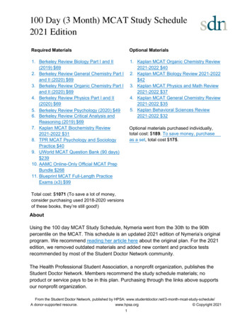
3Gel ElectrophoresisPurpose: Separation of proteins, DNA, or RNA based on size and/or chargeMacromolecules (proteins, DNA, or RNA) of interest are placed in the lanes of a gel. For proteins andsmall molecules of DNA and RNA, the gel will be polyacrylamide. For larger molecules of DNA ( 500 bp),the gel will be agarose. An electrical charge is placed across the gel. At the bottom is the positivelycharged anode and at the top is the negatively charged cathode. Keep in mind, since a voltage source isapplied to gel electrophoresis, it follows the same principles as an electrolytic cell. Negatively chargedmolecules will travel toward the anode. Because of the resistance of the gel, larger molecules will have aharder time moving and thus, the molecules will be separated by size with the smallest moleculestoward the bottom. The gel can then be stained for visualization, typically using Coomassie Blue dye. Alane will be loaded with a collection of molecules of a known size, called a ladder, which can used todetermine the size of the molecules being ran. There are several different applications of gelelectrophoresis:Native-PAGENative-PAGE is a polyacrylamide gel electrophoresis method for proteins that occurs under nondenaturing conditions. This method willseparate proteins by size while retainingtheir structure.SDS-PAGESDS-PAGE is a polyacrylamide gelelectrophoresis method for proteins thatoccurs under denaturing conditions to separate proteins by mass. Negatively-charged sodium dodecylsulfate (SDS) is added to the solution of proteins, denatures the proteins, and binds one SDS for everytwo amino acids, giving all proteins the same charge-to-mass ratio. Since all proteins have the samecharge-to-mass ratio, they are separated solely on mass, with the smallest proteins found toward thebottom of the gel. SDS will only interrupt non-covalent bonds, so if disulfide bridges are present in theprotein, they will not be broken. This is useful when analyzing proteins with multiple subunits.
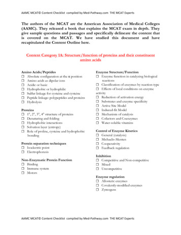
4Reducing SDS-PAGEReducing SDS-PAGE is exactly like SDS-PAGE, but with the addition of a reducing agent, like βmercaptoethanol, which will reduce disulfide bridges and result in a completely denatured protein.Isoelectric FocusingIsoelectric focusing is a gelelectrophoresis method that separatesproteins on the basis of their relativecontents of acidic and basic residues. Apolyacrylamide gel with a pH gradient(low pH on one side, high pH on theother) is used. When proteins migratethrough the pH gradient gel, they willtravel toward the anode until the areaof the gel with the pH that matches their isoelectric point (pI). When a protein is at its pI, it has a netcharge of zero and will not be attracted to the positively charged anode so it will not move.Western Blotting (Protein)Purpose: Detection of a specific protein in a sampleStep 1: Proteins from a sample are loaded into an SDS-PAGE gel and separated based on sizeStep 2: Proteins from the gel are transferred to a polymer sheet and exposed to a radiolabeled antibody(sometimes using two antibodies; one specific to the protein of interest and another radiolabeledantibody that binds to the first antibody) that is specific to our protein of interestStep 3: The polymer sheet is viewed used autoradiography. The protein of interest that is bound to theradiolabeled antibody will be visible.
5Southern (DNA) and Northern (RNA) BlottingPurpose: Detection of a specific DNA (Southern blot) or RNA (Northern blot) sequence in a sampleStep 1: The DNA strand of interest is exposed to restriction enzymes that cut the DNA strand intosmaller fragmentsStep 2: The newly cleaved strands of DNA are denatured using a solution of NaOH to create ssDNAstrandsStep 3: The single stranded cleaved strands of DNA undergo gel electrophoresis, separating them bysize. Smaller fragments will be found at the bottom of the gel. Polyacrylamide is used if the stands areless than 500 base pairs. Agarose is used if the strands are over 500 base pairs.Step 4: The DNA from the gel is transferred to a sheet of nitrocellulose paper and then exposed to a 32Pradiolabeled DNA probe that is complementary to our DNA of interest.Step 5: The nitrocellulose paper is then viewed using autoradiography to identify the strand of interest.NOTE: These methods are nearly identical for Southern and Northern blotting. The steps listed aboveare for Southern blotting, however, the only difference is that Northern blotting uses RNA, so steps 1and 2 are not done.
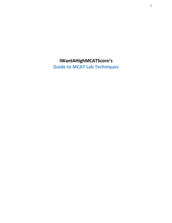
6DNA Sequencing (Sanger Dideoxynucleotide Sequencing)Purpose: Used to determine the sequence of nucleotides in a strand of DNAModified nucleotides, known as “dideoxynucleotides” (ddNTPs), are used in this method. ddNTPs aremissing the OH group on the 3’ carbon, thus they are unable to create a new 5’ 3’ phosphodiesterbond. This allows us to control the termination of replication.Step 1: The DNA strand of interest is denatured using an NaOH solution to create a ssDNA strand thatwe can use for replicationStep 2: The ssDNA strand of interest is added to a solution containing:1. A radiolabeled DNA primerthat is complementary to thegene of interest2. DNA polymerase3. All four dNTPs (dATP, dTTP,dCTP, dGTP)4. A very small quantity of asingle ddNTP (e.g., ddATP)This step is done once for each of thefour nucleotides in separate solutions.Step 3: Each solution containing aspecific dNTP and ddNTP are placed intheir own lane of a gel and ran undergel electrophoresis. The gel istransferred to a polymer sheet andautoradiography is used to identify thestrands in the gel.For each respective nucleotide, the insertion of a ddNTP will terminate replication and create variousstrands of different length that correspond to that specific nucleotide. Therefore, the gel can be readfrom bottom-to-top to determine the nucleotide sequence. The smaller the fragment, the further ittravels in the gel.
7ChromatographyPurpose: The separation of two or more molecules from a mixtureThere are several different types of chromatography that can be used for separating or analyzing amixture of two or more molecules based on their properties. Traditionally, there are two components tochromatography; a stationary phase, which is typically polar, and a mobile phase, which is typically nonpolar. Polar molecules are separated from a mixture by staying with the stationary phase, while nonpolar molecules stay with the mobile phase. However, if reverse-phase is specified, then the propertiesof the two phases are switched. This also changes based on the type ofchromatography used, as some methods use ligands or gel beads.Liquid ChromatographyIn liquid chromatography, silica is traditionally used as the stationaryphase while toluene or another non-polar liquid is used as the mobilephase.High-Performance Liquid Chromatography (HPLC)HPLC is a type of liquid chromatography that utilizes high pressures topass the solvent phase through a more finely-ground stationary phase,which increases the interactions between the molecules and thestationary phase, giving HPLC a higher resolving power. Molecules canthen be determined based on their absorbance and elution time as seenon the right. In normal phase HPLC, stationary phase is polar, mobilephase is nonpolar; in reverse phase HPLC, stationary is nonpolar, mobile is polar.Gas ChromatographyGas chromatography (also known as gas-liquid chromatography) is used to separate and analyzemolecules that can be vaporized. The mobile phase is an inert or unreactive gas, such as helium ornitrogen, while the stationary phase is a thin layer of liquid or polymer that surrounds the walls of atube. The stationary phase allows more polar molecules to elute slower, giving them a higher retentiontime.Gel-Filtration (Size Exclusion) ChromatographyGel-filtration chromatography (also known as size-exclusionchromatography) is used to separate molecules by size rather than polarity.Smaller molecules can enter the porous gel beads, allowing them to elutelater, while larger molecules that do not fit will elute faster. The gel beadscan be viewed as the stationary phase, while the solution in the column canbe viewed as the mobile phase.
8Ion-Exchange ChromatographyIon-exchange chromatography will separate proteins by their net charge. Thecolumn is filled with charged beads, either positive or negative. In cationexchange, negatively-charged beads are used which attract positively chargedproteins and negatively-charged proteins will elute first. In anion-exchange,positively-charged beads are used which attract negatively-charged proteinsand positively-charged proteins will elute first.Affinity ChromatographyAffinity chromatography will separate proteins based on their affinityfor a specific ligand. Beads that are bound to a specific ligand will beused and proteins with a high affinity for that ligand will bind to thebeads, allowing proteins with a low affinity to elute first. The highaffinity proteins are then eluted by increasing the concentration ofthe free ligand in the column, which competes for the active site ofthe bound proteins.Thin-Layer ChromatographyThin-layer chromatography consists of asmall sheet of medium that is coated in anadsorbent material, such as silica gel. Thepolar silica is the stationary phase. Themolecules of interest are added to thebottom of the sheet and the sheet isplaced in a non-polar liquid, such asheptane, until it reaches the origin. Themobile phase then travels up the plateusing capillary action, allowing themolecules to move with it if they arerelatively non-polar. The spots are thenvisualized using UV light.The relative distances traveled between the molecules is represented by the Rf value, which is measuredas the ratio of the distance the molecule traveled from the origin to the distance the solvent fronttraveled from the origin.
9DistillationPurpose: Used to separate two or more molecules from a solutionSimple DistillationSimple distillation is used to separate two molecules from a solution when their boiling points differ by25o C or greater.Fractional DistillationFractional distillation is used to separate two molecules from a solution when their boiling points differby less than 25o C.Vacuum DistillationVacuum distillation is used to separate two molecules from a solution when their boiling points are highand risk changing chemically.Polymerase Chain ReactionPurpose: Used to amplify a small quantity of DNA by severalorders of magnitudeStep 1: DNA strands and complementary DNA primers are heatedto 95o C for 15 seconds to separate the strands.Step 2: The solution is abruptly cooled to 54o C to allow theprimers to anneal to each ssDNA.Step 3: The solution is heated to 72o C and new complementarystrands are synthesized using Taq DNA polymerase.Step 4: The cycle is repeated until the desired quantity of DNA issynthesized.
10SpectroscopyPurpose: Used for structuraldetermination of molecules1H-NMR SpectroscopyThe application of nuclear magneticresonance regarding the 1H isotopewithin the molecules of a substance.Chemical shift: The chemical shift onthe x-axis (δppm) represents theamount of deshielding of electrons thatis caused by an adjacent heteroatom orpi bond.- 0 - 5 ppm Alkane region- 3 - 5 ppm Alkane with aheteroatom region- 5 - 7 ppm Alkene region- 6 - 8 ppm Aroma c region- 9 - 10 ppm Aldehyde region- 10 - 13 ppm Carboxylic acid regionIntegration: The integration of the peak determines the number of equivalent hydrogens a signalrepresents.Neighbors: The number of peaks determines the number of neighboring hydrogens that are 3 bondsaway. The number of peaks equals the number of neighbors 1.- Singlet No neighboring hydrogens- Doublet One neighboring hydrogen- Triplet Two neighboring hydrogens- Quartet Three neighboring hydrogens- Quintet Four neighboring hydrogens- Sextet Five neighboring hydrogens- Septet Six neighboring hydrogens- Mul plet Seven or more neighboring hydrogens
1113C-NMR spectroscopyThe application of nuclear magnetic resonance regarding the 13C isotope within the molecules of asubstanceChemical shift: The chemical shift on the x-axis (δppm) represents the amount of deshielding ofelectrons that is caused by an adjacent heteroatom or pi bond.- 0 - 70 ppm Alkane region- 90 - 120 ppm Alkene region- 110 - 160 ppm Aroma c region- 160 - 200 ppm Carbonyl regionIR SpectroscopyIR spectroscopy is a method thatis used to identify certainfunctional groups within amolecule. Only molecules thathave a dipole moment will showabsorbance. The x-axis is reportedin wavenumbers (reciprocalcentimeters) and the y-axis isreported in percent absorbance.Important regions:- 1700 - 1750 Carbonyls (sharp peak)- 1720 - 1740 Aldehydes- 1700 - 1725 Ketones- 1735 - 1750 Esters- 1700 - 1725 Carboxylic acids- 3200 - 3600 OH groups (broad peak)- 3300 - 3400 Amines- The number of peaks are relative to the number of hydrogens on the amine (e.g., 1oamines will have two peaks, 2o amineswill have one peak)UV-Vis SpectroscopyAs the number of conjugated pi bonds increase, theenergy gap between the highest occupied molecularorbital (HOMO) and lowest unoccupied molecularorbital (LUMO) decreases, which means that light of alower energy is absorbed. This results in light with alonger wavelength to be emitted (as seen on the right).If a molecule absorbs green light, we see it as red.
12AutoradiographyPurpose: To visualize the location of a radioactive substance in a molecule or structureIn the scope of the MCAT, autoradiography is used primarily as an imaging technique to identifyradiolabeled atoms that are in a molecule, often as part of a Southern, Northern, or Western blot. Theradiolabeled substance is placed in contact with a photographic emulsion containing silver halidecrystals. The radiation of the radiolabeled substance converts the silver halide crystals into metallicsilver, producing an image.X-Ray CrystallographyPurpose: To visualize the structure of a molecule, used often with proteinsIn x-ray crystallography, a crystalline molecule causes a beam of incident x-rays to diffract into manyspecific directions. A three-dimensional image can be constructed by measuring the different angles andintensities of the diffracted rays.ImmunoprecipitationPurpose: Used to purify proteins from a solutionIn immunoprecipitation, a protein of interest can be precipitated from a solution by adding beadconjugated antibody that is specific to the protein of interest. The antibody is bound to some sort ofsolid bead, which can be used for either extraction with a magnetic or by centrifuge.RadioimmunoassayPurpose: Used to determine the concentration of a protein of interest in a given sampleSimilar to ELISA, the wells of a plate are coated with the primary antibody that is specific to our proteinof interest. Next, the radiolabeled protein, typically containing a tyrosine residue labeled with 125I, isadded to the wells and binds to the primary antibody. The concentration of that radiolabeled protein isdetermined by counting the gamma emission. Next, a sample of the protein of interest in an unknownconcentration is added to the wells, where it competes for the active site on the primary antibody,displacing an amount of the radiolabeled protein. The concentration of the radiolabeled protein isdetermined again by counting the gamma emission. The difference in concentrations provides theconcentration of the protein of interest.
13Mass SpectrometryPurpose: Used to determine themolecular weight of a compoundand aid in determining the molecularstructureIn mass spectrometry, the sample isvaporized and subjected to ionizingconditions. The charged moleculecollides with an electron, resulting inthe ejection of an electron from themolecule, making it a radical. Thecharged radical can undergofragmentation or being detected.The x-axis represents themass/charge ratio (m/z), whichessentially just means the molecules mass using only the lowest isotopes of the atoms involved (e.g.,12C, 1H, 35Cl). The y-axis represents the intensity, or relative abundance of the molecule, usually given asa percentage.Base peak: The tallest peak. This does not always represent the actual intact molecule, as it maysometimes be a fragment of the molecule that is found in higher abundance.Molecular ion peak (M): The peak that represents the molecule. The m/z value of this peak representsthe molecular weight of the molecule.M 1 peak: The relative abundance of 13C in the molecule. Found in a relative abundance of 1.1%. So ifthere is a M 1 with a m/z value of 4.4, it means that there are 4 carbons present (4.4/1.1 4).M 2 peak: The relative abundance of either 37Cl or 81Br in the molecule. 37Cl will be found in a 3:1 ratiorelative to the M peak (e.g., if the M peak has a relative abundance of 90%, if the M 2 peak is at 30%, itmeans there is chlorine present in the molecule). 81Br will be found in a 1:1 ratio relative to the M peak(e.g., if the M peak has a relative abundance of 90%, if the M 2 peak is also at 90%, it means there isbromine present in the molecule).Fragments: There are three types of fragmentation that can occur, which can help identify the structureof the molecule. 1) Alkane fragmentation, 2) alcohol dehydration, and 3) alpha cleavage. These are mostlikely beyond the scope of the MCAT.
14Enzyme-Linked Immunosorbent Assay (ELISA)Purpose: Used to identify the concentrationof a molecule of interest in a given sampleELISA is an assay that uses primary antibodiesthat are specific to a molecule of interest andsecondary antibodies that are specific toprimary antibodies and are conjugated with afluorophore, so their presence can bemeasured via spectrophotometry. There aretwo different methods of ELISAs that areused.Indirect ELISAThe molecule of interest (in this case, considered an antigen) is coated to the wells of a plate. A primaryantibody that is specific for that antigen is added and washed to remove any unbound antibodies. Next,the secondary antibody is added. The secondary antibody is linked to an enzyme, often horseradishperoxidase (HRP), that will be converted into a fluorophore when reacted with an oxidizing agent, suchas hydrogen peroxide. This will cause a change in color, which can be detected usingphotospectrometry. The absorbance of the well is compared against a serial-diluted standard of knownconcentration, and based on a determined curve using the standards, the concentration of the moleculein the well can be determined.Sandwich ELISAThe primary antibody that is specific to our molecule of interest is coated to the wells of a plate. Next,the molecule of interest is added to the wells and then washed to remove any unbound molecules.Next, the secondary antibody is added. The secondary antibody is linked to an enzyme, oftenhorseradish peroxidase (HRP), that will be converted into a fluorophore when reacted with an oxidizingagent, such as hydrogen peroxide. This will cause a change in color, which can be detected usingphotospectrometry. The absorbance of the well is compared against a serial-diluted standard of knownconcentration, and based on a determined curve using the standards, the concentration of the moleculein the well can be determined.
15Edman DegradationPurpose: Used to sequence the amino acidresidues in a proteinEdman Degradation is conducted by removingone amino acid residue at a time from the Nterminus of the peptide. Phenyl isothiocyanate isadded to the N-terminus of a polypeptide, whichthen cyclizes and breaks off, leaving an intactpolypeptide that is shortened by one residue. ThePTH-amino acid hybrid can then be analyzedusing chromatographic techniques to identify theamino acid. This can be repeated until everyamino acid residue in the polypeptide isidentified. This method is limited in the fact thatit can only accurately identify polypeptides lessthan 50 residues.Gram StainingPurpose: Used to differentiate bacteria into two groups, gram-positive or gram-negative, based on thecontent of their cell wallThe sample of bacteria is heat-fixed to a slide and crystal violet dye is added. Iodide is added to thebacteria, which binds to the crystal violet dye and traps it in the bacterial cell. The bacteria is thenwashed with alcohol to remove any dye that was not taken up by the bacteria. Safranin is added as acounterstain, so that the bacteria that did not stain with crystal violet can be visualized.Gram-positive bacteria: Appears purple on the slide. Contains a thick peptidoglycan layer that takes upthe stain.Gram-negative bacteria: Appears pink on the slide. Contains a thin peptidoglycan layer sandwichedbetween two lipid bilayers that take up the safranin stain.
16Restriction Fragment Length Polymorphism (RFLP)Purpose: Used to identify differences in homologous DNA sequences based onthe length of fragments caused by restriction enzymesRFLP allows us to determine differences in two or more DNA sequences of thesame gene. Most often, this will be comparing a wild type (WT) gene with amutated version of the gene. The DNA strand is exposed to a restrictionenzyme, also called a restriction endonuclease. Restriction enzymes recognize aspecific palindromic sequence (the sequence is the same in the 5’ 3’ direc onin both strands) in the DNA and cleave both strands. Once the DNA sequence iscleaved with the restriction enzymes, it can be ran through gel electrophoresis.If a mutation has occurred, there may be a difference in the number and lengthof strands between the WT and mutant genes, as the restriction points maydiffer.Salting Out and DialysisPurpose: Purification of proteins in asolutionAdding salt to a solution containingproteins can allow you to selectivelyprecipitate out proteins. Salting out is dueto the competition between the salt ionsand the protein for water to keep theprotein in solution. The salt concentrationat which a protein will precipitate variesfor each protein.After a protein has been precipitated, thesalt can be removed via dialysis. Thesolution is placed in a dialysis bag andthen added to a hypotonic solution. Theions will pass through the semipermeablemembrane of the dialysis bag, while theprotein of interest is left inside.
17Reducing Sugar TestsPurpose: Used to identify the presence of a reducing sugar in asolutionA reducing sugar is any sugar that is capable of acting as areducing agent due to the presence of a free aldehyde or ketonefunctional group. All monosaccharides are reducing sugars, sincethey are capable of mutarotation, and therefore, will be found inthe open-chain form to some degree. However, not alldisaccharides, oligosaccharides, or polysaccharides are reducingsugars, since some glycosidic bonds are between two anomericcarbons (classified as a 1 2 bond), preven ng mutarota on.Sucrose is an example of a non-reducing disaccharide whilemaltose is an example of a reducing disaccharide (as seen in thefigure on the right). Certain reagents can test for the presence of afree aldehyde or ketone group in order to identify the presence ofa reducing sugar.Tollen’s TestTollen’s reagent tests for the presence of an aldehyde and can distinguish between aldoses and ketoses.Ketones do not react unless they are α-hydroxy-ketones. A positive Tollen’s test is characterized by theprecipitation of elemental silver.Tollen’s reagent consists of [Ag(NH3
4 Reducing SDS-PAGE Reducing SDS-PAGE is exactly like SDS-PAGE, but with the addition of a reducing agent, like β-mercaptoethanol, which will reduce disulf