Transcription
Physics of Nuclear MedicineYao WangPolytechnic Institute of NYU, Brooklyn, NY 11201Based on J. L. Prince and J. M. Links, Medical Imaging Signals andSystems, and lecture notes by Prince. Figures are from the textbook.
Lecture Outline Atomic structureRadioactive DecayDecay modesExponential decay lawStatistical properties of decayRadiotracersEL5823 Nuclear PhysicsYao Wang, Polytechnic U., Brooklyn2
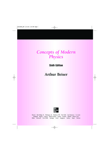
What is Nuclear Medicine Also known as nuclide imaging Introduce radioactive substance intobody Allow for distribution anduptake/metabolism of compound Functional Imaging! Detect regional variations ofradioactivity as indication ofpresence or absence of specificphysiologic function Detection by “gamma camera” ordetector array (Image reconstruction)From H. Graber, Lecture Note, F05EL5823 Nuclear PhysicsYao Wang, Polytechnic U., Brooklyn3
What is Nuclear Medicine Also known as nuclide imaging Steps:– Inject radio tracers into the body– The radio tracers undergo radioactive decay and generate gammy rays– A camera detect gamma rays from the radio tracer after a certain time Different physiological functions are imaged by using different radiotracers X-ray projection and tomography:– X-ray transmitted through a body from an outside source to a detector Nuclear medicine:– Gamma rays emitted from within a body– Emission computed tomography– Two popular method: Positron Emission Tomography (PET) Single photon emission computed tomography (SPECT)EL5823 Nuclear PhysicsYao Wang, Polytechnic U., Brooklyn4
Examples: PET vs. CT X-ray projection andtomography:– X-ray transmitted through abody from a outside source toa detector (transmissionimaging)– Measuring anatomic structure Nuclear medicine:– Gamma rays emitted fromwithin a body (emissionimaging)– Imaging of functional ormetabolic contrasts (notanatomic)From H. Graber, Lecture Note, F05 Brain perfusion, function Myocardial perfusion Tumor detection(metastases)EL5823 Nuclear PhysicsYao Wang, Polytechnic U., Brooklyn5
Atomic Structure An atom {a nucleus,electrons} nucleons {protons; neutrons} Nuclide: unique combination ofprotons and neutrons in anucleus mass number A # nucleons atomic number Z # protons # electrons An element is denoted by its Aand Z– Ex:EL5823 Nuclear Physics126C or C - 12Yao Wang, Polytechnic U., Brooklyn6
Stable vs. Unstable Nuclides Stable nuclides:– # neutrons # protons (A 2Z) when Z is small– # neutrons # protons when Z is large Unstable nuclides (radionuclides, radioactive atoms)– Likely to undergo radioactive decay, which gives off energy andresults in a more stable nucleusEL5823 Nuclear PhysicsYao Wang, Polytechnic U., Brooklyn7
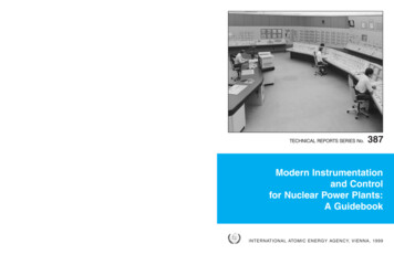
Line of StabilityStability depends on ratio Z:NEL5823 Nuclear PhysicsYao Wang, Polytechnic U., Brooklyn8
Isotopes, etc Isotopes: atoms with the same Z but different A– E.g. C-12 and C-11– Chemically identical Isobars: atoms with the same A but different Z– Different elements– Eg. Carbon-11 and boron-11 Isotones: atoms with the same number of neutrons butdifferent A Isomers: atoms with the same Z and A but with differentenergy levels (produced after gamma decay)EL5823 Nuclear PhysicsYao Wang, Polytechnic U., Brooklyn9
What is Radioactivity?EL5823 Nuclear PhysicsYao Wang, Polytechnic U., Brooklyn10
Decay ModesEL5823 Nuclear PhysicsYao Wang, Polytechnic U., Brooklyn11
Alpha Decay Alpha decay: the nucleus emits a Helium-4 particle(alpha particle)– Alpha decay occurs most often in massive nuclei that have toolarge a proton to neutron ratio. Alpha radiation reduces the ratioof protons to neutrons in the parent nucleus, bringing it to a morestable configuration.– mostly occurring for parent with Z 82From: lEL5823 Nuclear PhysicsYao Wang, Polytechnic U., Brooklyn12
Beta Decay Beta decay occurs when, in a nucleus with too manyprotons or too many neutrons, one of the protons orneutrons is transformed into the other. Mass number A does not change after decay, protonnumber Z increases or decreases. Beta minus decay (or simply Beta decay): A neutronchanges into a proton, an electron (beta particle) and aantineutrinoFrom: lEL5823 Nuclear PhysicsYao Wang, Polytechnic U., Brooklyn13
Positron Decay Also known as Beta Plus decay– A proton changes to a neutron, a positron (positive electron), and aneutrino– Mass number A does not change, proton number Z reducesFrom: lEL5823 Nuclear PhysicsYao Wang, Polytechnic U., Brooklyn14
Mutual Annihilation after Positron Decay The positron later annihilate a free electron, generate two gammaphotons in opposite directions– The two photons each have energy 511 KeV, which is the energyequivalent to the rest mass of an electron or positron– These gamma rays are used for medical imaging (Positron EmissionTomography), detected using a coincidence detection circuitEL5823 Nuclear PhysicsYao Wang, Polytechnic U., Brooklyn15
Gamma Decay (Isometric Transition) A nucleus (which is unstable) changes from a higher energy state toa lower energy state through the emission of electromagneticradiation (photons) (called gamma rays). The daughter and parentatoms are isomers.– The gamma photon is used in Single photon emission computedtomography (SPECT) Gamma rays have the same property as X-rays, but are generateddifferent:– X-ray through energetic electron interactions– Gamma-ray through isometric transition in nucleusFrom: lEL5823 Nuclear PhysicsYao Wang, Polytechnic U., Brooklyn16
Measurement of RadioactivityBq BequerelCi Curie:(orig.: activity of 1 g of 226Ra)Naturally occurring radioisotopes discovered 1896 by BecquerelFirst artificial radioisotopes produced by the Curie 1934 (32P)The intensity of radiation incident on a detector at range r from a radioactivesource isAEI 4πr 2A: radioactivity of the material; E: energy of each photonEL5823 Nuclear PhysicsYao Wang, Polytechnic U., Brooklyn17
Radioactive Decay Law N(t): the number of radioactive atoms at a given time A(t): is proportional to N(t)dNA λNdtλ : decay constant From above, we can deriveN (t ) N 0 e λtA(t ) A0 e λt λN 0 e λt The number of photons generated ( number of disintegrations)during time T isTT N A(t )dt λN 0 e λt dt N 0 (1 e λT )0EL5823 Nuclear Physics0Yao Wang, Polytechnic U., Brooklyn18
Half-Life Half-life is the time it takes for the radioactivity todecrease by ½.EL5823 Nuclear PhysicsYao Wang, Polytechnic U., Brooklyn19
Statistics of Decay The exponential decay law only gives the expected number of atomsat a certain time t. The number of disintegrated atoms over a short time t T1/2 aftertime t 0 with N0 atoms follows Poisson distributiona k e aPr{ N k} ; a λN 0 t ;k!λN 0 is called the Poisson rate.Strictly speakinga N 0 (1 e λ t )When λ t is small, e λ t 1 λ t , a N 0 λ tEL5823 Nuclear PhysicsYao Wang, Polytechnic U., Brooklyn20
Radiotracers: Desired Property Decay mode:– Clean gamma decay: do not emit alpha or beta articles– Positron decay: positron will annihilate with electrons to produce gammarays Energy of photon:– Should be high so that photons can leave the body w/ little attenuation– Hard to detect if the energy is too high– Desired energy range: 70-511 KeV Half-life– Should not be too short (before detector can capture) or too long (longerpatient scan time)– Minutes to hours desired Half-value-layer (HVL)– Thickness of tissue that absorbs half of the radioactivity produced– Should be around the dimension of the organ to be imaged Monoenergetic– Energy sensitive detectors can discriminate the primary photons fromscattered ones.EL5823 Nuclear PhysicsYao Wang, Polytechnic U., Brooklyn21
Decay Process Examplesα decay23892U 23490Th 24He, T1 2 4.5 109 yβ - decay23490Th 1023491Pa e ν e , T1 2 24.1 dn 11H e ν e , T1 2 10.6 mβ decay116C 115B e ν e , T1 2 20.38 m106C 105 B e ν e , T1 2 19.2 s158O 157 N e ν e , T1 2 122 sMost of these naturally occurringprocesses are not useful for medicalimaging applications, with too longHalf-time, too short HVL, too highenergy.They can be used asradiotherapeutic agents, if they canbe targeted to tumors, to destroydiseased tissue and stops thecancer from proliferating.e capture412041Ca e 19K ν e , T1 2 1 105 yEL5823 Nuclear PhysicsYao Wang, Polytechnic U., Brooklyn22
Radionuclides in Clinical Use Most naturally occurring radioactive isotopes not clinically useful(long T1/2, charged particle emission, alpha or beta decay) Artificial radioactive isotopes produced by bombarding stableisotopes with high-energy photons or charged particles Nuclear reactors (n), charged particle accelerators (Linacs,Cyclotrons)T 2.5d99Mo1/ 2 99 m Tc e νFrom H. Graber, Lecture Note, F05EL5823 Nuclear PhysicsYao Wang, Polytechnic U., Brooklyn23
The Technetium Generator Can be produced from an on-site generator– 99 Mo 99m Tc 99 Tc, Decay characteristics of 99m Tc:– half life 6.02h, E 140 KeV, HVL 4.6 cm99 mTcT1 / 2 6 h 99Tc γ (140 keV ) Used in more than 90% of nuclear imaging More detail: see handout [Webb, sec. 2.5]EL5823 Nuclear PhysicsYao Wang, Polytechnic U., Brooklyn24
Radiopharmaceuticals Radionuclide is bound to pharmaceuticals that is specific tometabolic activities (cancer, myocardial perfusion, brain perfusion) Gamma emitter––99mTc-Sestamibi(myocardial perfusion, cancer)99mTc-labeled hexamethyl-propyleneamine (brain perfusion) Positron emitters–11C,T1/2 20 min[12C (p,pn) 11C; 14N (p,α) 11C]: many organic compounds (binding to nerve receptors, metabolic activity)–13N,T1/2 10 min[16O (p,α) 13N; 13C (p,n) 13N]: NH3 (blood flow, regional myocardial perf.)–15O,T1/2 2.1 min[15N (p,n) 15O; 14N (d,n) 15O]: CO2 (cerebral blood flow), O2 (myoc. O2 consumption), H2O (myoc. O2consumption & blood perfusion)–18F,T1/2 110 min[18O (p,n) 18F; 20Ne (d,α) 18F]: 2-deoxy-2-[18F]-fluoroglucose (FDG, neurology, cardiology, oncology,metabolic activity)From H. Graber, Lecture Note, F05EL5823 Nuclear PhysicsYao Wang, Polytechnic U., Brooklyn25
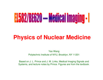
Common RadiotracersThyroid functionKidney functionMost commonly usedOxygen metabolismEL5823 Nuclear PhysicsYao Wang, Polytechnic U., Brooklyn26
Summary Nuclear medicine relies on radiation (gamma rays) generatedthrough radioactive decay Radioactive decay is the process when a unstable nuclide ischanged to a more stable one– Four modes of decay, generating alpha particles, beta particles,positrons and gamma rays respectively Radioactivity follows an exponential decay law, characterized bythe decay constant or the half-life Desired properties for radio tracers Common radiotracers in nuclear medicineEL5823 Nuclear PhysicsYao Wang, Polytechnic U., Brooklyn27
Reference Prince and Links, Medical Imaging Signals and Systems,Chap 7. “Guide to the Nuclear Wallchart”, Chap 3.http://www.lbl.gov/abc/wallchart/outline.html Recommended readings:– K. Miles, P. Dawson, and M. Blomley (Eds.), FunctionalComputed Tomography (Isis Medical Media, Oxford, 1997).– R. J. English, SPECT: Single Photon Emission ComputedTomography: A Primer (Society of Nuclear Medicine, Reston,VA, 1995).– M. Reivich and A. Alavi (Eds.), Positron Emission Tomography(A. R. Liss, NY, 1985).EL5823 Nuclear PhysicsYao Wang, Polytechnic U., Brooklyn28
Homework Reading:– Prince and Links, Medical Imaging Signals and Systems, Ch. 7.– Handouts from [Webb] Note down all the corrections for Ch. 7 on your copy ofthe textbook based on the provided errata. Problems for Chap 7 of the text book––––––P7.1P7.2P7.4P7.6P7.7 (assume the energy of the photons is E)P7.9EL5823 Nuclear PhysicsYao Wang, Polytechnic U., Brooklyn29
Physics of Nuclear Medicine Yao Wang Polytechnic Institute of NYU, Brooklyn, NY 11201 Based on J. L. Prince and J. M. Links, Medical Imaging Signals and Systems, and lecture notes b