Transcription
Understanding and Living with GlaucomaBetty hopes futuregenerations won’t have tolive with glaucoma.Dr. Goldberg’sbreakthrough researchcould mean theywon’t have to.Understanding and Living with Glaucoma cSUPPORTED BY AN EDUCATIONAL GRANT FROM AERIE PHARMACEUTICALS, INC.
Glaucoma is an eye disease thatgradually steals your vision. Usually,glaucoma has no symptoms in itsearly stages. But without propertreatment glaucoma can lead toblindness.The good news is that with regulareye exams, early detection, andtreatment, you can preserve yoursight. This guide will give you acomplete introduction to the factsabout glaucoma.d Glaucoma Research Foundation
2INTRODUCTION3UNDERSTANDING GLAUCOMA3Understand the Eye to Understand Glaucoma4How Glaucoma Affects the Eye6Who Gets Glaucoma?7Are There Symptoms?7When Should You Get Your Eyes Checked for Glaucoma?8DIFFERENT TYPES OF GLAUCOMA8Primary Open-Angle or Open-Angle Glaucoma9Primary Angle-Closure or Angle-Closure Glaucoma or Narrow-AngleGlaucoma10Other Types of Glaucoma Normal-Tension Glaucoma10 Secondary Glaucoma1012 DETECTING GLAUCOMA12How Is Glaucoma Diagnosed?12What To Expect During Glaucoma Examinations Tonometry12 Ophthalmoscopy12 Perimetry15 Gonioscopy16 Pachymetry1617TREATING GLAUCOMA17Treatment of Primary Open-Angle Glaucoma Selective Laser Trabeculoplasty17 Glaucoma Medications18 Incisional Surgeries21 Unapproved Treatments2525Treatment of Primary Angle-Closure Glaucoma26Treatment of Other Types of Glaucoma28FREQUENTLY ASKED QUESTIONS30 LIVING WITH GLAUCOMA30Working with Your Doctor32Responding to Vision Changes Due to Glaucoma32Questions For Your Doctor34Your Lifestyle Counts35Looking Ahead36APPENDIX36A Guide To Glaucoma Medications38GlossaryUnderstanding and Living with Glaucoma 1
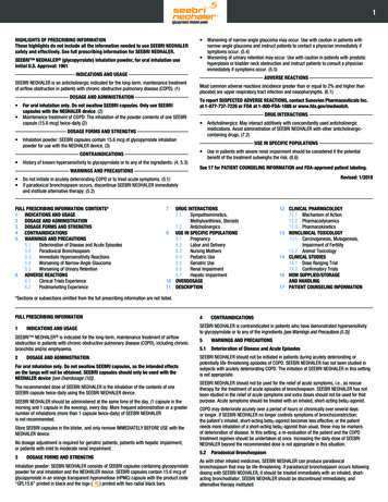
IntroductionIf you have been diagnosed with glaucoma, or are a glaucoma suspect, you probably havea lot of questions and some concerns. Coping with a long-term eye condition can seemoverwhelming, but you are fortunate compared to the many who have glaucoma and don’tknow it. You are now empowered to learn how you can preserve your vision and live anormal life as a glaucoma patient.When faced with a new diagnosis of glaucoma there is one question that is foremost in everypatient’s mind: “Will I go blind?” Thankfully, for most patients the answer is no. Glaucomatypically progresses very slowly over many years, and most people never lose vision if they seetheir eye doctor regularly and follow their treatment plan.Excellent glaucoma treatments are available that work to control eye pressure, the maincause of vision loss in glaucoma. These treatments include a wide range of eye dropmedications, laser treatments that are performed in the office, and many types of surgery.Newer treatments are continuously being developed and evaluated.Glaucoma is a chronic disease, and you are the most important part of your treatment.Working closely with their doctor, the vast majority of people with glaucoma will retain theirvision. The key to preserving your vision is speaking honestly with your doctor about yourdisease and its treatment.This guide will give you a complete introduction to the facts about glaucoma and how to workwith your doctor to manage your glaucoma.A HEALTHY EYERetinaCiliary bodyAngleAqueousIrisLensCorneaOptic nerveScleraOptic disc2 Glaucoma Research Foundation
Understanding GlaucomaGlaucoma is an eye disease that usually has no symptoms in its early stages. Without propertreatment, glaucoma can lead to blindness. The good news is that with regular eye exams,early detection, and treatment, you can preserve your sight.UNDERSTAND THE EYE TO UNDERSTAND GLAUCOMATo understand glaucoma, it is important to have an idea of how the eye works and thedifferent parts of the eye.Covering most of the outside of the eye is a tough white layer called the sclera. A clear thinlayer called the conjunctiva covers the sclera. At the very front of the eye is a clear surface, likea window, called the cornea that protects the pupil and the iris behind that window. The iris,a muscle, is the colored part of the eye that contracts and expands to let light into the eye. Atthe center of the iris is a hole (covered by the clear cornea) called the pupil, where light entersthe eye. The lens inside our eye focuses this light onto the back of the eye, which is called theretina. The retina converts the light images into electrical signals, and the retina’s nerve cellsand fibers carry these signals to the brain through the optic nerve. The optic disc is the area onthe retina where all the nerve fibers come together to become the optic nerve as it leaves theeye to connect to the brain.Healthy Eye DrainageThe front part of the eye is filled with a clear fluid (called aqueous humor) made by theciliary body. The fluid flows out through the pupil. It then reaches the eye’s drainage system,including the trabecular meshwork and a network of canals. The inner pressure of the eye(intraocular pressure or “IOP”) depends on the balance between how much fluid is madeand how much drains out of the eye. If your eye’s fluid system is working properly, then theright amount of fluid will be produced. Likewise, if your eye’s drainage system is workingproperly, then fluid can drain freely out to prevent pressure buildup. Proper drainage helpskeep eye pressure at a normal level and is an active, continuous process that is needed forthe health of the eye.Understanding and Living with Glaucoma 3
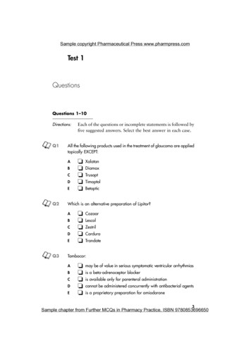
HOW GLAUCOMA AFFECTS THE EYEThe Optic DiscYou have millions of nerve fibers that run from your retina to form the optic nerve. Thesefibers meet at the optic disc. In most types of glaucoma, the eye’s drainage system becomesclogged so the intraocular fluid cannot drain. As the fluid builds up, it causes pressure to buildinside the eye, which can damage these sensitive nerve fibers and result in vision loss. As thefibers are damaged and lost, the optic disc begins to hollow and develops a cupped shape.Doctors can identify this cupping shape in their examinations.DEVELOPOMENT OF GLAUCOMAHEALTHY EYEEYE WITH GLAUCOMAOpticdiscFlow ofaqueoushumorDrainage canalblocked; build-upof fluidDrainagecanalOpticnerveIncreasedpressure damagesoptic nerveThe blockage of drainage canals at the front of the eye causes fluid to build up and increases eyepressure. This can lead to optic nerve damage at the back of the eye.KEYBlue arrows indicate the flow ofintraocular fluid/aqueous humor4 Glaucoma Research FoundationRed arrows indicate the direction of pressureof intraocular fluid/aqueous humor
Intraocular PressureA normal intraocular pressure (IOP) ranges between 12 and 22 mmHg (“millimeters ofmercury,” a measurement of pressure.) If the pressure remains too high for too long, the extrapressure on the sensitive optic disc can lead to permanent vision loss.Although high IOP is clearly a risk factor for glaucoma, we know that other factors alsoare involved because people with IOP in the normal range can experience vision loss fromglaucoma. Identifying these other factors is a focus of current research.AQUEOUS FLUID PRODUCTION AND DRAINAGE PATHWAYVision Loss in GlaucomaGlaucoma usually occurs in both eyes, but increased eye pressure tends to happen in one eyefirst. This damage may cause gradual visual changes and loss of sight over many years. Often,peripheral (side) vision is affected first, so the change in your vision may be small enoughthat you may not notice it. With time, your central vision may also be affected. Sight lostfrom glaucoma cannot be restored. However, early detection and treatment can preventvision loss and maintain remaining vision.Understanding and Living with Glaucoma 5
WHO GETS GLAUCOMA?Glaucoma affects people of all ages, from babies to teens to younger and older adults.ALTHOUGH EVERYONE IS AT RISK FOR GLAUCOMA, THOSE AT HIGHER RISK INCLUDEPEOPLE WHO: Are over 60 years old Use steroid medications Are of African, Asian, or Have high eye pressureHispanic descent Have a thin central cornea Have relatives with glaucoma Are very nearsighted (myopic) Have had an eye injuryor far-sighted (hyperopic)People who are of African, Asian, or Hispanic descent are at higher risk forglaucoma.6 Glaucoma Research Foundation
ARE THERE SYMPTOMS?In the most common form of glaucoma, Primary Open-Angle Glaucoma, buildupof fluid pressure happens very slowly. There are no uncomfortable or painful symptoms.Usually, the initial loss of vision is of side (peripheral) vision, and central vision is maintaineduntil late in the disease. Since we use our central vision for driving, reading and most tasks, thegradual loss of peripheral vision in glaucoma isn’t typically noticed until a significant amount ofvision has been permanently lost.In the less common kinds of glaucoma, symptoms can be noticeable.THESE SYMPTOMS CAN INCLUDE: Hazy vision Eye and head pain Nausea or vomiting The appearance ofrainbow-colored circlesaround bright lights Sudden sight lossYou should see your doctor if you experience any of the symptoms listed above.WHEN SHOULD YOU GET YOUR EYES CHECKED FOR GLAUCOMA?Early detection, through regular and complete eye exams, is the key to protecting your visionfrom damage caused by glaucoma. It is important to have your eyes examined regularly.You should get a baseline eye screening at age 40. Early signs of eye disease and changes invision may start to occur at this age. Your eye doctor will tell you how often to have followup exams based on the results of this screening. If you have diabetes, high blood pressure, ora family history of glaucoma, you should see an eye doctor now to determine how often tohave eye exams.Understanding and Living with Glaucoma 7
Different Types of GlaucomaThere are several types of glaucoma. The two main types are Open-Angle and Angle-Closure.Primary Open-Angle GlaucomaPrimary Open-Angle Glaucoma, the most common form of glaucoma and also called OpenAngle Glaucoma, is a lifelong condition that accounts for at least 90% of all glaucoma cases.In patients with Open-Angle Glaucoma, the angle in your eye where the iris (the coloredpart of their eye) meets the cornea (the clear window at the front of the eye) is as open as itshould be, but the eye’s drainage canals become clogged over time, similar to a clogged pipebelow the drain in a sink. This can result in the buildup of intraocular fluid and increased eyepressure that can damage the optic nerve.There are no early warning signs of Open-Angle Glaucoma. It develops slowly and sometimeswithout noticeable sight loss for many years. If Open-Angle Glaucoma is not diagnosed andtreated, it can cause gradual loss of vision. With regular eye exams, Open-Angle Glaucomamay be found early and usually responds well to treatment to preserve vision.FLUID PATHWAY IN OPEN-ANGLE GLAUCOMADrainage canalsIrisCorneaFluid flowOpen angleDrainage canalsdThe angle between the iris and cornea is open in Open-Angle Glaucoma. But the drainage canalsbecome clogged over time.8 Glaucoma Research Foundation
Primary Angle-Closure GlaucomaThe other main type of glaucoma is Primary Angle-Closure Glaucoma, also called NarrowAngle Glaucoma. In this type of glaucoma, the angle in many or most areas between theiris and cornea is closed, reducing fluid drainage and causing increased eye pressure. Thisincreased pressure leads to optic nerve damage and possible vision loss. There are also earlystages of the disease in which parts of the angle are closed, but the eye pressure may or maynot be high and the optic nerve is not yet affected.Eye pressure elevation in Primary Angle-Closure Glaucoma usually occurs gradually and hasno symptoms. Rarely the rise in pressure occurs rapidly—an acute attack of angle-closure. InAcute Angle-Closure Glaucoma, the intraocular pressure rises very quickly, causing noticeablesymptoms such as eye pain, blurry vision, redness, rainbow-colored rings (“haloes”) aroundlights, and nausea and/or vomiting.Acute Angle-Closure Glaucoma is a medical emergency. It can cause permanent visiondamage and requires immediate medical attention.FLUID PATHWAY IN ANGLE-CLOSURE GLAUCOMADrainage canalsIrisCorneaFluid flowClosed angleDrainage canalsdThe angle between the iris and cornea is narrow or closed in Angle-Closure Glaucoma. More of theangle may become closed over time.Understanding and Living with Glaucoma 9
OTHER TYPES OF GLAUCOMAMost other types of glaucoma are variations of the open-angle or angle-closure types. Theseglaucoma types can occur in one or both eyes.Normal-Tension GlaucomaNormal-Tension Glaucoma (NTG), also known as Low-Tension Glaucoma, is a form ofglaucoma in which damage occurs to the optic nerve even though the eye pressure is nothigh. The cause of Normal-Tension Glaucoma is unknown. Researchers are studying why someoptic nerves are damaged by these relatively low fluid pressures.People at higher risk for Normal-Tension Glaucoma have a family history of Normal-TensionGlaucoma, are of Japanese ancestry, or have a history of systemic heart disease such asirregular heart rhythm, have migraines, or low diastolic blood pressure.Secondary GlaucomaSecondary Glaucoma is any form of glaucoma that has an identifiable cause for increasedeye pressure that results in optic nerve damage and vision loss. For example, an eye injury,inflammation, and certain drugs may cause a Secondary Glaucoma. Secondary Glaucomaincludes Pigmentary Glaucoma, Congenital Glaucoma, Exfoliative Glaucoma, NeovascularGlaucoma, Uveitic Glaucoma, and Traumatic Glaucoma. The treatment for SecondaryGlaucoma depends on whether it is open angle or angle closure.Pigmentary GlaucomaPigmentary glaucoma is a form of Open-Angle Glaucoma that occurs when tiny pigmentgranules on the back of the iris break off into the aqueous humor and become trapped inthe eye’s drainage system. The pigment can block the eye’s drainage canals and lead to anincrease in eye pressure and damage to the optic nerve. This condition is more common inyoung, Caucasian, male patients who are near-sighted.Congenital GlaucomaCongenital Glaucoma occurs in babies when there is incorrect or incomplete development ofthe eye’s drainage canals before birth. This leads to increased eye pressure that can damage theoptic nerve. Symptoms of Congenital Glaucoma include unusually large eyes, excessive tearing,cloudiness of the cornea, and sensitivity to light. This is a rare condition that may be inherited.10 Glaucoma Research Foundation
Exfoliative GlaucomaExfoliative Glaucoma, also known as Pseudoexfoliative Glaucoma, occurs when a flaky,dandruff-like material peels off the outer layer of the lens within the eye. The material collectsin the angle between the cornea and iris and can clog the drainage system of the eye, causingeye pressure to rise. This is a type of Open-Angle Glaucoma that is known for causing moreepisodes of high pressure, more fluctuations, and higher peak pressures than other types ofglaucoma. It is associated with a gene and is more common in certain racial groups, includingpeople from Nordic countries, Russia, and India, Mediterranean populations, and others.Neovascular GlaucomaNeovascular Glaucoma is caused by the abnormal formation of new blood vessels on the irisand over the eye’s drainage canals. The new blood vessels block the eye’s fluid from exitingthrough the drainage canals, causing an increase in eye pressure. Neovascular Glaucoma isalways associated with other abnormalities, most often diabetes.Uveitic GlaucomaUveitic Glaucoma is a result of uveitis, an inflammation of the iris and other nearby structures(together called the “uvea”) in the eye. These structures become inflamed and disrupt fluiddrainage out of the eye, or the steroid medication used to treat the inflamed structures candamage the drainage canals and result in increased fluid pressure.Traumatic GlaucomaInjury to the eye may cause Traumatic Glaucoma. This form of Open-Angle Glaucoma canoccur immediately after the injury or develop many years later. It can be caused by bluntinjuries that bruise the eye (“blunt trauma”) or by injuries that penetrate the eye.Congenital glaucoma can often be corrected with surgery.Understanding and Living with Glaucoma 11
Detecting GlaucomaHOW IS GLAUCOMA DIAGNOSED?TO ACCURATELY AND SAFELY DIAGNOSE GLAUCOMA, YOUR EYE DOCTOR WILL CHECKFIVE FACTORS DURING YOUR EXAMINATION: TonometryA measure of the pressure inside the eye (intraocular pressure or IOP) OphthalmoscopyExamination of the shape and color of the optic nerve, also called a dilated eye exam PerimetryA test that measures your field of vision GonioscopyExamination of the angle in the eye where the iris meets the cornea PachymetryA measure of the thickness of the corneaWHAT TO EXPECT DURING GLAUCOMA EXAMINATIONSTonometryDuring tonometry, eye drops are used to numb the eye. Then a doctor or technician uses adevice called a tonometer to measure the eye pressure. A small amount of pressure is appliedto the eye by a small device. The average range for eye pressure is 12–22 mm Hg (“mm Hg”refers to millimeters of mercury, a scale used to record eye pressure.) The level of eye pressureat which glaucoma develops is not the same for everyone, and some people can getglaucoma even if their eye pressures are within the average range of 12–22 mm Hg.OphthalmoscopyEye drops are used to dilate the pupil so that the doctor can see into your eye with a speciallens to examine the shape and color of the optic nerve and note whether there is glaucomadamage. You may need to wait in a waiting room for your eyes to fully dilate. The doctor willuse a small device to shine light on and magnify the optic nerve. The doctor will check if theoptic nerve is cupped or not a healthy pink color, which may be a cause for concern.12 Glaucoma Research Foundation
Checking the eye pressure using tonometry.An ophthalmoscope helps your doctor see your optic nerve.Understanding and Living with Glaucoma 13
Various laser-based imaging technologies can also be used to examine the optic nerve andof these, Optical Coherence Tomography (OCT) is the most commonly used. OCT measuresthe reflection of laser light (much like ultrasound measures the reflection of sound) and candirectly measure the thickness of the nerve fiber layer. Imaging your optic disc over timeduring multiple visits can help detect progressive loss of optic nerve fibers.Healthy optic nerve14 Glaucoma Research FoundationOptic nerve in eye with glaucoma
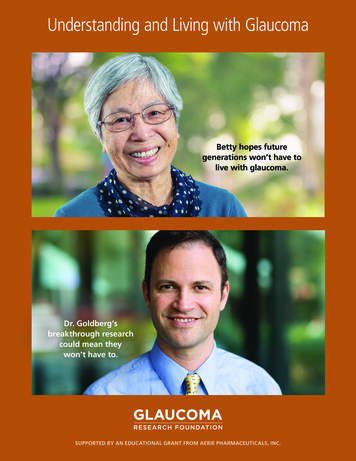
PerimetryPerimetry (or “visual field testing”) produces a map of your field of vision. This test will helpyour doctor determine whether your vision has been affected by glaucoma.During this test, you will be asked to look straight ahead and then press a button wheneveryou see a spot of light in your peripheral (side) vision. The spots of light will vary in intensitysuch that some are very easy to see and others are not visible even when vision is normal. Donot be concerned if there is a delay in seeing the light as it may be in or around your blind spot.This is perfectly normal and does not necessarily mean that your field of vision is damaged. Tryto relax, blink normally, and respond as accurately as possible during the test. Don’t worry ifyou feel you missed a spot as areas are automatically retested. Your test performance may varyif you are tested at the end of a long day.Your doctor may want you to repeat the test to see if the results are the same. After glaucomahas been diagnosed, visual field tests are usually done one to two times a year to check forany changes in your vision.Visual field within normal limitsVisual field outside normal limitsTest results from a normal visual field without vision loss (left) and a visual field with vision loss fromglaucoma (right). Darker gray and black areas represent loss of vision in a visual field. The optic discappears black in both fields since there is no vision there; this is normal.Understanding and Living with Glaucoma 15
GonioscopyGonioscopy is a diagnostic exam that helps determine whether the angle where the iris meetsthe cornea is open or closed. During the exam, eye drops are used to numb the eye, and a specialhand-held lens is gently placed on the eye for a few moments. This special lens includes a mirrorthat allows the doctor to see the angle between the iris and cornea to determine its status.Gonioscopy uses a special mirrored lens to see the angle between the irisand cornea.PachymetryPachymetry is a simple, painless test that measures the thickness of the cornea—the clearwindow at the front of the eye—using a probe that is gently placed on your eye. A thincornea is a risk factor for glaucoma. Corneal thickness also has the potential to influence eyepressure readings. For example, if a cornea is thicker than average, pressure readings with atonometer may be higher than the actual pressure and if a cornea is thinner pressure readingsmay be lower than actual. Using pachymetry, your doctor can better understand your eyepressure readings and develop a treatment plan that is right for you.WHY ARE THERE SO MANY DIFFERENT EXAMS AND TESTS?Diagnosing glaucoma is not always easy, and careful evaluation of the optic nerve continues to beessential to diagnosis and treatment. Doctors look at many factors before they make recommendationsabout your treatment. Their most important goal is to protect your sight. If your condition is particularlydifficult to diagnose or treat, you may be referred to a glaucoma specialist. A second opinion is alwayswise if you or your doctor become concerned about your diagnosis or your progress.16 Glaucoma Research Foundation
Treating GlaucomaGlaucoma is treated by lowering intraocular pressure, and this can be achieved with lasersurgery, medications, conventional (“incisional”) surgery, or a combination of these. Treatmentplans can differ depending on the type of glaucoma and the individual patient. Treatmentoptions are organized below by glaucoma type.TREATMENT OF PRIMARY OPEN-ANGLE GLAUCOMASelective Laser Trabeculoplasty (SLT)Selective laser trabeculoplasty (SLT), often a first-line treatment for Open-Angle Glaucoma, can alsobe effective in patients already on eye drops. SLT uses low levels of laser light to improvedrainage of intraocular fluid through the natural drainage pathway out of the eye. For mosteyes this improved drainage helps lower eye pressure and a single treatment’s effect lasts 2 to 3years, and sometimes longer. Since SLT leaves the drainage canals intact, it can be repeated if theinitial treatment was effective. Minor self-limited inflammation follows SLT and may aid in the eyepressure lowering effect.Some patients can have their eye pressure controlled with SLT alone. Others require glaucomamedications also or incisional surgery in the operating room.WHAT TO EXPECT: SLT is performed in an outpatient setting, frequently in the doctor’s office. Your doctor will use special eye drops to numb your eye. Then, your doctorwill have you sit at the laser and place a special lens with contact lens gel on youreye to help direct the laser light onto the drainage canals in your eye. You will hear a series of clicks and see bright lights, like a camera flash, as yourdoctor performs the procedure. You may feel a slight tingling sensation, but theprocedure is nearly painless. SLT takes about 5-10 minutes. At the end of the treatment, the lens is removed and your vision will be blurryuntil the thick contact lens gel is rinsed off. After the treatment, you should take it easy for a day and then return to yourdaily routine.Understanding and Living with Glaucoma 17
Glaucoma MedicationsOpen-Angle Glaucoma is often treated with medications that either help the eye’s fluid drainbetter or decrease the amount of fluid being made. Medications must be taken daily to keep eyepressure at a safe level. In most cases, medications can safely control eye pressure for many years.TIPS TO HELP STAY ON TRACK WITH YOUR MEDICATION ROUTINE: Make a schedule: Write down the name, dosage, and number of times your medication(s)should be taken each day. Use an alarm or smartphone to set reminders. There are many smartphone apps nowavailable. Schedule medications around daily routines like waking and mealtime. Rememberthat twice a day means every 12 hours, for example, 7 am and 7 pm. Put your medications and your schedule in a place where you will see them often,such as on the refrigerator door or above your desk at work. You may find color coding your medications will help you identify them easier. Take your medications with you when you go out. If you forget to use your eye drops, put them in as soon as you remember instead ofwaiting until the next scheduled time. Get back on your regular schedule for the next dose. Always check with your doctor if you are not sure about any part of yourmedication routine. You may want to demonstrate for your doctor how you put in youreye drops to be sure you are doing it most effectively.It is normal for your medication prescriptions to change over time. Changing medications doesnot necessarily mean that your glaucoma is getting worse. As your body begins to develop atolerance for a medication, it may slowly lose its effectiveness and may need to be replacedby a stronger version of the same drug or a different medication. Doctors often can return topreviously used medications after your body has had a chance to “forget” the old medication.Glaucoma medications most commonly are in the form of eye drops. It is important to putthem in your eye correctly.18 Glaucoma Research Foundation
HOW TO USE EYE DROPSBefore using eye drops, wash your hands. Sit down and tilt your head back, or lie down andlook at the ceiling.Make a pocket in your lower lid by pulling the lid down with yourindex finger.Look up. Squeeze one drop into the pocket in your lower lid. Toprevent the tip from getting dirty, don’t blink, wipe your eye, or touchthe tip of the bottle to your eye or face.Tip: If you are having trouble holding onto the bottle, try wrapping something (like a paper towel)around the bottle to make it wider. If your hands are shaking, try approaching your eye from theside so you can rest your hand on your face to help steady your hand.Tip: If you are having trouble getting the drop into your eye, lie down flat, face up, with youreye closed. Place the drop outside of the lid in the corner of your eye near your nose. As youopen your eye, the drop will roll in.Close your eye gently. Keep your eye closed for 2-3 minutes withoutblinking.Tip: If you are not sure the drop actually got into your eye, put in another. The eyelid can holdonly about one drop, so any excess will run out of your eye. It is better to have excess runoffthan to not have enough medication in your eye.If using two or more medications at the same time, wait 3-5 minutes before putting the next drop in your eye tomake sure the first drop has been absorbed. Repeat steps 1-4 for each eye and each medication you use.Understanding and Living with Glaucoma 19
Some manufacturers of glaucoma eye drop medications make helpful devices that canassist you in putting the drops in your eye. If you continue to have trouble despite thesesuggestions, ask your eye doctor if such a device might be useful to you.Most medications have some side effects, including effects on vision, eye comfort, andsometimes other parts of your body. In particular, older people with glaucoma should lookfor changes in behavior or mobility that may be a side effect of medications. If the sideeffects are very uncomfortable or last a while, your doctor may be able to prescribe adifferent medication. To make sure your glaucoma medications are not interacting withother medications you are taking, make sure to tell all of your doctors, including your familyphysician, about your glaucoma medications and any other drugs you may be taking,including aspirin, vitamins and natural remedies. Tell your doctors about any side effects youmay be experiencing or allergies you have.THE FOLLOWING ARE SOME OF THE POSSIBLE SIDE EFFECTS OF THE MAIN CLASSES OFGLAUCOMA MEDICATIONS: Prostaglandin AnalogsEye color change, darkening of eyelid skin, eyelash growth, droopy eyelids, sunken eyes,stinging, eye redness, and itching Beta BlockersLow blood pressure, slowed pulse rate, fatigue, shortness of breath Alpha AgonistsBurning or stinging, fatigue, headache, drowsiness, dry mouth and nose, allergic reaction Carbonic Anhydrase InhibitorsIn eye drop form—stinging, burning, eye discomfort; in pill form—tingling hands and feet,fatigue, stomach upset, memory problems, frequent urination Rho Kinase InhibitorsEye redness, deposits on cornea, stinging, small bleeds on the white of the eyeFor more information on possible side effects of medications, see Glaucoma Medication Guidein the Appendix. (Pages 36–37)20 Glaucoma Research Foundation
ON THE HORIZONNew forms of glaucoma drug delivery are being developed to improve medication treatmentoptions. One area of interest is “sustained-release” medication. Sustained-release medicationevenly releases a drug over a longer time. In this way, medications can be used weekly,monthly, or at even longer intervals. This would make the process of taking medications moreconvenient and efficient and potentially reduce side effects. There are many sustained-releaseoptions being researched and developed.In addition, new classes of drugs to treat glaucoma are being studied. Researchers are workingto find glaucoma medications with fewer side effects, ones that can be taken less often, anddrugs that are more responsive to the eye and so more effective at lowering eye pressure.Medications and Selective Laser Trabeculoplasty are important ways of treating Open-AngleGlaucoma. Medications may be used before SLT, with SLT, and after SLT to manage glaucoma.You and your doctor will decide together the best initial treatment approach
Coping with a long-term eye condition can seem overwhelming, but you are fortunate compared to the many who have glaucoma and don’t . treatment, glaucoma can lead to blindness. The goo