Transcription
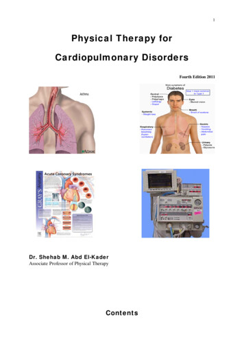
1Physical Therapy forCardiopulmonary DisordersFourth Edition 2011Dr. Shehab M. Abd El-KaderAssociate Professor of Physical TherapyContents
2SubjectsPage*Chronic Obstructive Pulmonary Disease4*Restrictive Lung Diseases18* Suppurative Lung Diseases27* Diabetes Mellitus31* Obesity40Role of physiotherapy in cardiothoracic surgery46* Complications following cardiothoracic surgery* Congenital heart diseases* Ischemic heart disease* Rheumatic fever* Heart failure* Cardiac Rehabilitation* Role of physiotherapy in intensive care unit49545961636888*
3CHEST PHYSICAL THERAPYDr. Shehab M. Abd El-KaderAssociate Professor of Physical TherapyChronic Obstructive Pulmonary DiseaseDefinitionChronic obstructive pulmonary disease is a general term that refers to a number ofchronic pulmonary conditions characterized by narrowing and obstruction of airways,
4increased retention of pulmonary secretions and structural deterioration of alveoli.This airflow limitation is progressive and not fully reversible.Several terms are used to describe obstructive lung disease they include:1- COLD : chronic obstructive lung disease.2- COAD: chronic obstructive airway dysfunction.3- COPD : chronic obstructive pulmonary disease.Diseases Classified as COPD1- Chronic bronchitis.2- Emphysema.3- Asthma.4- Other diseases such as cystic fibrosis and bronchiectasis usually lead to chronicobstructive dysfunction.Characteristics of patients with obstructive lung disease1- Patients exhibit persistent resistance of airflow, which causes prolonged andoften forced expiration.2- Vital capacity is decreased.3- Exercise tolerance is markedly diminished. Patients with COPD becomedyspneic with minimal physical exertion.General clinical problems1- Frequent episodes of shortness of breath “dyspnea on exertion”.2- Prolonged and labored expiration. Air gets trapped as airways narrow duringexpiration.3- Chronic accumulation of pulmonary secretions.4- Decreased endurance and exercise capacity.5- Associated postural defects.
5Figure (1): The loss of elastic recoil in lung tissue and the increased airway resistance decrease theexpiratory airflow in a patient with chronic obstructive pulmonary disease as compared with theexpiratory airflow in a normal subject.Presentation:Significant overlaps exist in signs and symptoms among the three major diseasesof airflow obstruction: asthma, chronic bronchitis and emphysema. The large overlaphas been long noted and well illustrated in Venn diagram fashion (Fig. 2).Figure (2): Schema of COPD.Classification of Severity
6For educational reasons, a simple classification of disease severity into four stages isrecommended (Table 1).Table 1. Classification of COPD by Severity.Stage0: At RiskI:COPDCharacteristics. normal spirometry. chronic symptoms (cough, sputum production)Mild . FEV1/FVC 70%II:ModerateCOPDIII: SevereCOPD. FEV1 80% predicted. with or without chronic symptoms (cough. sputumproduction). FEV1/FVC 70%. 30% FEV1 80% predicted (IIA: 50% FEV1 80% predicted)(IIB: 30% FEV1 50% predicted). with or without chronic symptoms (cough, sputumproduction, dyspnea). FEV1/FVC 70%. FEV1 30% predicted or FEV1 50% predicted plusrespiratory failure or clinical signs of right heartfailure.FEV1: forced expiratory volume in one second; FVC: forced vital capacity.PathophysiologyPathological changes in the lungs lead to corresponding physiological changescharacteristic of the disease, including mucus hypersecretion, Ciliary dysfunction,Expiratory airflow limitation, pulmonary hyperinflation, gas exchange abnormalities,pulmonary hypertension, and corpulmonale. They usually develop in this order overthe course of the disease.Respiratory muscles and COPD1- Diaphragm only contributes 30% (compared with its usual 65%) of the inspiratoryforce, while the accessory muscles play an increased role.2- The respiratory muscles may become fatigued and lung becomes hyperinflated.3-There is increased resistance of their airways and the hyperinflation. Thehyperinflation of the lung flattens the diaphragm, shortens the inspiratory muscles andplaces them at a mechanical disadvantage. In addition to the reduced efficiency of the
7inspiratory muscles, large amount of pressure work are required to overcome the highairway resistance.4-During maximal exercise, the respiratory muscles may utilize 35-40% (normal 1015%) of whole body oxygen consumption. More respiratory work is performed duringinspiration.5- About 25% of COPD patients are unable to maintain their nutritional status, asevidenced by weight loss. This nutritional depletion will increase mechanical and gasexchange impairment. In addition, loss of protein and lean body mass leads to skeletalmuscle and diaphragmatic weakness.Physiotherapy:Problems usuallyCOPD patients suffer from the following deficits:1- Dyspnea: Due to dysfunctional pulmonary mechanics, weak Ventilatorymuscles, poor diaphragmatic positioning for length – tension functioning,increased airway resistance and inadequate gas exchange.2- Accumulation of secretions.3- Decreased exercise tolerance: Due to general muscle weakness, poorendurance and inadequate nutritional status.Aims:1- Relief of dyspnea.2- Remove secretions.3- Improve exercise tolerance.Methods:I- Relief of dyspnea: Relaxed Positions:The first step towards self-help is positioning. It is an effective technique to reduceboth the symptoms of breathlessness and the work of breathing.
8 Breathing Retraining Exercises:Breathing exercises relieve dyspnea and improve gas exchange. The techniquesmost commonly taught are diaphragmatic breathing and pursed lips breathing or acombination of both.a- Diaphragmatic Breathing Exercise:Diaphragmatic breathing exercise increases the force of the diaphragm as aninspiratory muscle. It improves ventilation of small airways and bases of the lungs. Inaddition, it is often used in combination with pursed lips breathing and relaxationtechniques.b- Pursed lips breathing (PLB):Pursed lips breathing exercise prevents collapse of airways during expiration dueto maintain positive pressure in airways during expiration. In addition, prolongedexpiration leads to decrease air trapping and residual volume. In addition, it recruitsmore alveolar units at the lung base. Breathing Control Techniques:Breathing control techniques encourage deep breathing and to control dyspnea(shallow rapid breathing).Timing the breathing to steps works very well when walking or climbing stairs,e.g. one step to breathe in and two steps to breathe out, or one for each, or any rhythmor pattern that suits that particular individual. In addition, breathing control can beperformed via diaphragmatic and pursed lips breathing exercise, which encouragedeep breathing and control the dyspnea. Biofeedback and respiratory muscle training:Biofeedback teaches self-control over physiological functions and as a result,Ventilatory muscle training builds strength and endurance in the respiratory muscles.Applications:A-Incentive spirometry: The Goal of it’s application is to encourage patient to takedeep breathing which leads to reduction of breathlessness.
9B-Peak expiratory flow meter: which encourage patient to do full expiration ineach succeeding trial of expiration.C-The oximetry biofeedback augmented pursed lips breathing training: patients canuse pulse oximetry as a biofeedback guide to teach them to increase their oxygensaturation during performance of pursed lips breathing which relieves dyspneaand improves gas exchange, which result in improvement of oxygen saturation. Secretion clearance:A- Coughing:Patients are trained and encouraged to cough and clear secretions effectively. Asan alternative, the “huff” consists of a slow inspiration to total ling capacity, followedby huffs with the glottis open and may be effective. The multiple huffs are thought tominimize collapse of small airways, bronchospasm and fatigue.B- Chest physiotherapy:Postural drainage, percussion and chest wall vibration are clinically effective. Exercise:Muscle weakness both in skeletal and ventilatory muscles is common in COPDpatients. Strength training in specific muscle groups has enabled patients to morecomfortably and confidently perform their ADL. Hence, strength training may beadjunctive to endurance training.Guidelines for exercise prescription for patients with COPD:A) Flexibility exercise:Stretching of the major muscle groups of both upper and lower extremities.Flexibility/stretching considered as a part of the warm up before aerobic training andas part of the cool down after aerobic training.B) Aerobic Exercises:
101- Mode: Should incorporate Lange muscle groups that can be continuous andrhythmic in nature. Types of exercise include walking, cycling, rowing,swimming etc.2- Frequency: Recommended minimal frequency of training is three to five timesper week.3- Intensity: Minimal intensity 50% of peak VO2. Another approach is to exerciseat maximum limits tolerated by symptoms.4- Duration: Minimal recommended duration is 20 to 30 min. of continuousexercise.Specific Obstructive Pulmonary ConditionsA) CHRONIC BRONCHITIS AND EMPHYSEMA:Chronic bronchitis and emphysema are both classified as chronic obstructivepulmonary diseases (COPD). Since these diseases are often closely related and oftenseen in conjunction with each other, the underlying goals and principles of treatmentare similar.1- Chronic bronchitis – Clinical picture:a.Chronic bronchitis is an inflammation of the bronchi that causes anirritating and productive cough that lasts up to 3 months and recurs over atleast 2 consecutive years.b.This condition usually develops in heavy smokers.c.The pathologic changes that occur in chronic bronchitis are:1)An increase in the number of mucus-producing goblet cells in the lining ofthe bronchial tree2)A decrease in the number and action of the ciliated epithelial cells, whichmobilize secretions.3)A narrowing of airways because of chronic inflammation of the bronchialtree.d.General appearance of the patient:1)Cyanotic because of hypoxemia.2)Short of breath.3)Bloated because of venous stasis.
112- Emphysema – Clinical picture:a.Emphysema is a chronic inflammation, thickening, and destruction of therespiratory bronchioles and alveoli. These airways become scarred, distorted, andkinked, and the alveoli lose their elastic recoil, then weaken and rupture. As aresult, air remains trapped in the lungs. Over a period of years, severe chronicbronchitis and emphysema often lead to congestive heart failure and death.b.Emphysema is usually a condition secondary to chronic bronchitis. Although notas common, emphysema can also be a primary disease that can occur innonsmokers.c.The pathologic changes that occur in emphysema are:1) An over inflation of the lungs and formation of pockets of air known as bullae.This causes an increase in the air space in the lungs.2) Destruction of lung tissue and loss of area where effective gas exchange canoccur.d.General appearance of the patient:1) Similar to chronic bronchitis.2) Abnormal posture-forward head rounded and elevated shoulders.3) Clubbing of fingers.3- Clinical problems of chronic bronchitis and emphysema summarized:a.An increase in the amount and viscosity of mucus production.b.A chronic often productive cough.c.Attacks of shortness of breath (dyspnea).d.An abnormal breathing pattern with the most difficulty experienced duringexpiration which results in:1) Use of accessory musculature.2) Upper chest breathing.3) Poor exchange of air in the lower lobes.e.Changes in pulmonary function:1) Increased residual volume.2) Decreased vital capacityf.Decreased mobility of the chest wall – a barrel chest deformity develops.g.Abnormal posture-forward head and rounded shoulders.
12h.Decrease in general endurance, during daily activities.4- Treatment goals and plan of care:Treatment GoalsDecrease the amount and viscosity ofsecretions.Plan of Carea. Administration ofbronchodilators, antibiotics,and humidification therapy.If the patient smokes, he shouldbe strongly encouraged to stop.b.Remove or prevent the accumulationof secretions, (this is an important goalif emphysema is associated withchronic bronchitis or if there is anacute respiratory infection).b.c.Promote relaxation of the accessorymuscles of inspiration to decreasereliance on upper chest breathing anddecrease tenseness associated withdyspnea.c.d.Improve the patient’s breathing patternand ventilationd. Breathing exercisesRelaxed diaphragmatic breathing1Ba.Deep and effective coughpostural drainage to areaswhere secretions areidentifiedNote: Drainage positions mayneed to be modified if the patientis dyspneic in the head-downposition.Promote for relaxationrelaxed head – up position inbed; trunk, arms, and headare well supported- Sitting-leaning forward, restingforearms on thighs.- Sitting-leaning forward againstpillows on a table- Standing – leaning forward onan object, with hands on thighsor leaning backward against awall.- Relaxation exercises forshoulder musculatureActive shoulder shruggingfollowed by relaxation.Shoulder and arm circles.Horizontal abduction andadduction of the shoulders.
13Emphasize relaxed expiration; decrease therate of respiration and the use of accessorymuscles.Carry over controlled breathing tofunctional activities.with minimal upper chestmovement.Lateral costal breathing.Pursed lip breathing (careful toavoid forced expiration).Practice controlled breathingduring standing, walking,climbing stairs, etc.e.Minimize attacks of shortness of breath e.Have the patient assume arelaxed position, so the upperchest is relaxed and the lowerchest is as mobile aspossible.Emphasize relaxed diaphragmaticbreathing.Have the patient breathe out asrapidly as possible without forcingexpiration (Note: Initially, the rateof respiration will be rapid andshallow. As the patient getscontrol of his breathing, he willslow down the rate).Administer supplemental oxygenin a severe attack, if needed.f.Improve the mobility of the lowerthorax.f.Exercises for chest mobilityemphasizing movement ofthe lower rib cage.g.Improve postureg.Exercises to decreaseforward head and roundedshoulders.h.Increase exercise toleranceh.Graded endurance andconditioning exercises.B) ASTHMA:Asthma is an obstructive lung disease seen in young patients. It is related tohypersensitivity of the trachea and bronchi and causes difficulties with respiration arecause of bronchospasm and increased mucus production.
141- Clinical picture:a. The majority of patients with asthma are children.b. Asthmatic attacks involve severe shortness of breath when the patient comes incontact with a specific allergen. The patient has a very rapid rate of respirationand primarily used accessory muscles for breathing. There are audiblewheezes and rhonchi, and the patient feels severe tightness in his chest.c. Pathologic changes1) Severe spasm of smooth muscle of the bronchial tree.2) Narrowing of airways.3) Hypersecretion of mucus, which is usually sticky and therefore obstructivebecause of an increase in the size and number of goblet cells.4) Sever asthma over a prolonged number of years can lead to emphysema.d. General appearance of the patient:1) Chronically fatigued.2) Often thin.3) Poor posture – rounded shoulders and forward head.2- Clinical problems of asthma summarized:a. Severe attacks of shortness of breath.b. Cough – usually unproductive during an asthmatic attack, but productive later.c. Poor posture – rounded shoulders, forward head.3- Treatment goals and plan of care:Treatment Goalsa. Decrease bronchospasm.Plan of Carea.Removal of allergen (s);bronchodilators with IPPB.b. Minimize attacks of shortness ofb.Relaxation of upper chest andbreath and gain control ofaccessory muscles by positioningbreathing.Diaphragmatic breathing,emphasizing relaxed expiration.c. Mobilize and remove secretionsafter attack of shortness ofbreath.c.Humidification of secretions withaerosol therapy.Effective coughing.
15Postural drainage (after, not during, theasthmatic attack, as it may increasebronchospasm).d. Correct posture to decreased.Postural training.e.Avoid prolonged, vigorousrounded shoulders and forwardhead.e. Gradually increase exercisetolerance and endurance.physical activities.Encourage mild to moderate activitiesfor short periods, followed by rest.Use controlled breathing duringexertion.C) BRONCHIECTASIS:Bronchiectasis is an obstructive lung disease characterized by dilation of themedium-sized bronchioles, usually the fourth to the ninth generations, and repeatedinfections in these areas.1- Clinical picture:a. Severe infection of dilated obstructed bronchioles.b. Productive cough with purulent sputum and hemoptysis.c. Pathologic changes.1) Repeated infections of the lower lobes of the lungs.2) Destruction of ciliated epithelial cells in infected areas.d. If the infections are localized, a lobectomy may be indicated.2- Clinical problems of bronchiectasis summarized:a. Repeated infections of the affected lung area.b. Accumulation of purulent secretions.c. Productive cough.
163- Treatment goals and plan of care:Treatment Goalsa. Clear the airways of secretions.Plan of Carea. Effective, controlled coughpostural drainage BID to QIDduring acute episodes.b. Prevent recurrent infectionsb. Home program of posturaldrainage to be carried onthroughout life.4- Precautions:a. If mild hemoptysis (blood – streaked sputum) occurs, continue posturaldrainage, but omit percussion for at least 24 hours.b. If severe hemoptysis (hemorrhage) occurs, discontinue postural drainage untilfurther noticeD)CYSTIC FIBROSIS:Cystic fibrosis is a genetically based disease (autosomal recessive) whichinvolves malfunction of the exocrine glands, leading to abnormal secretions in thebody. The disease is characterized by a very high concentration of sodium in thesweat, diffuse lung disease, and malfunction of the pancreas. The disease must bemanaged throughout life with diet, medication, and preventive chest physical therapyas soon as any symptoms are noted in the young child.1- Clinical picture:a. These children are usually small for age because of mal-absorption of foods.b. The exocrine gland dysfunction leads to increased production of viscous mucus,which obstructs the airways. Chronic obstruction of the airways and pooling ofsecretions leave the child vulnerable to pulmonary infection.c. Prognosis for survival has improved in the past 20 years. The average patientnow survives into the late 20s or early 30s. The digestive involvement can bemanaged by diet; pulmonary complications are eventually the cause of death.2- Clinical problems of cystic fibrosis summarized:a. Increased production of viscous mucus throughout the lungs.b. Periodic pulmonary infections.
17c. Possible problems of compliance with a life – long regimen of home posturaldrainage and prevention of lung infections.3- Treatment goals and plan of care:Treatment Goalsa. Prevent accumulation of secretionsand pulmonary infection.b. Decrease viscosity of secretions.Plan of Carea. Daily home program of postural drainage,usually BID, if no acute pulmonaryproblems exist.b. Humidification therapy with mist tent orIPPB.c. Prevent use of accessory musclesof respiration.c. Diaphragmatic breathing and lateral costalexpansion.Daily practice and use of deep breathingduring postural drainage is important.Emphasize relaxed expiration sobronchospasm and air trapping does notoccur.d. Removal of secretions during anacute infection.d. Postural drainage QID or for longerperiods, as needed.Appropriate use of antibiotics.Note: The key to successful preventive treatment of cystic fibrosis over many years isa consistent home program of postural drainage. This requires a supportive andcooperative family atmosphere.
18Restrictive Lung DiseasesDefinitionAre group of diseases with different etiological factors, all characterized bydecrease in:1- Pulmonary ventilation.2- Lung expansion and deep breathing.3- Volume of air move in and out of the lung.AetiologyCan be classified in pulmonary and extrapulmonary causes.1- Pulmonary Causes1- Tumor2- Pneumonia3- Heart disease4- Atelectasis.5- Fibrotic lung disease.2- Extrapulmonary causes1- Pleural disease (pleural effusion).2- Chest wall Stiffness-Chest wall pain secondary to trauma or to pulmonary or cardiac surgery.-Postural deviations (scoliosis, kyphosis, ankylosing spondylitis).3- Respiratory muscle weakness-Neuromuscular disease (Muscular dystrophy, Parkinsonism, anteriorhorn cell disease).4- Insufficient excursion of the diaphragm because of obesity or ascities.PathomechanismAny of the previous causes affect mainly the lung expansion property and lead to:1- Decreased the lung compliance due to stiffness of the chest wall.2- Decrease all lung volumes and capacities.
193- Increase the work of breathing of respiratory muscles especially the diaphragm,with subsequent increase in the oxygen consumption and hence increase thedegree of hypoxia and hypoxemia.Clinical manifestationsSigns1- Tachypnea (increased respiratory rate)2- Hypoxemia ( oxygen tension in arterial blood) due to ventilation perfusionmismatching.3- Dry inspiratory rales due to opening of atelectatic alveoli at the end ofinspiration)4-Decrease breathing sounds5- Decreased lung volumes and capacities.Symptoms1- Dyspnea2- Cough3-Weight loss4- Muscle wastingGeneral clinical problems1- Dyspnea and inability to inspire deeply2- Decrease chest mobility and expansion.3- Postural deviations.4- Chest pain.5- General weakness and fatigue.Pulmonary Rehabilitation for RLD
20* General Goals:1- Relief dyspnea.2- Increase chest mobility and expansion.3- Correct postural defects4- Relieve pain.5- Improve exercise tolerance.This can be achieved by:1Respiratory exercises: nose ex, localized breathing exercise, deepbreathing exercise, exercise connected with respiration.Using of some devices as Triflow, incentive sprout using of weight inspiratory resistance exercise.2- Mobilizing exercises: active free through full ROM. Swinging exercise. Gym ex. using shoulder wheel, raw machine, parallel bar.3- Stretching exercises and positions: Stretching position phalanx and wring stretch position Pectoralis muscle and hip flexors stretching exercises.4- Pain relief modalities: any source of head especially moist heat, infrared,massage, TENS, didynamic currents or laser.5- Endurance exercises treadmill training, walking, bicyle ergometer andswimming exercises.Specific restrictive pulmonary conditionsI. Pleural Diseases1) Dry pleurisy:Definition:Inflammation of the pleura, of one or both sides with no detectable free exudates.Aetiology: Pneumonia the commonest cause. Pulmonary infarction. Bronchial carcinoma. Lung abscess. Pulmonary tuberculosis. Extension from a subdiaphragmatic abscess.Pathological changes:
211234-Pleural membrane becomes hyperaemic and red.Fibrin deposited on the inflamed membrane.Adhesions formed between both pleural layers.So respiratory movement is restricted causing pain.Clinical Features:Symptoms:1- Pleuritic pain: Pain that is maximal at the end of inspiration, it is worsened bydeep breathing and coughing. It may be referred to the anterior chest wall or –in the presence of diaphragmatic pleurisy – to the front of shoulder, or to theanterior chest wall.2- Difficulty of breathing.3- Dry cough.4- Bending toward the painful side.Signs:1- Rapid and shallow breathing pattern.2- Asymmetric breathing: limitation of chest movement on the affected side incases of diaphragmatic pleurisy.3- On palpation of chest wall: there is tenderness over the area of pleurisy.4- Pleural friction rub: which stimulates crepitations, yet is unaltered by coughing.5- Decreased the tactile vocal frimitius: due to limited air volume.6- On auscultation: there is a decreased vocal and breathing sound over theaffected side.Treatment:Medical:Antibiotics, anti-inflammatory, antipyretics and analgesics.Physical therapy:Aims:1- To relax the patient and improve respiration.2- To relieve the pain.3- To prevent the postural deformity.Methods:1- Rest in bed in proper supported alignment.2- Application of a moist heat.3- Bandage or strapping of the painful sides.4- Positioning of affected side to preventdeformity.2) Pleural Effusion:Accumulation of fluid in the pleural cavity as a result transudatation or exudationfrom the pleural surfaces.
22Aetiology:Transulates (hydrothorax): as in congestive heart failure, constrictive pericarditisand myxoedema.Exudates: fluid with a high protein content of 3 gm/100mL accumulates in thepleural space; it may occur due to bacterial pneumonia, pleural malignancyand T.B and collagen diseases as: rheumatic fever, rheumatoid arthritis.Clinical Feature:Symptoms:Acute symptoms onset: high fever, fatigue, dyspnea.Gradual, onset: toxemia, dull aching pain.Signs:1- Signs of the primary disease.2- Signs of the fluid in the pleural space:-Decreased or absent ribs movement on affected side.Displacement “shifting” of apex beat and usually trachea to opposite side(in large effusion).Stony dull percussion.Distant breath sounds: High-pitched bronchial breathing may be heardover upper margin of effusion.Pleural rub may be heard above fluid.Vocal resonance decreased or absent fremitus.Aegophony may be heard over upper margin of effusion.Treatment:1- Treatment of the primary cause.2- Build up the body resistance by proper diet.3- Aspiration of the excess pleural fluid to reduce dyspnea.4- Physical therapy treatment: Positioning: on the normal side to improve ventilation/ perfusion ratio, also ithelps the movement on the affected side and subsequently helps thedrainage. Breathing exercises: diaphragmatic and localized breathing exercises. Postural exercises: to maintain good posture and avoid chest wall unilateralcontracture. Aerobic exercises: as walking and up and down stairs to maintain physicalendurance and fitness.3) Empyema:
23Definition:Empyema is the presence of pus in the pleural cavity.Aetiology:1- Extension of infection from the lung as in T.B, Pneumonia, cancer or lungabscess.2- Extension of infection from the mediastinum or chest wall.3- Subdiaphragmatic abscess.4- General as septicemia or pyaemia.Clinical Feature:Symptoms:1- Those of the primary disease, usually pneumonia.23456-Fever, nigors, pleuritic pain and later loss of weight.Toxemia with swinging temperature.Insomnia.Chest pain.Sudden coughing of a large amount of sputum (pus), which may be bloodstained indicates the occurrence of a bronchopleural fistula.Signs:1- Clubbing fingers, developing is 2-3 weeks.2- Deformity of the chest wall.3- Restricted movement of the chest on the affected side.4- Scoliosis to the affected side.Treatment:Aim of treatment:1- Control of infection.2- Removal of pus.3- Obliteration of empyema space.Medical treatment: Appropriate antibiotics and analgesics.Surgical treatment: Repeated aspiration in case of thin pus.
24 Thoracoplasty.Physical therapy treatment:Aims: To re-expand the lung after aspiration. To prevent the deformity. To maintain adequate range of motion in the upper limbs and trunk. To relieve pain and anxiety. To reduce dyspnea and respiratory rate.Post-operative aims: To prevent pulmonary complications. To prevent circulatory complications. To prevent chest wall contracture and deformity. To improve lung expansion. To improve physical fitness.Physical therapy methods: Respiratory exercises. Circulatory exercises and early ambulation. Postural exercises. Endurance exercises. Heat application.4) Other forms of pleural disorders:Hemothorax:Haemorrhage into the pleural space.Chylothorax:Presence of white milky fluid in the pleural cavity.Pneumothorax:The presence of air in the pleural cavity.II.PneumoniaPneumonia is an inflammation of the lungs, characterized by consolidation andexudation and caused by a bacterial or viral infection.Classifications of pneumonia:1-By anatomical locationa- Bronchopneumonia.
25b- Lobar pneumonia.c- Segmental pneumonia.2- By causal organism:a- Viral pneumonia.b- Bacterial pneumonia.TreatmentGoals1- Control the infection.2- Maintain or improve ventilation.3- Mobilization of secretionsMethods1- Use of suitable antibiotics.2- Deep breathing and localized breathing exercises.3-Postural drainage with percussion and vibration to the4-Effective cough.affected areas.III. AtelectasisAtelectasis is a restrictive lung dysfunction in which lobes or segments of alobe have been collapsed.Clinical picture1- Absent breathing sounds over the collapsed lung area.2- Tachycardia and cyanosis.3- Decreased chest movement over the affected area.TreatmentGoals1- Reinflate collapsed areas of the lung2- Increase inspiratory capacity.Methods1- Postural drainage with percussion and vibration.2- Effective cough.3- Segmental breathing with emphasis over collapsed areas.Difference between COPD & RLDPathology0BResultin Obstruction to airflow Affect the gasexchange capability Difficulty inexpanding lungs. Cause a reductionin lung volumes.
26of lung.Work of Due tobreathinghyperinflation, gas exchange andDegenerativealveolar changes.Treatment & Mainly medicalprognosiswith goodprognosis. Due to lungcompliance andlung volume. Mainly surgicalwith badprognosis.Suppurative Lung Diseases Bronchiectasis. Cystic fibrosis Lung abscess Tuberculosis General Clinical problems of Suppurative lung diseases:1- Accumulation of purulent secretions and productive cough.2- Dyspnea and overuse of accessory muscles of respiration.3- Limitation of chest movement4- Reduced exercise tolerance.Aims:1234-Clear the lung fields.Improve strength and endurance of respiratory muscles.Maintain mobility of the shoulder girdle and thorax.Improve exercise tolerance.
27Methods:1- Clearing the lung fields:Postural Drainage: 2-3 times /day associated with assistive techniques asPercussion and Vibrations.2-Improve strength and endurance of respiratory muscles:Breathing exercises: Diaphr
Physical Therapy for Cardiopulmonary Disorders . Fourth Edition 2011 . Dr. Shehab M. Abd El-Kader Associate Professor of Physical Therapy . Contents . 2 Subjects Page *Chronic Obstructive Pulmonary Disease 4 *Restrictive Lung Diseases 18
