Transcription
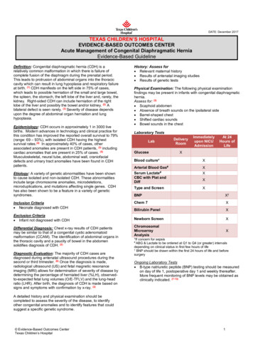
DATE: December 2017TEXAS CHILDREN’S HOSPITALEVIDENCE-BASED OUTCOMES CENTERAcute Management of Congenital Diaphragmatic HerniaEvidence-Based GuidelineDefinition: Congenital diaphragmatic hernia (CDH) is arelatively common malformation in which there is failure ofcomplete fusion of the diaphragm during the prenatal period.This leads to protrusion of abdominal organs into the thoraciccavity which can result in lung hypoplasia and respiratory failureat birth. (1) CDH manifests on the left side in 75% of cases,which leads to possible herniation of the small and large bowel,the spleen, the stomach, the left lobe of the liver and, rarely, thekidney. Right-sided CDH can include herniation of the rightlobe of the liver and possibly the bowel and/or kidney. (2) Abilateral defect is seen rarely. (3) Severity of disease dependsupon the degree of abdominal organ herniation and lunghypoplasia.Epidemiology: CDH occurs in approximately 1 in 3000 livebirths. Modern advances in technology and clinical practice forthis condition has improved the reported overall survival to 79%(range: 69 – 93%), with isolated CDH having the highestsurvival rates.(4) In approximately 40% of cases, otherassociated anomalies are present in CDH patients, (3) includingcardiac anomalies that are present in 25% of cases. (5)Musculoskeletal, neural tube, abdominal wall, craniofacialdefects and urinary tract anomalies have been found in CDHpatients.Etiology: A variety of genetic abnormalities have been shownto cause isolated and non-isolated CDH. These abnormalitiesinclude large chromosome anomalies, microdeletions,microduplications, and mutations affecting single genes. CDHhas also been shown to be a feature in a variety of geneticsyndromes.Inclusion Criteria Neonate diagnosed with CDHExclusion Criteria Infant not diagnosed with CDHDifferential Diagnosis: Chest x-ray results of CDH patientsmay be similar to that of a congenital cystic adenomatoidmalformation (CCAM). The identification of abdominal organs inthe thoracic cavity and a paucity of bowel in the abdomensolidifies diagnosis of CDH. (2)Diagnostic Evaluation: The majority of CDH cases arediagnosed during antenatal ultrasound procedures during thesecond or third trimester. (6) Once the diagnosis is made,radiological ultrasound (US) and fetal magnetic resonanceimaging (MRI) allows for determination of severity of disease bydetermining the percentage of herniated liver (%LH), observedto-expected fetal lung volumes (O/E-TFLV) and the lung-headratio (LHR). After birth, the diagnosis of CDH is made based onsigns and symptoms with confirmation by x-ray. (2)History: Assess for Relevant maternal history Results of antenatal imaging studies Results of genetic testsPhysical Examination: The following physical examinationfindings may be present in infants with congenital diaphragmatichernia.Assess for: (3) Scaphoid abdomen Absence of breath sounds on the ipsilateral side Barrel-shaped chest Shifted cardiac sounds Bowel sounds in the chestLaboratory TestsLabGlucoseDeliveryRoomImmediatelyupon NICUAdmissionAt 24Hours ofLifeXBlood culture*XArterial Blood Gas#Serum Lactate#CBC with Plat andDiffXXType and ScreenXXBNPX†Chem 7XBilirubin PanelXNewborn ScreenXChromosomalMicroarrayAnalysisX*If concern for sepsis#ABG & Lactate to be ordered at Q1 to Q4 (or greater) intervalsdepending on clinical status in first few hours of life†BNP should be drawn within the first 24 hours of life and beforesurgeryOngoing Laboratory Tests B-type natriuretic peptide (BNP) testing should be measuredon day of life 1, postoperative day 1 and weekly thereafter.More frequent monitoring of BNP levels may be obtained asclinically indicated. (7-13)A detailed history and physical examination should becompleted to assess the severity of the disease, to identifyother congenital anomalies and to identify features that couldsuggest a specific genetic syndrome. Evidence-Based Outcomes CenterTexas Children’s Hospital1
DATE: December 2017Imaging StudiesStudyChest X-rayincluding abdomenHead UltrasoundEchocardiogramTimingSTAT upon admission to NICUWithin 24 hours of admission unlessotherwise indicated; Upon arrival forhigh risk patients (see Appendix)Within 24-48 hours of admission unlessotherwise indicated; Upon arrival forhigh risk patients (see Appendix)Prenatal Assessments Depending on the gestational age at the time of CDHdiagnosis, it is recommended that a fetal echo be performedwithin a week or two after the diagnosis is made to rule outstructural heart disease, especially if Fetal trachealocclusion (FETO) is being considered. If FETO is not being considered, a fetal echo may beperformed at a convenient time, preferably between 24-32weeks gestation. The percentage of herniated liver (%LH) and observed-toexpected fetal lung volumes (O/E-TFLV) obtained from MRIshould be used to stratify CDH severity which can be usedin prenatal counseling to educate families on diseaseseverity and mortality risks. (14-26) Practitioners should measure and document %LH, O/ETFLV, the side of the defect, total lung volume (TLV), lungto-head ratio (LHR), observed-to-expected lung-to-headratio (O/E-LHR), stomach position and lung-to-thorax ratiofor risk stratification, research, benchmarking and outcomesreporting in CDH patients. (14-37) There should be a case-by-case determination of themanagement plan for CDH patients with comorbiditiesidentified prenatally. This should be preferably discussedwith the family prior to delivery and involve P-PACT ifnecessary. Limitations in care should be discussed in theantenatal visit.Postnatal Echo It is recommended that a postnatal echocardiogram beperformed within the first 24-48 hours after birth. It may beobtained earlier if the neonate exhibits a sub-optimalresponse to the usual therapy, especially if a prenatal echowas suboptimal or not performed. (38-42) A postnatal echo may be obtained electively, at a later,convenient time, if the neonate does not exhibit unduedifficulties in ventilation and oxygenation, especially if agood quality fetal echo was performed.Cardiac Cath – Acute Phase Cardiac cath is not necessary if left-to-right atrial and ductalflow is noted by echocardiography, as this would suggestadequate and favorable response to therapy. In the neonatal period, cardiac cath may be considered uponthe recommendation of a cardiologist, if the clinical course issuggestive of pulmonary venous abnormalities and otherless invasive imaging modalities are not appropriate. (38-42) Cardiac cath may be necessary if there is a need to performa transcatheter cardiac intervention (i.e., the need to stent aPDA in the presence of Coarctation) in a neonate deemednot to be a surgical candidate.Cardiac Cath – Chronic Phase with Persistent PulmonaryHypertension Cardiac cath should not be performed in a patient whodemonstrates normal RV function and left to right atrial andductal flow by echocardiography, as this would suggestadequate and favorable response to therapy. A cardiac cath may be considered, especially in thepresence of RV dysfunction (cor pulmonale) andpredominant right to left atrial and ductal level shunting, ifthe response to pulmonary vasodilator therapy is suboptimaland other pharmacologic options are being considered. (38-42) In the presence of severe pulmonary hypertension and RVfailure, cardiac cath is recommended if an intervention suchas an atrial septostomy or ductal stenting is beingconsidered. Consider cardiac cath if there is left ventricledysfunction present to better characterize the anatomy. A cardiac cath may be necessary to better delineate theanatomy when the patient has other anatomical issues thatare not able to be evaluated by echo.Other Imaging Tests Pulmonary venous abnormalities may be difficult to rule outby fetal or post-natal echocardiography in a patient withCDH. Additional imaging modalities, such as a contrastchest CT, cardiac MRI or angiography by cardiac cath maybe recommended by the clinician if echocardiography isunable to definitively rule out abnormalities of pulmonaryvenous return. This is especially true in the profoundlyhypoxemic neonate who does not respond to the usualmeasures, and in the presence of pure right to left shuntingin the atrial or ductal level. A small percentage of the CDH population may requireadditional imaging studies such as Ventilation Perfusion(V/Q) scan.Critical Points of EvidenceEvidence SupportsPrenatal Testing The percentage of herniated liver (%LH) and observed-to-expected fetal lung volumes (O/E-TFLV) obtained from MRI should be usedto stratify CDH severity which can be used in prenatal counseling to educate families on disease severity and mortality risks in CDHpatients. (14-26) – Strong recommendation, moderate quality evidence Practitioners should measure and document the %LH, O/E-TFLV, side of the defect, total lung volume (TLV), lung-to-head ratio(LHR), observed-to-expected lung-to-head ratio (O/E-LHR), stomach position and lung-to-thorax ratio for risk stratification, research,benchmarking and outcomes reporting in CDH patients. (14-37) – Strong recommendation, moderate quality evidence A pre-established objective criteria should be used to score the severity of cardiac lesions. The presence of a major cardiac anomalyis associated with an increased risk of mortality. (32,43-45) – Strong recommendation, low quality evidence Evidence-Based Outcomes CenterTexas Children’s Hospital2
DATE: December 2017 (46-49)The oxygenation index should continue to be used for management decisions in CDH patients.– Strong recommendation, lowquality evidence The Brindle Prediction Score (see appendix) should be used to educate families on the categorical (low, intermediate, or high)mortality risk in CDH patients. (14,50) – Strong recommendation, low quality evidenceInitial Management Clinicians may consider the use of surfactant in CDH patients with a history of Fetal Endotracheal Occlusion (FETO) or preterm birth( 37 weeks gestation). (5,51-57) – Weak recommendation, low quality evidenceo Remarks: The fetus that undergoes FETO can have a decrease in type II pneumocytes which can cause surfactant deficiency.Patients receiving this procedure during the prenatal period may benefit from postnatal surfactant administration. Response totherapy guides dosing of surfactant. Unless there were negative consequences to initial administration, clinicians should considera second dose. If the patient does not exhibit a positive response to the first two doses of surfactant, do not administer additionaldoses. (58,59)Laboratory Testing and Imaging B-type natriuretic peptide (BNP) testing should be measured on day of life 1, postoperative day 1 and weekly thereafter. Morefrequent monitoring of BNP levels may be obtained as clinically indicated. (7-13) – Strong recommendation, very low quality evidence Echocardiogram should be performed within 24 to 48 hours of life, one week after CDH repair, and within four weeks of discharge.Additional follow-up echos should be obtained every 1 to 2 weeks for patients with severe pulmonary hypertension until there isevidence of resolution with pulmonary pressures 1/2 systemic. (38-42) – Strong recommendation, very low quality evidenceSystemic Hypotension Management Titrate dopamine to manage systemic hypotension in CDH patients during the acute phase. (55,60-67) – Strong recommendation, lowquality evidenceECMO Administer a one-time dose of cefazolin within one hour of cannulation for ECMO in CDH patients. Do not routinely use continuedprophylactic antibiotics for patients on ECMO support. (68-70) – Strong recommendation, very low quality evidenceSurgical Repair In non-repaired CDH patients on ECMO, surgical repair of the diaphragm should be completed within 48 hours of ECMO cannulation.(1,5,55,63,64,66,67,71-78) – Strong recommendation, low quality evidence In CDH patients not requiring ECMO, surgical repair of the diaphragm should be performed when the patient is physiologically stabledefined as mean blood pressure normal for gestational age, preductal oxygen saturations between 85-95% on FiO2 below 50%,lactate below 3 mmol/L, urine output greater than 2 mL/kg and less than a 10% gradient between pre- and post-ductal saturations.(1,5,55,63,64,66,67,71-78) – Strong recommendation, low quality evidence In CDH patients that are not able to achieve a tension-free primary closure of the diaphragm, a patch repair should be completed withan appropriately sized domed patch. (76-79) – Strong recommendation, low quality evidence CDH patients with large defects should be repaired using an open surgical technique. (78,82-86) – Strong recommendation, moderatequality evidence The use of minimally invasive surgery (MIS) may be considered in patients that meet the following criteria: mean blood pressurenormal for gestational age without use of inotropes, pre-ductal oxygen saturations between 85-95% on FiO2 below 40% without use ofinhaled nitric oxide, lactate below 3 mmol/L, urine output greater than 2 mL/kg/hr, and less than a 5% gradient between pre- and postductal saturations. (1,5,55,63,64,66,67,71-78) – Weak recommendation, moderate quality evidenceEvidence Lacking/InconclusiveManagement Plan Determine management plan for severe cardiac lesions first before proceeding to establish plan of care for congenital diaphragmatichernia. – Consensus recommendationVentilation Strategy Patients with CDH should be initially ventilated with the conventional ventilator using the AC/VG mode with initial settings of PEEP 5-6cmH2O, TV 4-5 mL/kg, back-up rate 40 breaths/min, IT 0.3 seconds, and FiO2 adjusted for target preductal saturations of 80%.(5,55,63-67,78,87-91) – Strong recommendation, low quality evidence Tidal volume should be adjusted to meet optimal physiological monitoring parameters. (5,55,63-67,78,87-91) – Strong recommendation, lowquality evidence Clinicians should consider switching the mode of ventilation to HFOV for CDH patients that cannot achieve target PCO2 onconventional ventilation with PIP 28. (5,55,63-67,78,87-91) – Weak recommendation, low quality evidenceo Once on HFOV, increase MAP (to a max MAP of 17) and DeltaP as required to achieve physiological monitoring parameters. Clinicians should consider ECMO for CDH patients that cannot achieve saturations and/or blood gas targets with maximal HFOVsupport. (5,55,63-67,78,87-91) – Weak recommendation, low quality evidence Patients with CDH should have the following targets for physiological monitoring parameters: pre-ductal oxygen saturations 80% forthe first two hours of life. Thereafter, pre-ductal saturations should be 85%; pH 7.20; pCO2 50 to 70; and pO2 40 to 90. (5,55,63-67,78,8791) – Strong recommendation, very low quality evidence Patients with CDH should be initially ventilated with 100% FiO2. – Consensus recommendation Evidence-Based Outcomes CenterTexas Children’s Hospital3
DATE: December 2017 Pre-ductal saturations should be targeted at 70% for the first ten minutes after birth; increasing to 80% for the first two hours of life.Thereafter, pre-ductal oxygen saturations should be 85%. – Consensus recommendationVenous Access and Fluid Management Peripheral venous access should be established in the delivery room for CDH patients. – Consensus recommendation Umbilical lines and elective/non-emergent procedures should be completed in the NICU. – Consensus recommendation An umbilical venous catheter (UVC) and an appropriately placed umbilical arterial catheter (UAC) should be initially inserted in CDHpatients with a need for central venous access. If correct UVC position cannot be achieved, a temporary low-lying UVC can be initiallyutilized until a sufficient alternative is available. (102-103) – Strong recommendation, low quality evidence A peripherally inserted central catheter (PICC) and an arterial line should be placed by the NICU Vascular Access Team (VAT) inCDH patients with a need for long-term central venous access once stabilization occurs. – Consensus recommendation Initial fluid intake for CDH patients should be 65 mL/kg/day. – Consensus recommendationPulmonary Hypertension Management In infants with congenital diaphragmatic hernia with an oxygenation index (OI) 25, preductal saturations 90% on 100% FiO2 or agradient between pre- and postductal saturations 10%, consider the use of inhaled nitric oxide to improve oxygenation. (5,38,55,6367,78,104-109) – Weak recommendation, low quality evidence In infants with congenital diaphragmatic hernia, consider the use of sildenafil for continued clinically significant pulmonaryhypertension beyond the acute phase as evidenced by a preductal/postductal saturation difference of 10 points, in those infants whofail weaning from inhaled nitric oxide or to facilitate weaning from nitric oxide. (5,38,55,63-67,78,110,111) – Weak recommendation, low qualityevidence In infants with congenital diaphragmatic hernia requiring VA ECMO, inhaled nitric oxide should be discontinued once VA ECMOsupport is initiated and restarted when recruiting the lungs in preparation for discontinuing ECMO. (55, 112-114) – Strongrecommendation, very low quality evidence In patients on VV ECMO, consider the use of inhaled nitric oxide throughout ECMO run. (55, 112-114) – Weak recommendation, very lowquality evidenceo Remarks: Response to inhaled nitric oxide should be assessed in all users within one hour of initiation. Responders will exhibit a 5% increase in preductal saturations, an increase in PaO2 by 10 torr (obtained from the same pre- or post-ductal source) or adecrease in pre/post ductal saturation gradient to 10%. Inhaled nitric oxide should be weaned based upon patient classificationas responder or non-responder.Systemic Hypotension Management Consider administration of hydrocortisone (1mg/kg IV every 8 hours) in CDH patients on a dose of greater than or equal to 10mcg/kg/min of dopamine with continued systemic hypotension. (55,63-67,115-117) – Weak recommendation, very low quality evidence Consider the use of epinephrine in CDH patients on maximum dopamine dose and hydrocortisone. (55,63-67,118) – Weakrecommendation, very low quality evidenceo Remarks: If the patient has vasopressor refractory hypotension, an echo should be obtained and the treated according to findings.Evidence AgainstPrenatal Testing The Brindle Prediction Score (see appendix) should not be used for individual patient decisions regarding the use of ECMO or othermedical management. (14,50) – Strong recommendation, low quality evidence The SNAP II Score, Wilford Hall/Santa Rosa Clinical Prediction Formula, Congenital Diaphragmatic Hernia Study Group (CDHSG)formula, the best PaCO2 at 1 hour of life and 24 hours of life, and the highest preductal O2 saturation value during the first 24 hours oflife should not be used to predict survival or make decisions about treatment options for CDH patients. (46,119-130) – Strongrecommendation, low quality evidenceInitial Management Surfactant should not be routinely used in the non-FETO term CDH patient at birth. (5,51-57) – Strong recommendation, low qualityevidence Information obtained from near infrared spectroscopy (NIRS) readings should not be used for patient care management. (131) – Strongrecommendation, low quality evidencePulmonary Hypertension Management Prostaglandin E (PGE) should not be routinely used in the acute phase of CDH treatment. (5,38,55,63-67,78,132,133) – Strongrecommendation, very low quality evidence Epoprostenol should not be routinely used for the treatment of pulmonary hypertension in neonates with congenital diaphragmatichernia. (5,38,55,63-67,78,134-136) – Strong recommendation with very low quality evidenceImaging Cardiac cath is not recommended in the acutely presenting CDH neonate in whom congenital heart disease has been ruled out byechocardiography. (38-42) – Strong recommendation, very low quality evidenceVentilation Strategy Heliox should not b
ratio (LHR). After birth, the diagnosis of CDH is made based on signs and symptoms with confirmation by x-ray. (2) A detailed history and physical examination should be completed to assess the severity of the disease, to identify other congenital anomalies and to identify features that could

