Transcription
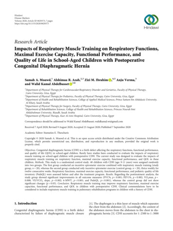
HindawiDisease MarkersVolume 2020, Article ID 8829373, 7 pageshttps://doi.org/10.1155/2020/8829373Research ArticleImpacts of Respiratory Muscle Training on Respiratory Functions,Maximal Exercise Capacity, Functional Performance, andQuality of Life in School-Aged Children with PostoperativeCongenital Diaphragmatic HerniaSamah A. Moawd,1 Alshimaa R. Azab,2,3 Zizi M. Ibrahim ,4,5 Anju Verma,3and Walid Kamal Abdelbasset 3,61Department of Physical Therapy for Cardiovascular/Respiratory Disorder and Geriatrics, Faculty of Physical Therapy,Cairo University, Giza, Egypt2Department of Physical Therapy for Pediatrics, Faculty of Physical Therapy, Cairo University, Giza, Egypt3Department of Health and Rehabilitation Sciences, College of Applied Medical Sciences, Prince Sattam bin Abdulaziz University,Al-Kharj, Saudi Arabia4Department of Physical Therapy for Surgery, Faculty of Physical Therapy, Cairo University, Giza, Egypt5Department of Rehabilitation Sciences, College of Health and Rehabilitation Sciences, Princess Nourah bintAbdulrahman University, Riyadh, Saudi Arabia6Department of Physical Therapy, Kasr Al-Aini Hospital, Cairo University, Giza, EgyptCorrespondence should be addressed to Walid Kamal Abdelbasset; walidkamal.wr@gmail.comReceived 7 April 2020; Revised 9 August 2020; Accepted 23 August 2020; Published 7 September 2020Academic Editor: Stamatios E. TheocharisCopyright 2020 Samah A. Moawd et al. This is an open access article distributed under the Creative Commons AttributionLicense, which permits unrestricted use, distribution, and reproduction in any medium, provided the original work isproperly cited.Objectives. Congenital diaphragmatic hernia (CDH) is a birth defect affecting the respiratory functions, functional performance,and quality of life (QOL) in school-aged children. Rarely have studies been conducted to evaluate the impacts of respiratorymuscle training on school-aged children with postoperative CDH. The current study was designed to evaluate the impacts ofrespiratory muscle training on respiratory function, maximal exercise capacity, functional performance, and QOL in thesechildren. Methods. This study is a randomized control study. 40 children with CDH (age: 9-11 years) were assigned randomlyinto two groups. The first group conducted an incentive spirometer exercise combined with inspiratory muscle training (studygroup, n 20), whereas the second group conducted only incentive spirometer exercise (control group, n 20), thrice weekly fortwelve consecutive weeks. Respiratory functions, maximal exercise capacity, functional performance, and pediatric quality of lifeinventory (PedsQL) were assessed before and after the treatment program. Results. Regarding the posttreatment analysis, thestudy group showed significant improvements in all outcome measures (FVC%, p 0:001; FEV1%, p 0:002; VO2max, p 0:008; VE/VCO2 slope, p 0:002; 6-MWT, p 0:001; and PedsQL, p 0:001), whereas the control group did not showsignificant changes (p 0:05). Conclusion. Respiratory muscle training may improve respiratory functions, maximal exercisecapacities, functional performance, and QOL in children with postoperative CDH. Clinical commendations have to beconsidered to include respiratory muscle training in pulmonary rehabilitation programs in children with a history of CDH.1. IntroductionCongenital diaphragmatic hernia (CDH) is a birth defectcharacterized by failure of diaphragmatic muscle closure[1]. The diaphragm is a thin layer of muscle which separatesthe chest from the abdomen [2]. Accordingly, the content ofthe abdomen moves from the abdomen to the chest in diaphragmatic hernia [3]. CDH accounts for 1 : 2500 to 1 : 3000
2live births [4]. Approximately 80-85% of CDH affects the leftside of the diaphragm [5]. According to the site of the diaphragmatic hernias, it is classified into three types: the Bochdalek hernia, the Morgagni hernia, and the central hernia[6]. The Bochdalek hernia occurs in the posterior-lateral partof the diaphragm and affects 70% of the cases, the Morgagnihernia occurs in the anterior part of the diaphragm andaffects 25–35% of the cases, and the central hernia occursin the central part of the diaphragm and affects 2-5% ofthe cases [6].Around 50-60% of CDH occur without any other congenital anomalies [7], while 40-50% occur with other congenitalanomalies such as craniofacial, ocular, cardiovascular, centralnervous system, skeletal, and genitourinary anomalies [8].Although there is no distinct reason for CDH, it may be associated with multiple genetic factors in concert with environmental and nutritional issues [9–11]. CDH and its relatedmalformation threaten life with a mortality rate ranging from10 to 50% [12]. Surviving children often present with severecomplications such as pulmonary disorders, cardiovasculardiseases, microstructural changes in lung gastrointestinal disease, gastroesophageal reflux, and recurrent episodes of lowerrespiratory tract infections [1, 13–15].In addition to the previous complication, children withCDH suffer from significant reductions in forced expiratoryvolume in 1 second (FEV1), forced vital capacity (FVC),FEV1/FVC, maximum midexpiratory flow, peak expiratoryflow, and exercise capacity with a significant increase in theratio of residual volume/total lung capacity [16].CDH has health and economic influences on childrenand their families. The financial cost of intensive utilizationof the health system by the surviving CDH children is continuously increasing with reduced productivity of families caring for these children and a marked reduction in quality oflife (QOL) for CDH children and their families [17].Recently, with a continued existence rate, longstanding evaluations have been considered in children withCDH, and this is where the QOL plays a key role in thisissue [18]. The World Health Organization stated thatQOL is a wide-range concept impacted in multiple dimensions by the individual’s physical, psychological, social,and personal status and their association with relevantattributes of their surroundings [19]. A previous studyconcluded that the malformation severity of CDH mayaffect QOL negatively [20]. A recent study has found thatexercise training improves respiratory functions and QOLin asthmatic children [21].Different modalities of physical therapy have been evaluated in children with different respiratory disorders includingaerobic exercise training [21], chest physical therapy [22],and inspiratory muscle training [23]. However, rarely havestudies evaluated the impacts of respiratory muscle trainingon respiratory function, exercise capacity, functional performance, and QOL in school-aged children with postoperativeCDH. Regarding that, our study was conducted to evaluatethe impacts of the respiratory muscle training on respiratoryfunctions, maximal exercise capacity, functional performance, and QOL in children with postoperative CDHhypothesizing that respiratory muscle training could have aDisease Markersuseful impact on respiratory functions, exercise capacity,and QOL among those children.2. Materials and Methods2.1. Study Design. Between April 2019 and January 2020, thisrandomized controlled clinical trial was conducted. It wasaccomplished at the outpatient physiotherapy clinic at PrinceSattam bin Abdulaziz University. The ethical clearance wasattained from the local institutional review board of the physiotherapy department (No. RHPT/018/055). All procedureswere fulfilled in accordance with the ethical standards ofthe 1964 Declaration of Helsinki and its updates.2.2. Subjects. Forty children with postoperative CDH wererecruited from the pediatric surgical departments of the KingKhalid Hospital and other referral hospitals in Al-Kharj,Saudi Arabia. Participants were included in the study if theirages ranged from 9 to 11 years, had BMI between 20 and25 kg/m2, were diagnosed with CDH-associated respiratorydistress within the first days of life (high-risk CDH), and haveundergone an operation immediately after birth. Childrenwho have a paraesophageal diaphragmatic defect, a diaphragmatic eventration, were unable to tolerably conductthe study procedures, or had any other serious anomalieswere excluded.2.3. Randomization and Blinding. From the forty-four children assessed for eligibility, three did not meet the inclusioncriteria of the study and one of the parents had refused toenroll his child in the study without a specific reason. Utilizing SPSS version 22 (IBM Corp., Armonk, NY, USA), fortychildren were randomized before initiating the study procedures into the study group that conducted a program ofinspiratory muscle training combined with incentive spirometer exercise, and the control group that conducted a program of incentive spirometer exercise alone. The parents orcaregivers of the children were informed of the proceduresof the study, and they were instructed to sign written consentforms before initiating the study procedure. The examinerwas blinded to the group assignments. Figure 1 demonstratesthe CONSORT flowchart of the study.2.4. Sample Size Estimation. Using VO2max as the primaryoutcome in the study, the sample size has been estimated. Aprior study has evaluated the effect of aerobic exercise onexercise capacity in school-aged children and approved thesignificant difference of the mean VO2max of 5 mL/kg/minat least [21]. Based on this difference and the study objectiveto realize 80% power with type I error of 0.05, the presentstudy required thirty-four children for the two study groups.Hence, the study included forty children to account for the20% dropout.2.5. Outcome Measures. Respiratory functions, maximalexercise capacity, functional performance, and quality oflife were assessed before and immediately after treatmentby an independent researcher who was not aware of thegroup treatment.
Disease Markers3Screening/participationAssessed for eligibility (n 44)Excluded (n 4)Not meeting inclusion criteria (n 3)Declined to participate (n 1)Randomized (n 40)AllocationAllocated to the control group (n 20)(i) Received control program (n 20)(ii) Did not receive control program (n 0)Allocated to the study group (n 20)(i) Received study program (n 20)(ii) Did not receive study program (n 0)Follow-upLost to follow-up (n 0)Discontinued the study program (n 0)Lost to follow-up (n 0)Discontinued the control program (n 0)AnalysisAnalyzed (n 20)(i) Excluded from analysis (n 0)Analyzed (n 20)(i) Excluded from analysis (n 0)Figure 1: The flow diagram of the study participants through the trial.2.5.1. Respiratory Functions. The respiratory functions wereevaluated utilizing Spirolab III (SDI Diagnostics Inc., USA),the tests were conducted at a defined time between09:00 am and 13:00 pm to limit diurnal variety. Using the spirometer, the youngsters were instructed to breathe 2-3 discretionary tidal breaths in and out, and to breatheprofoundly (quick and profound) with lips fixed firmlyaround the mouthpiece. At that point, they were instructedto blow air through the mouthpiece as quickly as could beallowed and maintain blowing until no air is left to breatheout. Amid instructions, a few expressions could be utilized,for example, “sucking on a straw” for a profound motivation,“smothering birthday candles” for a powerful lapse, and“continue blowing and continue blowing” to finish exhalinguntil no air is left. The test was repeated three times toachieve reliable findings. The PC screen was displayed tothe children frequently during the test to motivate the children to keep on performing the test [24–26]. The predictedvalues of forced vital capacity (FVC%) and forced expiratoryvolume in one second (FEV1%) were recorded before andafter twelve weeks of the study program.2.5.2. Maximal Exercise Capacity. The maximal exercisecapacity was evaluated through a cardiopulmonary exercisetest (CPET) using the Bruce treadmill test. Parents or children’s caregivers attended the test procedures. The Bruce testprotocol comprised 3 min stages of increasing speed andintensity on a treadmill. Before initiating the test, the children were allowed to familiarize themselves with the mouth-piece and treadmill for 3 min. Each child was encouraged tocontinue until the point of severe fatigue. Heart rate andoxygen saturation were monitored using finger pulse oximetry. The maximal oxygen uptake (VO2max) and the ratio ofminute ventilation (VE) to carbon dioxide production(VCO2) were recorded before and after twelve weeks of thestudy program [27, 28].2.5.3. Functional Performance. Functional performance wasmeasured using the 6 min walk test (6-MWT). It is a validand reliable modality to examine functional performance.Before conducting the 6-MWT, children and their parentswere informed and educated about the purpose of the testand were shown the start and endpoints and instructed alsoto avoid hopping, running, or jumping during the test. Eachchild was instructed to walk through a 50-meter straight corridor over a period of six minutes, while the examiner closelyfollowed them with a stopwatch. The distance in meterswithin the 6-MWT was recorded before and after twelveweeks of the study program [29].2.5.4. Quality of Life. Quality of life was assessed using a 23item pediatric quality of life inventory (PedsQL). This instrument was validated to assess children and adolescents withacute or chronic diseases [30, 31]. The PedsQL questionnaireincludes 4 domains (physical, social, emotional, and schoolfunctions). It provides child self-report for children aged 8years with a 5-point Likert scale (0 means never, and 4 meansalmost always). Each response scores 0%, 25%, 50%, and
4Disease MarkersTable 1: Demographic data of the study children.Study group (n 20)Control group (n 20)p valueAge (years)10:1 0:79:8 0:80.215Gender, boys/girls (n)11/9137:5 5:913/7135:8 5:40.518VariablesHeight (cm)0.347Weight (kg)42:6 4:241:8 4:70.573BMI (kg/m2)23:2 3:823:5 3:60.799Duration of hospitalization (days)52:4 7:549:8 7:20.2715 (25)15 (75)8 (40)12 (60)0.311Medications, n (%)Low/moderate dose of inhaled corticosteroidLong-acting β2-agonist low/moderate dose of inhaled corticosteroidSignificant level at p 0:05. BMI: body mass index.Table 2: Intra- and intergroup changes of outcome variables preand posttreatment.Study group(n 20)Control group(n 20)p valuePre-78:5 9:878:9 10:50.891Post-93:2 7:485:8 11:30.019 0.0010.053Pre-72:3 8:572:7 9:20.887Post-82:6 7:277:4 8:80.0470.0020.107Pre-40:3 7:440:7 8:10.871Post-49:1 7:943:5 8:40.0360.0080.290Pre-31:6 7:130:9 6:80.752Post-24:5 5:828:7 6:20.033p value6-MWT0.0020.291Pre-421:3 38:7438:4 44:50.203Post-488:5 42:8454:6 51:20.028 0.0010.292Pre-75:41 12:572:84 11:90.509Post-82:73 11:273:52 11:70.015 0.0010.632VariablesFVC (% pred.)p valueFEV1 (% pred.)p valueVO2max (mL/kg/min)p valueVE/VCO2 slopep valuePedsQL (overall score)p valueFVC: forced vital capacity; FEV1: forced expiratory volume in one second;VO2max: maximal oxygen uptake; VE/VCO2: minute ventilation/carbondioxide production; 6-MWT: six-minute walk test; PedsQL: pediatricquality of life inventory.100% regarding ranges from never to always. The responsesto the 23 items create a mean score for the 4 domains and amean score for overall PedsQL [32].2.6. Intervention. The study group was recruited to conduct aprogram of inspiratory muscle training combined with anincentive spirometer exercise. The control group wasrecruited to conduct the same incentive spirometer training.2.6.1. Incentive Spirometer Training. Incentive spirometertraining was conducted using a flow-centered incentive spirometer (Triflow II, Respirogram, India). The device is madeup of plastic material; it has three balls connected to a tube,and it also has a mouthpiece. Each child was informed tosit quietly in a relaxed position for some time and concentrate on his/her breathing. Consequently, each child wasinstructed to take a deep breath and to hold the flow-basedincentive spirometer by one hand, while the other hand holdsthe mouthpiece and the tube. Children were asked to take 3-4easy and slow breaths, then they were asked to inhale throughthe spirometer slowly and maximally to make the ball in thecylinder rise as high as possible, after that inspiration washeld for at least 2-3 seconds before normally exhaling without the mouthpiece. These steps were repeated 5 times, andeach child was informed to take a rest for about 60 seconds.This procedure was repeated for a total of 30 min thrice/weekfor 12 consecutive weeks [33].2.6.2. Inspiratory Muscle Training. Inspiratory muscle training was conducted using a Threshold IMT Breathing trainer(Respironics, Cedar Grove, NJ, USA). Inspiratory resistancewas adjusted through a spring-loaded valve presented in thedevice. Each session lasted for 30 min through six inspiratorycycles. Each cycle lasted for 4 min of resisted respiration. Therest period was 60 s after each cycle, which is aimed atimproving the strength of the respiratory muscles. Duringthe last cycle, each child was encouraged to breathe as frequently as possible with the aim of improving muscle endurance. Throughout the exercise training, the threshold loadwas 40% of the maximal inspiratory pressure estimated during the child’s assessment before starting the exercise session.The inspiratory muscle training was conducted thrice/weekfor 12 consecutive weeks [23].2.7. Statistical Analysis. SPSS for Windows (V.22, IBM Corp.,Armonk, NY, USA) was utilized for analyzing the collecteddata. Descriptive analysis was performed using means
Disease 52025Control groupStudy groupFigure 2: The percent of changes in the two groups posttreatment.standard deviations (SD). The Shapiro-Wilk test was utilizedfor assessing the normal distribution of the data. The Studentt-test was used in inferential statistics. The intergroup changeswere analyzed utilizing the unpaired t-test, whereas theintragroup changes were analyzed utilizing the paired t-test.The level of significance was considered at p 0:05.3. ResultsForty CDH children (24 boys and 16 girls) completed thestudy program. The study group comprised 20 children (11boys and 9 girls), whereas the control group comprised 20children (13 boys and 7 girls). As detailed in Table 1, demographic data (age, gender, height, weight, and BMI) showedno significant intergroup difference (p 0:05). The meanvalues of the outcome measures including respiratory functions, maximal exercise capacity, function performance, andQOL showed no significant intergroup pretreatment differences (FVC%, p 0:891; FEV1%, p 0:887; VO2max, p 0:871; VE/VCO2 slope, p 0:752; 6-MWT, p 0:203; andPedsQL, p 0:509) as detailed in Table 2.Regarding the posttreatment analysis intragroup, thestudy group showed significant improvements in the studyoutcome measures (FVC%, p 0:001; FEV1%, p 0:002;VO2max, p 0:008; VE/VCO2 slope, p 0:002; 6-MWT,p 0:001; and PedsQL, p 0:001), whereas the controlgroup showed no significant changes (FVC%, p 0:053;FEV1%, p 0:107; VO2max, p 0:290; VE/VCO2 slope, p 0:291; 6-MWT, p 0:292; and PedsQL, p 0:632) as presented in Table 2.The percent of changes in the study group (FVC% 18:7%;FEV1% 14:2%; VO2 max 21:8%; VE/VCO2 slope 22:4%;6 MWT 15:9%; and PedsQL 9:7%) versus the controlgroup (FVC% 8:7%; FEV1% 6:4%; VO2 max 6:9%; VE/VCO2 slope 7:1%; 6 MWT 3:7%; and PedsQL 0.93%)are expressed in Figure 2. For intergroup analysis, significant differences were illustrated in all outcome measuresposttreatment (FVC%, p 0:019; FEV1%, p 0:047;VO2max, p 0:036; VE/VCO2 slope, p 0:033; 6-MWT,p 0:028; and PedsQL, p 0:015) which favours the studygroup as detailed in Table 2.4. DiscussionThis randomized control study is aimed at evaluating theimpacts of respiratory muscle training on respiratory functions,
4Department of Physical Therapy for Surgery, Faculty of Physical Therapy, Cairo University, Giza, Egypt 5Department of Rehabilitation Sciences, College of Health and Rehabilitation Sciences, Princess Nourah bint Abdulrahman University, Riyadh, Saudi Arabia 6Department of Physical Therapy