Transcription
Table of ContentsCover ImageCopyright PageDedicationPrefaceTo Our StudentsAcknowledgments1. Cellular Pathology2. Acute and Chronic Inflammation3. Tissue Renewal and Repair: Regeneration, Healing, and Fibrosis4. Hemodynamic Disorders, Thromboembolic Disease, and Shock5. Genetic Disorders6. Diseases of Immunity7. Neoplasia8. Infectious Diseases9. Environmental and Nutritional Diseases10. Diseases of Infancy and Childhood11. Blood Vessels12. The Heart13. Diseases of White Blood Cells, Lymph Nodes, Spleen, and Thymus14. Red Blood Cell and Bleeding Disorders15. The Lung16. Head and Neck17. The Gastrointestinal Tract18. Liver and Biliary Tract19. The Pancreas20. The Kidney21. The Lower Urinary Tract and Male Genital System22. The Female Genital Tract23. The Breast24. The Endocrine System25. The Skin26. Bones, Joints, and Soft-Tissue Tumors27. Peripheral Nerve and Skeletal Muscle28. The Central Nervous System29. The Eye30. Clinical Pathology31. Final Review and Assessment
Copyright 2009 Elsevier Inc. All rights reserved.Copyright PageAll rights reserved. No part of this publication may be reproduced or transmitted in any form or by any means, electronic ormechanical, including photocopying, recording, or any information storage and retrieval system, without permission in writingfrom the publisher. Permissions may be sought directly from Elsevier's Health Sciences Rights Department in Philadelphia, PA,USA: phone: ( 1) 215 239 3804, fax: ( 1) 215 239 3805, e-mail: healthpermissions@elsevier.com. You may also completeyour request on-line via the Elsevier homepage (http://www.elsevier.com), by selecting ‘Customer Support’ and then ‘ObtainingPermissions’.NoticeNeither the Publisher nor the Authors assume any responsibility for any loss or injury and/or damage to persons orproperty arising out of or related to any use of the material contained in this book. It is the responsibility of the treatingpractitioner, relying on independent expertise and knowledge of the patient, to determine the best treatment andmethod of application for the patient.The PublisherPrevious editions copyrighted 2005, 2000ISBN: 978-1-4160-4930-2Acquisitions Editor: William SchmittDevelopmental Editor: Christine AbshirePublishing Services: Mary StermelDesign Direction: Ellen ZanollePrinted in China
DedicationTo our students, for constant challenge and stimulation
PrefaceThis book is designed to provide a comprehensive review of pathology through multiple-choice questions with explanations ofthe answers. The source materials are the eighth editions of Kumar, Abbas, Fausto, Aster Robbins and Cotran PathologicBasis of Disease (PBD8) and Kumar, Abbas, Fausto, Mitchell Robbins Basic Pathology (BP8). It is intended to be a usefulresource for students at a variety of levels in the health sciences.In keeping with recommended question writing style for licensing examinations, we have included single best answerquestions, most with a clinical vignette, followed by a series of homogenous choices. This approach emphasizes anunderstanding of pathophysiologic mechanisms and manifestations of disease in a clinical context. We have incorporatedrelevant laboratory, radiologic, and physical diagnostic findings in the questions to emphasize clinicopathologic correlations.Though this adds to the extent of individual questions, the thoroughness reinforces learning, as a review should. For eachquestion, the correct answer is provided with a brief explanation of why this answer is “correct” and why the other choices are“incorrect.” Each answer is referenced by page numbers to both Robbins and Cotran Pathologic Basis of Disease andRobbins Basic Pathology (both the current 8th and the previous 7th editions) to facilitate and encourage a more completereading of topics targeted for further review. Pathology is a visually oriented discipline; hence, we have used full-color imagesin many of the questions. The illustrations are taken mainly from the Robbins textbooks, so students can reinforce their study ofthe illustrations in the texts with questions that utilize the same images.The revisions in this third edition reflect new topics and new understanding of disease processes reflected in the 8th editions ofthe Robbins textbooks. The questions are intentionally written to be fairly difficult, with the purpose of “pushing the envelope”of student understanding of pathology. We are pushing it further by the addition of a final “integrative review” section, with anew set of clinical pathology questions emphasizing laboratory medicine, as well as a comprehensive final examination withquestions drawn from topics covered in the entire book.Mastery of this book will better prepare the student for further challenges. Many of the questions require a “two-step” process:first, interpreting the information presented to arrive at a diagnosis, and then solving a problem based on that diagnosis. Thisreinforces the clinical reasoning skills needed in delivery of health care. We must hasten to add that no review book is asubstitute for textbooks and other course materials provided by individual instructors within the context of a curriculum. Thisbook should be used after a thorough study of Robbins and Cotran Pathologic Basis of Disease and/or Robbins BasicPathology and course materials. Finally, we hope that students and instructors will find this review book to be a useful adjunctto the learning of pathology.Edward C. Klatt, MD and Vinay Kumar, MBBS, MD, FRCPath
To Our StudentsAlthough medical knowledge has increased exponentially over the past 100 years, the desire to learn and apply this knowledgeto the service of others has not changed. The study and practice of healing arts require persistence more than brilliance. Bycontinuing as a lifelong student, it is possible to become a better health practitioner with the passage of time. Use this book tofind where you are on the pathway to excellence and be inspired to continue down that path. We provide a means to explorethe broad sea of knowledge in pathology within the welcoming environment of this book.Common mistakes made by students in answering questions result from failure to read and analyze information carefully by (1)inappropriate use of a single finding as an exclusionary criterion, and (2) ignoring important diagnostic information. Medicine ismostly analogue, not digital, and the information you obtain is applicable across a continuum of probability. Four key elementsare involved in arriving at the best answer: (1) read the question thoroughly, (2) define the terms (use your vocabulary), (3)rank order possible answers from common to uncommon, and (4) recognize key diagnostic information that differentiates theanswers.There are no magic formulas for academic achievement. The most important thing you can do is to spend some time each dayin a learning process. Learning requires modification of synaptic interfaces at the dendritic level in the brain, and for learning tooccur, there are a finite number of synaptic modifications that can be established per unit time, above which totalcomprehension is reduced. Increasing the rate or length of information delivery diminishes the efficiency of learning. Lack ofbreak periods and “all nighters” presage onset of diminished performance, particularly when least desirable—during anexamination. There is also decay of learning over time, with inevitable random loss of data elements. The key branch points inlearning where review with reinforcement can reduce data loss occur at 20 to 40 minutes (transfer to intermediate memory) andat 24 to 48 hours (transfer to long-term memory) following initial learning.We hope, therefore, that this review will be useful not only in preparing for examinations but also for courses you take and foryour career. It is our sincere hope that this review book will make you a better health practitioner in your chosen career.Edward C. Klatt, MDVinay Kumar, MBBS, MD, FRCPath
AcknowledgmentsWe are very grateful to Christine Abshire, our Developmental Editor, and William Schmitt, Executive Editor, Medical Textbooks,Elsevier Science, for their support of this project. We are grateful to our families and colleagues for graciously accepting thisadditional demand on our time.Edward C. Klatt, MDVinay Kumar, MBBS, MD, FRCPath
Robbins & Cotran Review of PathologyPg. 11. Cellular PathologyPBD7 Chapter 1: Cellular Adaptations, Cell Injury, and Cell DeathPBD8 Chapter 1: Cellular Responses to Stress and Toxic Insults: Adaptation, Injury, and DeathBP7 Chapter 1: Cell Injury, Adaptation, and DeathBP8 Chapter 1: Cell Injury, Death, and Adaptation1 A 17-year-old boy infected with hepatitis A experiences mild nausea for about 1 week and develops very mild scleralicterus. On physical examination, he has minimal right upper quadrant tenderness. Laboratory findings include a serumAST of 68 U/L, ALT of 75 U/L, and total bilirubin of 5.1 mg/dL. The increase in this patient's serum enzyme levels mostlikely results from which of the following changes in the hepatocytes? (A) Autophagy by lysosomes (B) Clumping of nuclear chromatin (C) Defects in the cell membrane (D) Dispersion of ribosomes (E) Swelling of the mitochondria2 A 16-year-old boy sustained blunt trauma to the abdomen when the vehicle he was driving struck a bridge abutment athigh speed. Peritoneal lavage shows a hemoperitoneum, and at laparotomy, a small portion of the left lobe of the liver isremoved because of the injury. Several weeks later, a CT scan of the abdomen shows that the liver has nearly regainedits size before the injury. Which of the following processes best explains this CT scan finding? (A) Apoptosis (B) Dysplasia (C) Fatty change (D) Hydropic change (E) Hyperplasia (F) Hypertrophy (G) Metaplasia3 On a routine visit to the physician, an otherwise healthy 51-year-old man has a blood pressure of 150/95 mm Hg. If hishypertension remains untreated for years, which of the following cellular alterations would most likely be seen in hismyocardium? (A) Atrophy (B) Hyperplasia (C) Metaplasia (D) Hemosiderosis (E) Hypertrophy
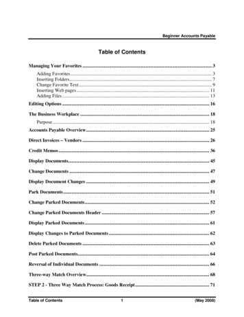
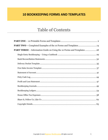
Robbins & Cotran Review of PathologyPg. 24 A 72-year-old man died suddenly from congestive heart failure. At autopsy, the heart weighed 580 g and showed markedleft ventricular hypertrophy and minimal coronary arterial atherosclerosis. A serum chemistry panel ordered before deathshowed no abnormalities. Which of the following pathologic processes best accounts for the appearance of the aorticvalve seen in the figure? (A) Amyloidosis (B) Dystrophic calcification (C) Lipofuscin deposition (D) Hemosiderosis (E) Fatty change5 A 69-year-old woman has had transient ischemic attacks for the past 3 months. On physical examination, she has anaudible bruit on auscultation of the neck. A right carotid endarterectomy is performed. The curetted atheromatous plaquehas a grossly yellow-tan, firm appearance. Microscopically, which of the following materials can be found in abundance inthe form of crystals that produce long, cleft-like spaces? (A) Glycogen (B) Lipofuscin (C) Hemosiderin (D) Immunoglobulin (E) Cholesterol6 A 38-year-old woman experienced severe abdominal pain with hypotension and shock that led to her death within 36hours after the onset of the pain. From the gross appearance of the mesentery, seen in the figure at the bottom of theprevious column, which of the following events has most likely occurred? (A) Hepatitis B virus infection (B) Small intestinal infarction (C) Tuberculous lymphadenitis (D) Gangrenous cholecystitis (E) Acute pancreatitis7 In an experiment, cells are subjected to radiant energy in the form of x-rays. This results in cell injury caused byhydrolysis of water. Which of the following cellular enzymes protects the cells from this type of injury? (A) Phospholipase
(A) PhospholipaseRobbins & Cotran Review of PathologyPg. 3 (B) Glutathione peroxidase (C) Endonuclease (D) Lactate dehydrogenase (E) Protease8 A 47-year-old woman has had worsening dyspnea for the past 5 years. A chest CT scan shows panlobular emphysema.Laboratory studies show the PiZZ genotype of α1-antitrypsin (AAT) deficiency. A liver biopsy specimen examinedmicroscopically shows abundant PAS-positive globules within periportal hepatocytes. Which of the following molecularmechanisms is most likely responsible for this finding in the hepatocytes? (A) Excessive hepatic synthesis of AAT (B) Retention of poorly folded AAT in the endoplasmic reticulum (C) Decreased catabolism of AAT in lysosomes (D) Inability to metabolize AAT (E) Impaired dissociation of AAT from chaperones9 A 68-year-old woman suddenly lost consciousness; on awakening 1 hour later, she could not speak or move her rightarm and leg. Two months later, a head CT scan showed a large cystic area in the left parietal lobe. Which of the followingpathologic processes has most likely occurred in the brain? (A) Fat necrosis (B) Coagulative necrosis (C) Apoptosis (D) Liquefactive necrosis (E) Karyolysis10 A 30-year-old man sustains a left femoral fracture in a skiing accident, and his leg is placed in a plaster cast. After theleg has been immobilized for several weeks, the diameter of the left calf has decreased. This change is most likely toresult from which of the following alterations in the calf muscles? (A) Aplasia (B) Hypoplasia (C) Atrophy (D) Dystrophy (E) Hyalinosis11 An experiment analyzes cells for enzyme activity associated with sustained cellular proliferation. Which of the followingcells is most likely to have the highest telomerase activity? (A) Endothelial cells (B) Germ cells (C) Neurons (D) Neutrophils (E) Erythrocytes
Robbins & Cotran Review of PathologyPg. 412 A 32-year-old man experiences “heartburn” and gastric reflux after eating a large meal. After many months ofsymptoms, he undergoes upper gastrointestinal endoscopy, and a biopsy specimen of the esophageal epithelium i
This approach emphasizes an understanding of pathophysiologic mechanisms and manifestations of disease in a clinical context. We have incorporated relevant laboratory, radiologic, and physical diagnostic findings in the questions to emphasize clinicopathologic correlations. Though this adds to the extent of individual questions, the thoroughness reinforces learning, as a review should. For .