Transcription
Electrophoresis 1999, 20, 1907 19151907Marianne FilletIsabelle BechetVeronique PietteJacques CrommenSeparation of nonsteroidal anti-inflammatory drugsby capillary electrophoresis using nonaqueouselectrolytesDepartment of Analytical,Pharmaceutical Chemistry,Institute of Pharmacy,University of Li ge,Li ge, BelgiumThe aim of the present work was to investigate the separation of nonsteroidal antiinflammatory drugs (NSAIDs: niflumic acid, flufenamic acid, piroxicam, alclofenac, tiaprofenic acid, flurbiprofen, suprofen, ketoprofen, naproxen, indomethacin, carprofen,indoprofen, sulindac) in capillary electrophoresis (CE) using completely nonaqueoussystems. The influence of different parameters such as nature and proportion oforganic solvent (methanol, acetonitrile, 2-propanol), apparent pH (ranging from 7 to 9)and temperature (ranging from 25 to 40oC) on selectivity and migration times werestudied systematically in an uncoated fused-silica capillary. A nonaqueous electrolytemade of 50 mM ammonium acetate 13.75 mM ammonia in methanol proved to resolve11 NSAIDs at 25oC and 13 NSAIDs at 36oC, both within 13 min and without a modifierbesides the methanol itself. The same buffer containing 30% acetonitrile provides asatisfactory separation for 13 NSAIDs within 14 min at 25oC.Keywords: Capillary electrophoresis / Nonaqueous systems / Anti-inflammatory drugsCapillary electrophoresis (CE) has been used increasingly as separating analytical method over the last tenyears. So far, the majority of CE separations have beenrealized in aqueous media. Organic solvents have beenused extensively in micellar electrokinetic chromatography (MEKC) [1 4] and in capillary electrochromatography(CEC) [5, 6], most often to improve the separation ofhydrophobic compounds. The use of nonaqueous electrolyte has gained attention in various publications becauseof the better solubility of hydrophobic analytes [7 23].Already in 1984, a separation in an acetonitrile buffer containing hydrochloric acid and tetraethylammonium perchlorate was described by Walbroehl, for the analysis ofquinoline derivatives [7]. Benson et al. used nonaqueouselectrolytes containing ammonium acetate and acetic acidin methanol for the CE separation of hydrophobic metabolites of the antitumor drug pyrazoloacridine [8] and of theH2 antagonist mifentidine [9, 10]. Tamoxifen and itsmetabolites have been separated using a mixture ofmethanol and acetonitrile (50:50) containing ammoniumacetate [11]. Sahota and Khaledi [12] reported a separation of peptides in formamide.Correspondence: Dr. Marianne Fillet, Department of Analytical, Pharmaceutical Chemistry, Institute of Pharmacy, University of Li ge, CHU B-26, Avenue de l Hopital,1, B-4000 Li ge 1,BelgiumE-mail: marianne.fillet@ulg.ac.beFax: 32-4-3664347Abbreviation: NSAID, nonsteroidal anti-inflammatory drug WILEY-VCH Verlag GmbH, 69451 Weinheim, 1999Bjùrnsdottir and Hansen have obtained important selectivity changes by using different organic solvents (formamide, N-methylformamide, N,N-dimethylformamide,dimethylsulfoxide, methanol, and acetonitrile) as separation media for cationic compounds, such as tricyclic antidepressives [13] and opium alkaloids [14], that would bedifficult to resolve in aqueous buffers.A tris(hydroxymethyl)aminomethane-acetate buffer inmethanol has been described for the separation of aromatic and aliphatic acids [15] and N-methylformamide forthe separation of carboxylic acids [16]. Two papers havereported the analysis of inorganic anions in nonaqueoussystems, using dimethylformamide [17] and methanol[15] as organic solvents. The potential of nonaqueouselectrolyte in CE for the separation of hydrophobic soluteshas also been studied by Salimi-Moosavi and Cassidy[18] for a series of alkanesulfonates (C2 C16), alkylsulfates (C8 C18) and linear alkyl benzenesulfonates. Various acidic compounds (pharmaceuticals, including chiralseparation of NSAIDs, dyes or surfactants) have beenstudied using running electrolyte containing differentratios of methanol and acetonitrile [20, 22].Nonaqueous media extended the application range of CEto the analysis of compounds of poor solubility in water,and also improved the selectivity of the separation ofcompounds, which are characterized by similar electrophoretic mobilities in aqueous media. Organic solventsfor separation media in CE are selected according to theirdielectric constant, their viscosity, their UV-absorbance,and by their capability to increase hydrophobic interac0173-0835/99/0909-1907 17.50 .50/0CE and CEC1 IntroductionEL 3508
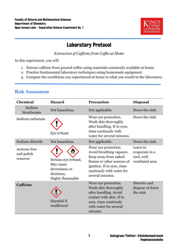
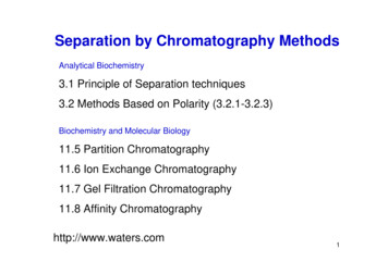
1908M. Fillet et al.tions between the solvent and the analytes, leading to significant changes of the selectivity of analytical separations. Solvents with high dielectric constants and low viscosity are the most suitable for the composition of buffermedia [19].Another interesting aspect of nonaqueous electrolytes isthe complete evaporability of these separation media,which is highly suited to the coupling between CE andmass spectrometry [8 10]. CE with aqueous buffers containing additives (such as cyclodextrins, cellulose derivatives, surfactants) is not as convenient to mass spectrometric detection. Moreover, nonaqueous electrolytes canbe prepared with volatile electrolytes, such as ammoniumacetate, ammonia, or acetic acid. Applications of suchmedia coupled to mass spectrometry were discussed byTomlinson et al. [8 10].The aim of the present work was to investigate the separation of nonsteroidal anti-inflammatory drugs (NSAIDs)in CE using methanol, acetonitrile, and a mixture of bothas nonaqueous medium. NSAIDs are acidic compoundscharacterized by low solubility in water; some have similarcharge densities, which makes their separation difficult inaqueous systems. The influence of different parameterssuch as apparent pH (*pH) and temperature of the buffermedium were also studied.2 Materials and methods2.1 ApparatusAll experiments were performed on a Spectraphoresis1000 CE instrument (SpectraPhysics, San Jose, CA,USA) equipped with an automatic injector, an autosampler, a variable wavelength UV-visible absorbance detector (190 800 nm) and a temperature control system (15 60oC). An IBM PS/2 Model 90486 was used for instrument control and data handling. Electropherograms wereprinted on a Laserjet 4 printer (Hewlett-Packard, Avondale, PA, USA). A capillary cartridge was obtained fromSpectraPhysics and fused-silica capillaries were providedby Polymicro Technologies (Phoenix, AZ, USA). The pHand apparent pH (noted *pH) of running buffers weremeasured by means of a model Delta 345 pH meter witha glass electrode from Mettler (Halstead, UK).2.2 Chemicals and reagentsMethanol of HPLC grade (maximum 0.02% of water) wasobtained from Acros (Geel, Belgium). Ammonium acetateof analytical grade was purchased from Merck (Darmstadt, Germany), as were ammonia, acetic acid and ben-Electrophoresis 1999, 20, 1907 1915zylic alcohol of analytical grade. Water was of Milli-Qquality (Millipore, Bedford, MA, USA). Piroxicam, indomethacine, niflumic acid, flufenamic acid, ketoprofen,suprofen, sulindac, carprofen, indoprofen, flurbiprofenand naproxen were obtained from Sigma Chemicals (St.Louis, MO, USA). Alclofenac was obtained from Continental Pharma (Brussels, Belgium) and tiaprofenic acidfrom Roussel (Brussels, Belgium). All drugs were used asreceived without further purification. The chemical structures and the pKa values of these compounds are given inFig. 1. All solutions were filtered using polytetrafluoroethylene (PTFE) membranes of 0.22 mm from Schleicher &Schuell (Dassel, Germany).2.3 Electrophoretic techniqueElectrophoretic separations were carried out withuncoated fused-silica capillaries having 50 mm internal diameter and 44 cm length (37 cm to the detector). Beforeeach injection, the capillary was treated successively withalkaline solutions (1 M NaOH, 0.1 M NaOH), water andrunning buffer. At the beginning of each working day, thecapillary was rinsed with running buffer for 10 min. Between each injection, the capillary was successivelyrinsed with water and methanol for 2 min (about fourvolumes of capillary), and with buffer for 3 min (about sixvolumes of capillary). The optimal separation buffer forNSAIDs consisted of 50 mM ammonium acetate 13.75 mM ammonia in different organic solvents, preferably methanol. The applied voltage was 25 kV (detectorat the anode end of the capillary) for methanolic buffersand 25 kV (detector at the cathodic end) for aqueous buffers. UV detection was performed at 280 nm. This wavelength was found to be a good compromise for the detection of the NSAIDs tested. Injections were made inhydrodynamic mode for a period of 2 s (corresponding to5.3 nL). The capillary was thermostated at 25oC, unlessotherwise stated. Test mixtures of NSAIDs were preparedin methanol, at a concentration of ca. 4 10 5 M (20 mg/mL) each. The migration order was determined by injection of individual solutions of each NSAID at the sameconcentration and by spectral comparison. The electroosmotic flow was measured with a neutral marker (0.01%solution of benzylic alcohol). The resolution (Rs) and platenumber (N) were calculated according to the standardexpressions based on the peak width at half height [24].The asymmetry factor (As) was determined using theexpression: As b/a where a is the distance between theperpendicular from the peak maximum to the leadingedge of the peak at one-tenth of the peak height and b isthe distance between the perpendicular from the peakmaximum to the trailing edge of the peak at one-tenth ofthe peak height.
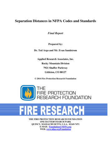
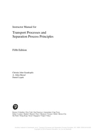
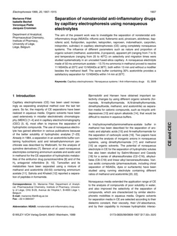
Electrophoresis 1999, 20, 1907 19153 Results and discussion3.1 Organic solventThe influence of the nature and the concentration oforganic solvents was studied first. As can be seen in Fig.CE of nonsteroidal anti-inflammatory drugs19092, the addition of methanol, ethanol and 2-propanol to therunning electrolyte significantly reduced the EOF. Acetonitrile does not have as strong an influence, probably dueto its higher dielectric constant and the lower viscosity ofthe mixture. Methanol was also found to provide importantFigure 1. Chemical structures of NSAIDs and their pKavalues. (1) Alclofenac (pKa 4.6); (2) Carprofen (pKa 4.3);(3) Flufenamic acid (pKa 3.9); (4) Flurbiprofen (pKa 4.1);(5) Indomethacin (pKa 4.5); (6) Indoprofen (pKa 5.8);(7) Ketoprofen (pKa 4.0); (8) Naproxen (pKa 4.2); (9) Niflumic acid; (10) Piroxicam (pKa 6.3); (11) Sulindac(pKa 4.7); (12) Suprofen (pKa 3.9); (13) Tiaprofenic acid(pKa 3.0).
1910M. Fillet et al.Electrophoresis 1999, 20, 1907 1915Figure 2. Influence of organicsolvent concentration on electroosmotic mobility (meo). Buffer:75 mM glycine adjusted topH 9.1 with triethanolaminecontaining an organic solvent indifferent proportions (0 40%).Voltage, 15 kV. Detection wavelength, 200 nm. Neutral marker,0.01% solution of benzylic alcohol in water. Organic solvents:(&) acetonitrile; (*) methanol;( ) ethanol; ( ) 2-propanol.Table 1. Electroosmotic mobilities and NSAID effectivemobilities in aqueous and nonaqueous systemsEffective mobilities (mep 10 5) cm2/VsAqueous system (1)Nonaqueous system (2)AlclofenacFlufenamic acidNiflumic acidTiaprofenic amIndomethacinSulindac 28.1 27.0 26.8 26.5 26.4 26.4 25.8 25.5 24.0 22.1 21.3Niflumic acidFlufenamic acidPiroxicamAlclofenacTiaprofenic rofenSulindac 26.0 24.2 22.8 22.2 21.6 21.3 20.7 20.1 19.6 18.8 17.6Electroosmotic mobilities (meo 10 5) cm2/VsAqueous system (1)Nonaqueous system (2)meo63.0meo7.5Buffer: 50 mM ammonium acetate 13.75 mM ammonia(1) in water or (2) in methanolOther conditions as described in Section 2.3changes in selectivity, compared to other solvents tested.In a completely methanolic system, the cathodic electroosmotic flow was strongly decreased in comparison to theanalogous aqueous system (cf. Table 1), because ofchanges in the dielectric properties and the viscosity ofthe system. The electroosmotic mobility of methanolicmedium was lower than the effective mobilities ofNSAIDs, so that these anionic compounds could notreach the detector when the latter was located at thecathodic side of the capillary. Effective mobilities of mostNSAIDs in nonaqueous systems were slightly reducedcompared to those obtained in aqueous systems (Table1), whereas electroosmotic mobility was strongly decreased (meo 63 10 5 cm2/Vs in aqueous systems andmeo 7.5 10 5 cm2/Vs in methanolic systems).Consequently, the electroosmotic flow is too low to transport anionic NSAIDs towards the cathode located at thedetector side, as is the case in aqueous systems [25].Therefore, in methanolic systems the polarity of the electrical field had to be reversed and the NSAIDs weredetected at the anodic end of the capillary. The electroosmotic flow then moves in the opposite direction.If the reduction of the electroosmotic flow was the onlyeffect when methanol was used as separation medium, areverse migration order of NSAIDs should be observed,compared to that in aqueous systems. However, Table 1shows several significant differences in the migrationorder of NSAIDs between the two systems. For example,sulindac exhibits the lowest effective mobility in both systems, while alclofenac, which should be the first migratingpeak in methanolic systems (because of its higher effective mobility), appears as the fourth peak in methanolicelectrolyte. Piroxicam appears among the analytes with alower effective mobility in the aqueous system, while ithas one of the highest effective mobilities in the nonaqueous system. In the aqueous buffer, a series of compounds(flufenamic acid, niflumic acid, tiaprofenic acid, flurbiprofen, naproxen, carprofen, and ketoprofen) exhibit similarelectrophoretic mobilities and are difficult to resolve undersuch conditions. As shown in Table 1, the range of effective mobilities of NSAIDs is larger in the methanol bufferthan in the aqueous buffer, so that greater differe
cal, Pharmaceutical Chemistry, Institute of Pharmacy, Universi-ty of Liłge, CHU B-26, Avenue de l'Hopital,1, B-4000 Liłge 1, Belgium E-mail: marianne.fillet@ulg.ac.be Fax: 32-4-3664347 Abbreviation: NSAID, nonsteroidal anti-inflammatory drug Electrophoresis 1999, 20, 1907–1915 1907