Transcription
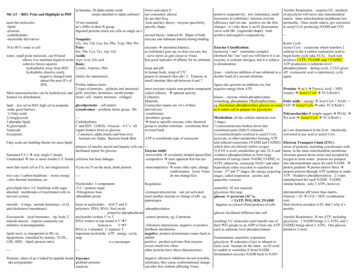
MCAT – BIO: Print and Highlight in PDFmost bio e derivativesin humans, 20 alpha amino acidsamine attached to alpha carbonyl10 are essential.aa’s differ in their R group.digested proteins reach our cells as single aa’sNonpolar:Gly, Ala, Val, Leu, Iso, Phe, Tryp, Met, Pro70 to 80 % water is cellPolarSer, Thr, Cys, Tyr, Asp, Gluwater, small polar molecule, can H-bondAcidicallows it to maintain liquid at room Aspr Acid, Glu acidcohesive forces squeezeBasic:Lysine, Arginine, Histhydrophobic away from H20hydrophilic dissolve easily-negative charged ends (italics for mnemonic)attract the posi H’s ofProline induces turns.H20.2 types of proteins – globular and structural.Most macromolecules can be hydrolyzed, and glob: enzymes, hormones, memb pumpsstruct: cell / matrix structure. collagen.formed via dehydration.lower activation Enot consumed, altereddo not alter Keqlock-and-key theory / enzyme specificity.specific shape.second theory: induced fit. Shape of bothenzyme and substrate altered during binding.enzymes Æ saturation kinetics.as [substrate] goes up, so does rxn rate, butcurve slows as gets closer to Vmax.Km good indicator of affinity for its substratetemp and pH.in human body, temp of 37Cpepsin in stomach likes ph 2. Trypson, insmall intestine likes ph between 6 and 7.most enzymes require non-protein componentcalled cofactor. Æ optimal activity.Cofactors:Minerals,Coenzymes (many are vit’s of theirderivatives)-cosubstrates-prosthetic groups.Æ bind to specific enzyme, txfer chemicalgroup to another substrate. cosubstrate thenreverted back.positive cooperativity. low [substrate], smallincreasees in [substrate] increase enzymeefficiency and rxn rate. positive are the firstchanges. it’s why there is an 02 dissociationcurve with Hb. (sigmoidal shape). bothpositive and negative cooperativity.Aerobic Respiration – requires O2. productsof glycolysis will move into mitochondrialmatrix. inner mitochondrial memberate lesspermeable. Once inside matrix, pyr convertedto acetyl CoA producing NADH and CO2--Krebs CycleAcetyl CoA – coenzyme which transfers 2carbons to the 4 carbon oxaloacetic acid tobegin krebs cycle (aka TCA). Each turnproduces 1ATP, 3NADH, and 1 FADH2.ATP production is substrate-levelphosphorylation. during cycle, 2 CO2 givenlyase – catalyzes addition of one substrate to a off. oxaloacetic acid is reproduced, cycledouble bond of a second substrate.again.Enzyme Classification:memorize “-ase” sometimes complexchemical has “ase” and you will know it is anenzyme, it contains nitrogen, and it is subjectto denaturation.ligase also governs an addition rxn, butrequires energy from ATP.Proteins Æ aa’s Æ Pyruvic Acid NH3(waste) Æ Acetyl CoA Æ TCA/Kreb’skinase – enzyme which phosphorylatessomething, phosphatase DEphosphorylates.Fatty acids energy Æ Acyl CoA NAD eg, hexokinase phosphorylates glucose as soon FAD Æ Acetyl CoA Æ enter TCA/Kreb’sglycolproteins – cell matrixlipid – low sol in H20, high sol in nonpolaras it enters cell to prepare for glycolysis.cytochromes – prothetic heme group. Hbmake good barriersPolysaccharidesÆ simple sugars Æ PGAL Æ1) Fatty acidsMetabolism: all the cellular chemical rxns--Pyr acid Æ Acetyl CoA Æ TCA/Kreb’s2) triaglycerols3 stagesCarbohydrates3 phsopho lipids1) macromolecules broken down intoC and H20. C(H20). Glucose – 6 C’s. all4) glycolipidsconstituent parts (little E released)sugars broken down to glucose.aa’s are deaminated in the liver. chemically5)steroids2) constituent parts oxidized to acetyl CoA,-2 anomers, alpha (trans) and beta (cis)converted to pyr acid or acetyl CoA.6) terpenespyruvate, or other metabolites forming ATPAnimals eat Alpha. Bacteria break BetaATP is cosubstrate type of coenzymeand reduced coenzymes (NADH and FADH2) Electron Transport Chain (ETC)Fatty acids are building blocks for most lipidswhich does not directly utilize oxygenabsence of insulin, neural and hepatic cells use --series of proteins, including cytochromes with3) if O2 is avail, metabolites go into TCA and heme, in the inner mitochondrial membrane.facilitated txport for glucose.Enzyme inhib:electrons passed down series and accepted by-irreversible Æ covalently bonded (penicillin) oxidative phosphorylation to form largeSaturated FA’s Æ only single C-bondsamounts of energy (more NADH, FADH2, or oxygen to form water. protons are pumped-competitive Æ raise apparent Km but notUnsaturated Æ one or more double C-C bonds cellulose has beta linkagesVmaxATP); otherwise, coenzyme NAD and other into intermembrane space for each NADH. Æif you see N on the mcat, think proteinproton gradient Æ proton motive force Æ-noncompetitive Æ some other spot, changemost fats reach cell as FA, not triaglycerolsbyproducts either recycled or expelled asconformation. lower Vmax waste. 2nd and 3rd stages, the energy acquiring propels protons through ATP synthase to make---ATP. Oxidative phosphorylation. 2-3 atpsdo not change Kmtria’s are 3 carbon backbone – stores energystages, called respiration. aerobic andmanufactured for each NADH. FADH2--also thermal insulation, etc.anaerobic versions.Nucleotides: 3 componentssimilar fashion. only 2 ATPs, however.Regulation:-5-C / pentose sugarglycolipids have 3-C backbone with sugaranaerobic: 02 not required.-Nitrogenous baseintermembrane pH lower than matrix.-zymogen/proenzyme – not yet activated.attached. membranes of myelenated cells inglycolysis first step.-phosphate groupGlucose 02 Æ CO2 H20 (combustionneed another enzyme or change of pH. eg,nervous systemglucose Æ pyruvate (3C’s).rxn)pepsinogen. 2ATP, PO3, H20, 2NADHbases in nucleotides – AGCT and Ufinal electron acceptor is 02, that’s why it’ssteroids – 4 rings. include hormones, vit D,happens in cytosol (fluid portion) of cellspolymers: DNA, RNA, Nucl-acidsaerobic-phosphorylationand cholesterol (membrane)joined by phosphodiesterglucose facilitated diffusion into cell.nucleotides written 5’ to 3’Aerobic Respiration: 36 net ATP, including-control proteins, eg, G proteinsEicosanoids – local hormones – bp, body T,DNA written so top strand is 5’Æ3’glycolysis. 1 NADH brings 2-3 ATPs, and 1smooth muscle. Aspirin commonly useresulting 3-C molecules each transfer one of-Allosteric interactions: negative or positiveinhibitor of prostaglandins.their PO3 groups to an ADP to form one ATP FADH2 brings about 2 ATPs. One glucosebottom is3’Æ5’produces 2 turns.feedback mechanism.each in substrate level phosphorylation.RNA is 1-stranded. U replaces T.negative: product downstream comes back tolipids insol, so transported in Hb viaimportant nucleotide: ATP. energy. cyclicinhibitlipoproteins. classified by density, VLDL,Fermentation: anaerobic respiration.amppositive: product activates first enzyme.LDL, HDL. (lipid::protein ratio).glycolysis Æ reduction of pyr to ethanol oris a messenger.occurs much less often.lactic acid. humans do the latter. no 02 availother proteins have these characteristics---or unable to assimilate E from NADH.--fermentation recycles NADH back to NAD negative allosteric inhibitors do not resembleProteins: chain of aa’s linked by peptide bonds Enzymessubstrates, they cause conformational change.aka polypeptidesglobular proteinscan alter Km without affecting Vmax.catalysts
Genesgene – series of n-tides. codes for singlepolypeptide, or mRNA, rRNA, or tRNA.Eukary’s have more than 1 copy of somegenes. Prokary’s only have 1 copy of each.one gene; one polypeptide. exception: posttranscriptional processing RNA.as well5Æ3. 5 is upstream, 3 downstream.“reading DNA like paddling upstream”5 steps of replication:1) helicase unzips double helix2) RNA polymerase builds a primer3) DNA polymerase assembles leading andlagging strands4) Primers are removed5) Okazaki fragments joinedGenome: entire DNA sequence of organism.process of replication: semidiscontinuousonly 1% of genome codes for proteinhuman DNA differs only at about 0.08%.Small variation Æ big difference.Central Dogma: DNA transcribed to RNA,translated to aa’s for proteinDNA Æ RNA Æ Protein.(same for all organisms)4 bases of DNA:-Adenine (purine) – two ring-Guanine (purine) – two ring-Cytosine (pyrimidine) – one ring-Thymine (pyrimidine) – one ringtelomeres: ends of eukaryotic chromosomalDNA. protect from chromosomal erosionRNAcarbon 2 not deoxygenatedsingle strandeduracil instead of thyaminecan move through the nuclear pores3 types-mRNA: delivers DNA code for aa’s tocytosol for protein manufacturingoperator promoter genes operoneg, lac operon. codes for enzymes to allow Ecoli to import and metabolize lactose whenlow glucose. low glucose, high cAMP,activates CAP, activates promoter. operatordownstream, too. Allows for repression viabinding to a protein, allolactose (inducer).Genetic code: mRNA nucleotides.code is degenerative. more than one set of 3nucleotides can code for a single amino acid.but 1 and only 1 aa, so unambiguous.start codon is AUGstop codons UAA, UAG, and UGA.64 possible combinations of the bases20 possible amino acids.if protein contains 100 aa’s, then 20 100possible sequences.initial mRNA sequence called primarytranscript. processed by addition of n-tides,deletion of n-tides, modification of n-bases. 5’RNA n-tides written 5’Æ3’end capped with GTP. 3’ end poly A tail toprotect from exonucleasesTranslation: mRNA directed protein synthesis.mRNA the template. tRNA carries n-tidesprimary txscript cleaved into introns, exonssnRNPs (snurps) recognize, form spliceosome, complementary for codon, called anticodon.rRNA with protein make up ribosome, whichcut off introns. only 30,000 genes, butis the site of translation.120,000 proteins possible bc of splicing.introns::exons 24::1small subunit, large subunit. ribosomesrequire nucleolus for their origin.denatured DNA – heat Æ separated strands.tRNA posessing 5’-CAU-3’ anticodonmore C3G pairs, higher Tmsequesters methionin and enters at P site.DNA-RNA hybridizationLarge subunit joins (initiation). next tRNArestriction enzymes cut DNA at certainsequences, usually palindromic. leave DNA enters A site. translocation. tRNA shifts,moves to E site.with sticky end so they can reconnect.initiation, elongation, and termination.recombinant DNA.translocation – segment of DNA from 1chromo inserted into anotherinversion – orientation reversedtransposons can excise themselves and insertthemselves elsewhereforward mutation – changing organism awayfrom original statebackward – back to original stateoriginal state called wild typeCancerproto-oncogenes – stimulate normal growth incells. can be converted to oncogenese – genesthat cause cancer, by UV radiation, chemicals,or simple random mutations. Mutagens thatcause these called carcinogensDNA is 5 ft for each cell. wrapped tightlyaround globular proteins, histones. 8 histoneswrapped in DNA – nucleosome. wraps intocoils, supercoils, entire complex calledchromatin.somatic cell: 46 double stranded DNAmolecules. chromosome. 46 chromosomesbefore replication, 46 after replication.txlation begins on free floating ribosome.duplicates referred to as sister chromatids.DNA library – use a vector in a bacterium,signal peptide can transport polypeptide to-rRNA: combines w/ proteins to formDiploid means cell as 23 homologous pairs.then reproduce bacterium. active gene, turnribosomes Æ protein synthesis.blue with x-gal. some bacteria wont take up, lumen. SRP can carry entire ribosome towards sex cells haploid.ERso introduce lac-z with your inserted vector.In DNA, two strands run antiparallel boundDNA is produced by replicationstages of cell’s lifeintroduce X-gal and the right ones will turntogether by H-bond. Double stranded. hMutationsonly in nuc and mito matrix1) G1 – first growthbluebonding Æ base pairing.any alteration that is not recombinationRNA by transcription2) S – Synthesiscomplementary strands Æ double helixalso in cytosolone way to find gene in library – hybridization gene mutation – sequence of n-tides in a single 3) G2 – second growth phase4) M – mitosis / meiosisradioactive labeled comp sequence of desired geneeach groove spirals once around double helixchromosomal mutation – structure is changed 5) C – cytokinesistranscription: starts w/ initiation. promoter.DNA fragment (probe). cDNA product –for every 10 base pairs.RNA polymerase. promoter is upstream from mRNA produced by the DNA. lacks intronsdiameter of double helix is 2 nanometerssomatic vs. germ cell mutationsgene.(good).latter more serious1-3 called interphase.remember: Ntide made of pentose sugar, P03replication: transcription bubble, elongationin G1 – regions of heterochromatin have beenbetter cloning: polymerase chain rxn (PCR).group, nitrogenous base.mode. strand transcribed: template orunwound into euchromatin, RNA synth andfast way to clone dna. heating and annealing. point mutation – single n-tide changedbase pair mutation – AT to GC, vice versaantisense. other strand is coding. RNA poly, primers hybridize. polymerase replicates.protein synth very active. G1 checkpoint Æ Spairings: AT, GCmissense – bp mutation in aa sequence oflike DNA poly, reads in the 3Æ5 direction,stage, if ratio of cytoplasm to DNA is high2 H bonds in A-T, 3H bonds in C-Ggenebuilding new RNA to be made 5Æ3enough.southern blotting: ID target fragments of“A2T, C3G”may or may not be seriousno proofreading mechanism. slower. rate of known DNA in large pop of DNA. DNAGzero is nongrowing state. neurons, livereg, sickle cell anemiaerror is higher. not hereditary errors. end iscells.cleaved into restriction fragments. separatedDNA replication: semi-conservativeinsertion or deletion Æ frameshift mutation G2 checkpoint – Mitosis promoting factorcalled termination. Coding strand resembles by size in gel elctrophoresis. large movesnew dbl strand created Æ has one new onemultiples other than 3.RNA transcript.(MPF)slower than small. gel denatures DNAold.sometimes nonframeshift and still functionalfragments. probe hybridizes w/ and marksusually frameshift is non-functionalreplication doesn’t distinguish genes.Mitosis, nuclea
MCAT – BIO: Print and Highlight in PDF most bio molecules: -lipids -proteins -carbohydrates -nucleotide derivatives 70 to 80 % water is cell water, small polar molecule, can H-bond allows it to maintain liquid at room cohesive forces squeeze hydrophobic away from H20 hydrophilic dissolve easily -negative charged ends attract the posi H’s of
