Transcription
Stroke Rehabilitation Clinician Handbook 20202. Brain Reorganization, Recovery and Organized CareRobert Teasell MD, Norhayati Hussein MBBS MRehabMed, Mitchell Longval BScTable of Contents2.1 Important Principles of Recovery . 22.1.1 Neurological Recovery . 22.1.2 Time Course of Recovery . 22.2 Mechanisms of Neurological Recovery . 32.2.1 Processes of Neurological Recovery . 32.2.2 CNS Reorganization (Later Recovery)42.3 Predictors of Stroke Recovery . 62.3.1 Stroke Severity as Predictor . 62.3.2 Impact of Age onRecovery/Rehabilitation . 72.3.3 Hemorrhagic versus Ischemic Stroke 82.4 Measures of Functional Outcome . 92.4.1 Motor Function . 112.4.2 Balance . 112.4.3 Cognition . 132.4.4 Stroke Severity . 142.4.5 Community Reintegration . 16Organized Stroke Care – InterdisciplinaryCare/Team . 162.5 Efficacy of Stroke RehabilitationOrganized Care. 162.5.1 Stroke Rehabilitation Programs . 162.5.2 Reviews of Stroke Rehabilitation UnitEfficacy . 172.5.3 Acute Stroke Rehabilitation Units . 182.5.4 Combined Acute and RehabilitationUnits . 202.5.5 Subacute Rehabilitation . 232.5.6 Mobile Stroke Teams . 272.5.7 Meta-Analyses of Combined Results. 29Elements of Stroke Rehabilitation . 332.6 Earlier Therapy is Better. 332.6.1 Benefit of Early Therapy in Animals 332.6.2 Clinical Evidence for Early Therapy . 342.7 Intensity: More is Better . 362.7.1 Intensity of Physiotherapy andOccupational Therapy . 362.7.2 Intensity of Aphasia Therapy PostStroke . 412.7.3 Weekend Therapy and OtherInnovative Approaches to Increase TherapyIntensity . 432.7.4 Inactive and Alone . 442.7.5 Time Accountability: TheCollaboration Evaluation of Rehabilitationin Stroke Across Europe (CERISE) Trial . 442.7.6 Caregiver-Support of IntensiveTherapy . 452.8 Task-Specific Treatment . 462.8.1 Stroke Rehabilitation Must Be TaskSpecific . 462.8.2 Therapy Philosophies . 472.8.3 BobathApproach/Neurodevelopmental Technique(NDT) . 482.9 Outpatient Therapy . 492.9.1 Importance of Outpatient Therapy 492.9.2 Outpatient Stroke Rehab Therapy . 502.9.3 Hospital vs. Home-Based Therapy . 512.9.4 Early Supported Discharge (ESD) . 522.9.5 Canadian Best Practice GuidelineUpdate 2015 . 55References . 57Stroke Rehabilitation Clinician Handbookwww.ebrsr.compg. 1 of 64
Stroke Rehabilitation Clinician Handbook 20202.1 Important Principles of Recovery2.1.1 Neurological RecoveryRecovery after a stroke is associated with cortical reorganization. Motor recovery is a complex processcombining:1. Neurological or Spontaneous Recovery. Recovery of impairment or normal way of moving asmeasured by Fugl-Meyer score or 3D Kinematics (restoration of normal movement patterns).2. Functional Recovery. Recovery of tasks or activities often through learned compensatorymovements (new movement patterns) as measured by the ARAT, Barthel Index or even the FIM.Both involve changes to the remaining motor cortex and this relationship is not fully understood.Spontaneous or Intrinsic Neurological RecoveryNeurological recovery is defined as recovery of neurological impairments. These are determined primarilyby the site and extent of the stroke. As a general rule, the severity of the initial deficit is inverselyproportional to the prognosis for recovery. The majority of neurological recovery occurs within the first 13 months. Afterwards recovery may occur much more slowly for up to one year. The course of recoveryis a predictable phenomenon; it is initially very rapid and then negatively accelerates as a function of time(Skilbeck et al. 1983). Skilbeck et al. (1983) studied 92 stroke survivors with a mean age of 67.5 years(range 36-89) at final assessment, either 2 or 3 years after stroke. The majority of recovery was reportedwithin the first 6 months, with continued but non-statistically significant recovery after 6 months. Thistype of recovery is still largely if not completely independent of rehabilitationand is discussed further lateron.Functional or Adaptive RecoveryFunctional deficits are often referred to as disabilities and are measured in terms of functions such asactivities of daily living. Functional recovery is defined as improvement in mobility and activities of dailyliving; it has long been known that it is influenced by rehabilitation. This recovery depends on the patient'smotivation, ability to learn and family supports as well as the quality and intensity of therapy. Functionalrecovery is highly influenced by neurological recovery but is not dependent on it.2.1.2 Time Course of RecoveryPeak neurological recovery from stroke occurs within the first one to three months. A number of studieshave shown that recovery may continue at a slower pace for at least 6 months; with up to 5% of patientscontinuing to recover for up to one-year. This is especially true with patients who are severely disabledat the time of initial examination (Bonita & Beaglehole 1988, Duncan et al. 1992, Ferrucci et al. 1993, KellyHayes et al. 1989, Wade et al. 1983, Wade et al. 1987).Progress towards recovery may plateau at any stage of recovery with only a very small percentage ofthose with moderate to severe strokes (about 10%) achieving “full recovery”. The return of motor poweris not synonymous with recovery of function; function may be hampered by the inability to perform skilledcoordinated movements, apraxias, sensory deficits, communication disorders as well as cognitiveimpairment. Functional improvements may occur in the absence of neurological recovery (Duncan & MinStroke Rehabilitation Clinician Handbookwww.ebrsr.compg. 2 of 64
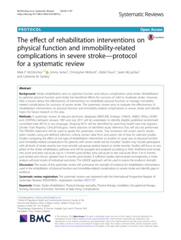
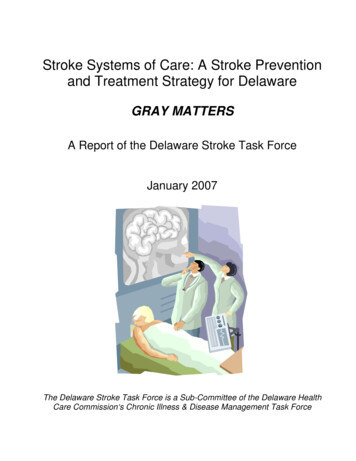
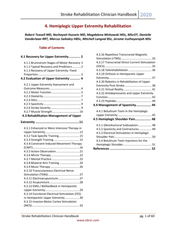
Stroke Rehabilitation Clinician Handbook 2020Lai 1997, Nakayama et al. 1994). Functional recovery (the ability to do activities despite limitations) andimprovement in communication may continue for months after neurological recovery is complete.2.2 Mechanisms of Neurological RecoveryNeurological recovery is defined as recovery of neurological impairments and is often the result of anumber of factors listed below.2.2.1 Processes of Neurological RecoveryProcesses leading to initial clinical improvement occur independent of behaviour or stimuli, althoughthere is some concern about too aggressive very early mobilization (Bernhardt 2015). Processes whichmay account for neurological recovery include: 1) Resolution of post-stroke edema; 2) Reperfusion of theischemic penumbra; 3) Resolution of diaschesis.Post-Stroke EdemaEdema surrounding the lesion may disruptnearby neuronal functioning. Some of the earlyrecovery may be due to resolution of edemasurrounding the area of the infarct (Lo 1986) andas the edema subsides, these neurons mayregain function. This process may continue forup to 8 weeks but is generally completed muchearlier (Inoue et al. 1980). Cerebralhemorrhages tend to be associated with moreedema, which take longer to subside, but whichmay in turn be associated with a more dramaticrecovery.Reperfusion of the Ischemic PenumbraReperfusion of the ischemic penumbra isanother local process which can facilitate early recovery. A focal ischemic injury consists of a core of lowblood flow which eventually infarcts (Astrup et al. 1981, Lyden & Zivin 2000), surrounded by a region ofmoderate blood flow, known as the ischemic penumbra (Astrup et al. 1981, Lyden & Zivin 2000), which isat risk of infarction but is still salvageable. Reperfusion of this area causes affected and previously nonfunctioning neurons to resume functioning with subsequent clinical improvement.The AVERT (2015) trial, looking at very early mobilization of the acute stroke patient, raised concernsabout the penumbra and worsening or extension of the stroke.Stroke Rehabilitation Clinician Handbookwww.ebrsr.compg. 3 of 64
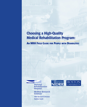
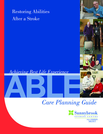
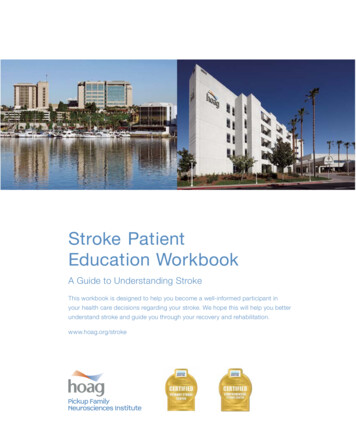
Stroke Rehabilitation Clinician Handbook 2020Lesion with Ischemic PenumbraReperfusion of IschemicPenumbraDiaschesisDiaschesis is a state of low reactivity or depressed function as a result of a sudden interruption of majorinput to a part of the brain remote from the site of brain damage. With injury to one area of the brain,other areas of brain tissue are suddenly deprived of a major source of stimulation. Nudo et al. (2001)noted that diaschesis occurs early after injury and is an inhibition or suppression of surrounding corticaltissue or of cortical regions at a distance that are interconnected with the injury core. The reversibilitymay be partially due to the resolution of edema, which may account for a portion of spontaneous recovery(Nudo et al. 2001). Neuronal function may return following the resolution of diaschisis, particularly if theconnected area of the brain is left intact. This is particularly true of non-cortical structures after corticalinjury (Lo 1986).Proportional Recovery of Upper Limb ImpairmentOne recent controversial concept has been that within 6 months after a stroke, upper limb impairmentresolves by fixed proportion. Fixed proportion argues that 70% of each patient’s maximum possibleimprovement occurs regardless of the initial impairment (for instance, as measured by the Fugl-Meyerscore), but only for those with relatively intact corticospinal (motor) tract functioning (Prabhakaran et al.2008). This holds true for patients across all ages and countries with different rehabilitation services(Byblow et al. 2015).As mentioned, proportional recovery or resolution of upper extremity impairment post stroke isdependent on corticomotor tract integrity. Motor evoked potentials (MEPs) early post stroke isassociated with recovery outcomes (Stinear 2010). Irreversible structural damage to the corticospinaltract severely limits recovery of upper limb movement (Stinear 2010, Stinear et al. 2007). This recovery isunaffected or minimally affected by rehabilitation therapy. 3D kinematics in subacute and chronic strokesurvivors have shown that motor recovery associated with rehabilitation is driven more by adaptive orcompensatory learning strategies. Most of the tests that we use clinically only assess a patient’s ability toaccomplish a certain function or task; we do not tend to measure function.2.2.2 CNS Reorganization (Later Recovery)Neurological reorganization plays an important role in the restoration of function. It can extend for amuch longer period of time than local processes, such as the resolution of edema or reperfusion of theStroke Rehabilitation Clinician Handbookwww.ebrsr.compg. 4 of 64
Stroke Rehabilitation Clinician Handbook 2020penumbra, and is of particular interest because it can be influenced by rehabilitation training. Nudo(2003), based on animal research, has suggested that changes occurring during motor learning (i.e.synaptogenesis and increases in synaptic strength), are likely the same type of changes that occur duringthis part of recovery from stroke. This has been well shown after small, focal lesions in the motor cortexwhere the same principles of motor learning and development of functional connections are occurring inadjacent, undamaged tissue.In Normal IndividualsCramer (2003) notes that, “in normal right-handed persons, performance of a unilateral motor task by theright hand is associated with activation that is largely contralateral, with brain activity ipsilateral to theactive hand being small by comparison (Kim et al. 1993). In contrast, there is greater ipsilateral activationfor movements by the left hand.”In Individuals Post StrokeNudo (2003) reports that neuroplasticity post-stroke (with damage to the motor cortex as an example) isbased on three main concepts:1. In normal (non-stroke) brains, acquisition of skilled movements is associated with predictablefunctional changes within the motor cortex.2. Injury to the motor cortex post-stroke results in functional changes in the remaining cortical tissue.3. After a cortical stroke, these two observations interact so that reacquiring motor skills is associatedwith functional neurological reorganization occurring in the undamaged cortex (Nudo 2003).Mechanism of ReorganizationCramer (2003) noted that after a stroke in humans, movement of the affected hand resulted in threepatterns of cortical reorganization that were not mutually exclusive of each other and which may occurconcomitantly:1. A greater degree of bilateral motor cortex activity was seen with recruitment of the motor networkof the ipsilateral (unaffected hemisphere; (Cramer 2003). In fact, there is widespread areas of corticalhyperactivity occurring days post stroke and dimishing within months of the stroke onset. Non-stroke(less affected) hemisphere cortical activity decreases over months post stroke in those patients whoshow a good motor recovery but not in those who do not show a good motor recovery.2. There was increased recruitment of secondary cortical areas such as supplementary motor area (SMA)and pre-motor cortex in the contralateral (affected) hemisphere (Cramer 2003). Recruitment alongthe cortical rim of the infarct was seen (Cramer 2003). Reorganization of the brain after a stroke isdependent not only on the lesion site, but also on the surrounding brain tissue and on remotelocations that have structural connections with the injured area. Following a stroke, brainreorganization in response to relearning motor activities, involves primarily the contralateral(affected) hemisphere. Reorganization in response to training occurs along the cortical rim of theinfarction with increased recruitment of secondary cortical areas such as supplementary motor areaand pre-motor cortex in the contralateral (affected) hemisphere.3. Ipsilateral cortical involvement is more prominent early on; however, persistence of ipsilateral corticalinvolvement is generally associated with larger strokes and a poorer recovery.Therefore, we can note that reorganization of cortex post stroke is dependent on the lesion site but alsoon remote brain areas with structural connections with the damaged area of the brain. Motor recoveryis largely dependent on intact cortex adjacent to the infarct pointing out the importance of preservingpenumbral areas.Stroke Rehabilitation Clinician Handbookwww.ebrsr.compg. 5 of 64
Stroke Rehabilitation Clinician Handbook 20202.3 Predictors of Stroke RecoveryAlexander (1994) noted the two most powerful predictors of functional recovery were initial strokeseverity and age. Stroke severity is by far the most predictive factor.2.3.1 Stroke Severity as PredictorThe best predictor of stroke outcome is initial clinical assessment of stroke severity. This correlates withthe length of time to maximal neurological and functional recovery.Garraway et al. (1985, 1981) first proposed the concept of 3 bands of stroke patients based upon strokeseverity during the acute phase:1. Mild Strokes: Few deficits, early FIM score (1st 5-7 days) 80, Stineman et al. (1998) defined asmotor FIM 62; rehab gains limited by “ceiling” effect.2. Moderately Severe Strokes: Moderate deficits, conscious with significant hemiparesis, early FIM40-80 or motor FIM 38-62; make marked gains in rehab and 85% discharged to community.3. Severe Strokes: Severe deficits, unconscious at onset with severe paresis or serious medicalcomorbidity, early FIM 40 or motor FIM 37; slower improvement, unlikely to achievefunctional independence (unless young) and smallest likelihood of community discharge.Time Course for Recovery Depends on Initial Severity of ImpairmentsJorgensen et al. (1995c, 1995d) studied 1,197 acute stroke patients in what is referred to as theCopenhagen Stroke Study. Impairments were classified using the Scandinavian Neurological Stroke Scale(SSS) and functional disability was defined according to the Barthel Index (BI). Neurological recoveryoccurred on average two weeks earlier than functional recovery. In surviving patients, the bestneurological recovery occurred within 4.5 weeks in 80% of the patients, while best ADL function wasachieved by 6 weeks. For 95% of the patients, best neurological recovery was reached by 11 weeks andbest ADL function within 12.5 weeks. Jorgensen and associates (1995b) reported that best walkingfunction was reached within four weeks for patients with mild paresis of the affected lower extremity, sixStroke Rehabilitation Clinician Handbookwww.ebrsr.compg. 6 of 64
Stroke Rehabilitation Clinician Handbook 2020weeks for those with moderate paresis and 11 weeks for severe paralysis. Consequently, the time courseof both neurological and functional recovery was strongly related to both initial stroke severity andfunctional disability. Jorgensen et al. (1995a, 1995b, 1995c), found two-thirds of all stroke survivors havemild to moderate strokes and are able to achieve independence in ADL.Impairment and Neurological Recovery of Stroke Patients in the Copenhagen Stroke StudyCategory (SSS)Admission 1 Discharge 2 SurvivalWeeks to 80%Weeks to 95%(%)Best Neurological Best NeurologicalRecovery 3Recovery 3Very Severe (0-14)19%4%38%1013Severe (15-29)14%7%67%915Moderate (30-44)26%11%89%5.510.5Mild/No (45-58)41%78%97%2.56.51Percentage patient distribution on admission, grouped by stroke severity sub groups, as measured bySSS (scores range from 0-58 points).2Percentage distribution of survivors (79% of initial group) after completion of stroke rehabilitation.3Neurological recovery as measured by SSS.Disability and Outcome of Stroke Patients in the Copenhagen Stroke StudyCategory (BI)Discharge 1SurvivalWeeks to 80% BestWeeks to 95% Best(%)Functional Recovery 2Functional Recovery 2Very Severe (0-20)14%501117Severe (25-45)6%921516Moderate (50-70)8%9769Mild (75-95)26%982.55No (100)46%1Percentage patient distribution on discharge, grouped by stroke severity sub groups, as measured byBarthel Index.2Functional recovery as measured by Barthel Index.Based on these observations one can safely conclude that the initial severity of the stroke is inverselyproportional to the final functional outcome, with the majority of patients who suffer mild strokesdemonstrating no or only mild disabilities, while the majority of patients suffering very severe strokes stillexperience severe or very severe deficits even after the completion of rehabilitation.2.3.2 Impact of Age on Recovery/RehabilitationRecovery is more rapid and occurs to a greater extent in younger individuals with a stroke. This correlateswith decline in ability to form neurological connections with aging. There is also a small but significanteffect of age on functional recovery.Impact of Age in Animal StudiesIn rats, the duration of motor impairment post brain lesion increases with age (Brown et al. 2003). Theregenerative response of neurons and glial cells, though largely preserved with age, appears to be delayedor occurs at a diminished rate the older the animal (Popa-Wagner et al. 1999, Whittemore et al. 1985).Reactive neuronal synaptogenesis declines (Scheff et al. 1978), sprouting responses are less robustStroke Rehabilitation Clinician Handbookwww.ebrsr.compg. 7 of 64
Stroke Rehabilitation Clinician Handbook 2020(Schauwecker et al. 1995, Whittemore et al. 1985) and synaptic replacement rates diminish (Cotman &Anderson 1988). Generally recovery is more rapid and occurs to a greater extent in a younger animal. Thiscorrelates with a decline in the rate of formation of new neuronal connections or synaptogenesis in olderanimals. Older animals do improve post-stroke but it takes longer and occurs to a lesser extent.Impact of Age in Clinical StudiesIn a cohort study of 2219 patients, Kugler et al. (2003) studied the effect of patient age on early strokerecovery. The authors found that relative improvement decreased with increasing age: patients youngerthan 55 years achieved 67% of the maximum possible improvement compared with only 50% for patientsabove 55 years (p 0.001). They also found that age had a significant but relatively small impact on thespeed of recovery with younger patients demonstrating a slightly faster functional recovery (p 0.001).The authors concluded that although age had a significant impact it nevertheless was a poor predictor ofindividual functional recovery after stroke and could not be regarded as a limiting factor in therehabilitation of stroke patients. However, younger patients did demonstrate a more complete recovery.In conclusion, in humans, age has a small but significant effect on the speed and completeness of recovery.However, because older stroke patients do recover, albeit at a slower rate, and the overall impact of ageis relatively small, age in and of itself is a poor predictor of functional recovery after stroke.2.3.3 Hemorrhagic versus Ischemic StrokeApproximately 10% of all strokes are due to intra-cerebral hemorrhage (Andersen et al. 2009, Kelly et al.2003, Paolucci et al. 2000). Hemorrhagic strokes have been associated with more severe neurologicaldeficits and are generally thought to have a higher mortality rate. The apparent poorer outcome amongpatients with hemorrhagic stroke was attributed to greater initial stroke severity compared to patientswith ischemic stroke (Jørgensen et al. 1995a). Patients with hemorrhagic strokes have lower functionalscore upon admission to rehabilitation but tend to fare better in terms of functional gains and achievehigher outcome efficiency scores when compared to those with ischemic strokes. Hemorrhagic strokesare usually admitted to rehabilitation later than ischemic strokes because of greater initial severity.Lipson et al (2005) studied medical records of 819 consecutive patients with strokes and found that thosewith a hemorrhagic stroke were admitted to rehabilitation at a significantly later date post stroke withmedian of 30 days (IQR 15-77) compared to ischemic stroke with median of 18 days (IQR 10.39; p 0.0001).Kelly et al (2003) reported that although the total admission FIM score was lower in patients withhemorrhagic compared to ischemic (51 vs 59, p 0.0001), there was no significant difference in totaldischarge FIM score between the two groups (79.1 hemorrhagic vs 82.3 ischemic, p 0.2). Patients withICH gained more FIM points during rehabilitation then ischemic strokes (28 vs 23.3, p 0.002).Hemorrhagic stroke patients with the most severely disabling strokes had significantly greater recoverythan ischemic strokes of similar severity.Paolucci et al (2003) matched patients on the basis of initial stroke severity, age and onset to admissiontime and found that patients with hemorrhagic strokes demonstrated higher outcome scores at dischargewhen compared to ischemic strokes. Hemorrhagic patients showed a probability of high therapeuticresponse on the BI at approximately 2.5 times greater than that of ischemic stroke. The authors attributedthe greater gains in hemorrhagic strokes to better neurological recovery associated with resolving braincompression.Stroke Rehabilitation Clinician Handbookwww.ebrsr.compg. 8 of 64
Stroke Rehabilitation Clinician Handbook 20202.4 Measures of Functional OutcomeCategoryStrokeseverityActivities ofDaily LivingMotorfunctionRationaleThese outcomemeasures assessed theseverity of one’s strokethrough a globalassessment of amultitude of deficits astroke survivor mayexperience.These outcomemeasures assessedperformance and level ofindependence in variouseveryday tasks.These outcomemeasures covered grossmotor movements and aseries of generalimpairment measureswhen using the upperextremities.Individual Assessment Tools Canadian Neurological Scale (CNS) Modified Rankin Scale (MRS) National Institutes of Health Stroke Scale (NIHSS) Oxford Handicap Scale Scandinavian Stroke Scale (SSS) Adelaide Activities Profile4-point ADL ScaleAssessment of Motor and Process Skills (AMPS)Barthel Index (BI)Canadian Occupational Performance Measure (COPM)Frenchay Arm Test (FAT)Frenchay Activities Index (FAI)Functional Independence Measure (FIM)Hamrin Activity IndexInstrumental Activity MeasureKatz Index of Independence in Activities of Daily LivingLawton Instrumental Activities of Daily Life ScaleLondon Handicap ScaleMotor Assessment Scale (MAS)Nottingham Extended Activities of Daily LivingNottingham Leisure QuestionnaireNottingham Stroke Dressing Assessment (NSDA)Older Adults Resources and Services – Activities ofDaily Living ScaleRivermead Activities of Daily LivingStroke Impact Scale (SIS)Action Research Arm Test (ARAT)B. Lindmark Motor AssessmentFugl-Meyer Assessment (FMA)Jebsen-Taylor Hand Function Test (JTHFT)Motor Club Assessment (MCA)Motor Status Scale (MSS)Motricity Index (MI)Nine Hole Peg Test (9HPT)Profiles of RecoveryRivermead Motor Assessment (RMA)Rivermead Mobility Index (RMI)Rivermead Motor Assessment (RMA)Stroke Rehabilitation Assessment of Movement(STREAM)Wolf Motor Function Test (WMFT)Stroke Rehabilitation Clinician Handbookwww.ebrsr.compg. 9 of 64
Stroke Rehabilitation Clinician Handbook 2020Ambulationand mobilityThese outcomesmeasures assessedambulatory abilitiesduring distance-based ortimed walking exercisescommonly.BalanceThese outcomemeasures assessedpostural stability, andboth static and dynamicbalance.CognitionThese outcomemeasures assessed anindividual’s overallcognitive processingcapability factoring inmultiple domains.These outcomemeasures assessedspeech and languageoutcome measures.Speech andlanguageSpasticityMentalHealthQuality ofLifeThese outcomemeasures assessedchanges in muscle tone,stiffness, andcontractures.These outcomemeasures assesspsychiatric dysfunctionin a number of mentalhealth relateddimensions.These outcomemeasures assessed anindividual’s overallquality of life and theirperception of it,generally compared totheir preinjury status. 10-Metre Walk Test5-Meter Walk Test6-Minute Walk TestFunctional Ambulation CategoryGait SpeedDynamic Gait IndexRivermead Mobility Index (RMI)Step Length (SL)Walking Speed (WS)Berg Balance ScaleTrunk Impairment Scale (TIS)Activities-Specific Balance Confidence ScaleFunctional Reach TestPostural Assessment Stroke Scale (PASS)Timed Up & Go Test (TUG)Stair Climb Test (SCT)Dizziness Handicap InventoryThe Barrow Neurological Institute Screen for HigherCerebral FunctionsShort Portable Mental Status QuestionnaireMini Mental Status Examination (MMSE)Cognitive Test 50 Aachen Aphasia TestAction Communication TestFrenchay Aphasia Screening TestFunctional Communication ProfileReinvang’s Aphasia TestWestern Aphasia BatteryModified Ashworth Scale (MAS) General Health QuestionnaireBeck Depression Inventory (BDI)Geriatric Depression Scale (GDS)Hospital Anxiety and Depression Scale (HADS)Montgomery-Asberg Depression Rating Scale Australian Quality of LifeDartmouth co-op chartsEuroQol Quality of Life (EQ-5D)Life Satisfaction IndexMedical Outcome Trusts’ Short Form Health Survey(SF-36 or SF-12)Nottingham Health Profile Stroke Rehabilitation Clinician Handbookwww.ebrsr.compg. 10 of 64
Stroke Rehabilitation Clinician Handbook 2020 Pearlman’s Quality of Life ScaleSatisfaction with stroke care questionnaireSickness Impact ProfileBrief Assessment of Social Engagement McMasterFamily Assessment DeviceReintegration to Normal Living Index (RNLI)Subjective Index of Physical and Social OutcomeCommunityThese outcomeReintegration measures assess anindividual’s ability to reintegrate into their community and socialbehaviours.CaregiverThese outcome Caregiver Strain IndexBurdenmeasures assess thelevel of burden forcaretakers of strokesurvivors.Length ofAssessed how long a patient was admitted to a stroke unit or outpatient service.stayMortalityAssessed a patient’s mortality.2.4.1 Motor FunctionFugl-Meyer Assessment (FMA)FMA is an impairment measure used to assess locomotor function and control, including balance,sensation, and joint pain in patients post-stroke. It consists of 155 items, with each item rated on a threepoint ordinal scale. The maximum motor performance score is 66 points for the upper extremity, 34 pointsfor the lower extremity, 14 points for balance, 24 points for sensation, and 44 points each for passive jointmotion and joint pain, for a maximum of 266 points that can be attained. The measure is shown to havegood reliability and construct validity (Nilsson et al. 2001, Sanford et al. 1993).Walking Speed (WS)Walking speed is a measure that simply evaluates how quickly a stroke patient can walk and comparesthat to an age-matched baseline score. This measure consists of the patient walking a set distance (usually10-15m) with a trained clinician timing them. The patient’s time is then compared to the average agematched score in nonstroke patients. This measure has been shown to have good reliability and validity(Himann et al. 1988, Jordan et al. 2007).2.4.2 BalanceBerg Balance ScaleStroke Rehabilitation Clinician Handbookwww.ebrsr.compg. 11 of 64
Stroke Rehabilitation Clinician Handbook 2020The Berg Balance Scale is a 14-item scale that measures balance ability and control while sitting andstanding. Each item is ranked on a 4-point scale for a total score of 56. The measure is shown to havehigh interr
Stroke Rehabilitation Clinician Handbook 2020 Stroke Rehabilitation Clinician Handbook pg. 4 of 64 www.ebrsr.com Diaschesis Diaschesis is a state of low reactivity or depressed function a