Transcription
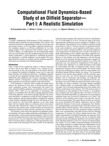
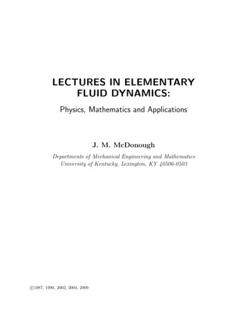
12100J. Phys. Chem. 1996, 100, 12100-12107Vibrational Dynamics of Carbon Monoxide at the Active Sites of Mutant Heme Proteins†Jeffrey R. Hill and Dana D. Dlott*School of Chemical Sciences, UniVersity of Illinois at Urbana-Champaign, Urbana, Illinois 61801C. W. RellaHansen Experimental Physics Laboratory, Stanford UniVersity, Stanford, California 94305Kristen A. PetersonDepartment of Chemistry and Biochemistry, New Mexico State UniVersity, Las Cruces, New Mexico 88033Sean M. Decatur, Steven G. Boxer, and M. D. Fayer*Department of Chemistry, Stanford UniVersity, Stanford, California 94305ReceiVed: February 22, 1996; In Final Form: April 2, 1996XPicosecond mid-IR pump-probe measurements of vibrational relaxation (VR) of CO bound to the activesites of wild-type and mutant myoglobins (Mb) reveal that an approximately linear relationship exists betweenthe protein matrix-induced CO frequency shift and the VR rate. This relation parallels a similar linearrelationship seen in a series of heme model compounds where Fe was replaced by Ru and Os. The VR rateof CO in the Mb is sensitive only to the magnitude of protein-induced carbonyl frequency shifts and apparentlyis not sensitive to the specific details of how the shift is induced, e.g., hydrogen bonding to CO or electrostaticinteractions in the heme pocket. CO VR is insensitive to substantial changes in protein structure that do notaffect the CO vibrational frequency. These observations suggest that the mechanism of carbon monoxideVR in heme proteins such as Mb occurs by through-π-bond anharmonic coupling, which, as shown in priorwork, is also the dominant coupling in the model compounds. The experiments indicate that differing proteinstructures influence VR of CO bound at the active site not by opening and closing channels for vibrationalenergy flow from CO to the protein but by affecting the rate of energy flow from CO to heme. The rates aredetermined by the extent of back-bonding, which determines the magnitude of through-π-bond anharmoniccoupling between CO and heme. The back-bonding and, therefore, the extent of anharmonic coupling areinfluenced by the electric fields in the heme pocket, which likely differ in the proteins studied here.IntroductionIn this paper, the relationships between protein structure andprotein dynamics at the active site are investigated by usingpicosecond infrared experiments to probe the vibrationalrelaxation (VR) of a CO ligand bound at the active site ofmyoglobin (Mb) mutants. Myoglobin stores dioxygen at anactive site located at the central Fe atom of the prosthetic groupprotoheme (iron(II) protoporphyrin IX).1 Although bare protoheme alone can bind dioxygen, in biology protein is neededto influence the chemical reactivity of the active site by resistingoxidative degradation and increasing the relative affinity foroxygen versus poisonous carbon monoxide.1 Many studies havebeen made of the interactions between the protein and a boundCO ligand at the active site, using techniques including X-raycrystallography,2-4 13C NMR,5-7 and mid-infrared (mid-IR)spectroscopy.8-10 Changes in the mid-IR carbonyl stretchingspectrum induced by altering the protein structure by geneticengineering have been used to investigate the influence ofprotein structure on bound ligands at the active site.8,10,11Although conventional mid-IR absorption spectroscopy is apowerful technique, it is not capable of revealing the fastvibrational dynamics of ligands bound to the active site.* Corresponding authors.† This paper is dedicated to Prof. Robin Hochstrasser on the occasion ofhis 65th birthday.X Abstract published in AdVance ACS Abstracts, June 15, 1996.S0022-3654(96)00541-2 CCC: 12.00Recently, mid-IR pump-probe experiments were used toinvestigate the vibrational relaxation (VR) of vibrationallyexcited carbon monoxide bound to the active site of Mb12-14and heme model compounds.15 VR is used here to denote theloss of vibrational energy from a vibrationally excited CO toits surroundings.16 These studies showed that VR of CO boundto heme occurs on the time scale of a few tens of picoseconds12,13,15 and that VR is little affected by temperature inthe 10-300 K range.14 The lack of temperature dependenceindicates the initial VR step does not involve significant couplingto vibrations with frequencies less than 400 cm-1.14,16 Thecarbonyl VR rate can be influenced by protein structure.14 Forexample, two different conformers of Mb-CO were seen in ref14 to have significantly different VR rates. Studies of carbonylVR in model heme compounds showed that VR could beinfluenced by changing the porphyrin structure14 or by substituting different metal atoms for Fe.15 The model compound workrevealed that VR of carbonyls bound to heme was qualitativelydifferent from VR in previously studied metal carbonyls suchas W(CO)6.17 These differences arise because heme is a largearomatic organic molecule with an extensive delocalizedπ-electron system.In the present work, we investigate how protein structureinfluences VR at the active site of Mb by using a series ofgenetically engineered mutants. Since the cloning and expression of human18 and sperm whale Mb,19 the use of mutant Mbproteins has become widespread.9 Figure 1, taken from a recent 1996 American Chemical Society
CO Vibrational DynamicsJ. Phys. Chem., Vol. 100, No. 29, 1996 12101measurements, for the first time open up the possibility ofunderstanding the details of mechanical energy transfer at theactive site of a protein and how protein structure can influencemolecular dynamics at the active site.Figure 1. Schematic diagram of the active site of wild-type myoglobin(WT Mb), adapted from Yang and Phillips.3 H64(alt), the alternateposition (the up position), is found to dominate at pH 4 and below.The down position for H64 is thought to be representative of the activesite structure of the A1 conformer and the up position representative ofthe A0 conformer.high-resolution X-ray structure of Mb-CO,3 shows the structureof Mb in the vicinity of the active site. The recombinantproteins used here involve single-point mutations at three ofthe most important and highly conserved1 amino acid residuesshown in Figure 1: distal histidine H64, valine V68, andproximal histidine H93. Spectroscopic studies of mutant MbCO complexes indicate that some mutations strongly influencecarbonyl stretching transitions in the mid-IR by affecting theheme-Fe-CO bonding.8-10 However, the detailed relationshipsbetween mid-IR spectra and carbonyl bonding in wild-type (WT)and in mutants have been the subject of lively discussion anddebate (reviewed recently in ref 9).Studies of vibrationally excited CO have been used toinvestigate dynamics in many condensed-phase systems, including CO dissolved in monatomic and diatomic liquids,20,21crystalline CO,22 organometallic complexes in solution containing CO bound to a single metal atom or a cluster of metalatoms,17,23-25 CO bound to clean metal surfaces,26-28 CO boundto heme proteins,12,13,15,29 and CO bound to model compounds.15Although the mechanical dynamics of such complicated systemsordinarily are quite difficult to treat theoretically, CO introducesseveral useful simplifications. CO is a diatomic molecule thatbinds to a vast variety of sites, and its electronic structure iswell understood.30 CO vibrational relaxation can be investigatedusing the techniques of molecular dynamics31,32 because VRoccurs on a suitable (sub-nanosecond) time scale. In contrastto simulations of picosecond time scale photoinduced processesin Mb such as photodissociation33,34 or vibrational cooling,35carbonyl VR occurs solely on the ground electronic potentialsurface, and simulations need not invoke ad hoc assumptionsabout the nature of heme electronic transitions. The VR ofexcited CO can also be studied by exact quantum dynamicscalculations, which are presently capable of modeling the shorttime behavior of a diatomic molecule weakly coupled toharmonic baths of arbitrary complexity. This possibility isespecially intriguing in light of recent, highly accurate calculations of the vibrational density of states of a protein.36An initial excitation of CO produced by mid-IR absorptionwill be localized on CO. Picosecond time scale relaxation ofCO vibrational excitation in heme systems occurs via weakanharmonic coupling to other vibrational states of the system,16resulting in a transfer of the CO vibrational energy to heme,the heme protein, and the solvent. By using the techniques ofsynthetic chemistry and site-directed mutagenesis, it is possibleto produce a wide variety of tailored Mb structures and analogs.Modified Mb and models, combined with mid-IR dynamicsMaterials and MethodsThe preparation and characterization of recombinant mutanthuman Mb proteins has been discussed previously in ref 18,and the preparation of sperm whale Mb cavity mutants wasdiscussed in ref 37. All Mb samples were dissolved in a mixtureof 50:50 glycerol and phosphate buffer at pH ) 7. Measurements were made at ambient temperature. In the mid-IR, allthe proteins studied exhibit one dominant protein conformer,as indicated by the presence of a single intense carbonylstretching transition, although a few of the proteins evidencedminor conformers, as discussed in ref 10. The present sensitivityof the pump-probe apparatus permitted measurements only onthe dominant conformer of the mutant proteins.Mid-infrared pump-probe experiments were performed atthe Stanford Free Electron Laser Center.14,17 The pump pulseswere 1.5 ps in duration, with an energy of 150 nJ and a spectralFWHM of 7.3 cm-1. For each pump pulse, a pair of adjacent15 nJ probe pulses from the free-electron laser was used. Thefirst pulse of the pair arrives at the unperturbed sample 85 nsbefore the pump pulse. It is used as an intensity reference forthe second pulse. The second pulse, which detects pumpinduced absorption changes, reaches the sample within a variabletime window of the pump pulse. This window can be variedover a 1 ns range by using a computer-controlled optical delayline. Because of the high Q of the free-electron laser opticalcavity, the two adjacent probe pulses are highly correlated inamplitude. Use of these highly correlated pulses to determinepump-induced absorbance changes makes this method insensitive to laser fluctuations. The probe intensities were measuredusing a mid-IR detector with a time response faster than the 85ns interpulse spacing, two gated integrators, and a computerwith analog-to-digital converters. The pulses were selected fromthe free-electron laser high-repetition-rate output using fastacoustooptic modulators38 and were brought to a common focusof 100 µm diameter on the sample. Experiments were performed with pump and probe polarizations parallel and withthe probe pulse at the magic angle. Careful measurements weremade to ensure that the vibrational decay constants were not afunction of the laser repetition rate or power and that the sampleremained chemically stable during data acquisition.A brief description of the proteins used in this work follows.Wild-Type Sperm Whale and Horse Heart Myoglobin(WT SW, WT HH). The structure in the vicinity of the activesite, which is quite similar for both WT proteins, is shown inFigure 1. The mid-IR spectrum of WT provides evidence forat least four conformers, denoted A0-A3, in order of decreasingcarbonyl stretching frequency.39 Under normal conditions inWT, A1 is the dominant conformer, A0 is a minor presence,and A2-A3 are hardly present.39 At low pH, the relativepopulation of A0 increases relative to that of A1. It has beensuggested by several authors40 that, among other differences,the A1 state is characterized by H64 down in the heme pocketand A0 is characterized by H64 up out of the pocket. A newhigh-resolution X-ray structure3 of Mb-CO at normal and lowpH shows that H64 can be made to move out of the pocketbelow pH 4, when the imidazole ring becomes fully protonated.The structures of the active site with H64 down and up [the upstate is labeled H64(alt)] are shown in Figure 1. Because therelative population of A0 increases as pH is decreased,39,41 thesetwo active site structures are thought to be representative ofthe A0 (up) and A1 (down) conformers.
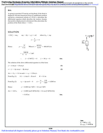
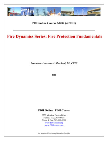
12102 J. Phys. Chem., Vol. 100, No. 29, 1996Hill et al.TABLE 1: Vibrational Lifetimes of CO Bound to HemeProteins Measured in This WorksampleWT (SW) Mb-13CO, A1 conformerH93G(Im) (SW), A1 conformerV68N (human), A0 conformerH64V (human),a A0 conformerH64L (human), A0 conformercarbonyl stretch carbonyl stretchfrequency (cm-1) lifetime (ps)1901 ((1)1946 ((1)1917 ((1)1967 ((1)1967 ((1)17 ((2)17 ((1)10.5 ((2)21 ((2)24.5 ((1)a Some preliminary experiments were run using H64V (SW) protein.These data gave the same CO stretching frequency and essentially thesame decay constant as the H64V (human) protein.Figure 2. Pump-probe decay data obtained with CO bound to humanH64V and V68N proteins. The CO stretching frequencies are 1967and 1917 cm-1, respectively. The smooth exponential decay curvesthrough the data give vibrational relaxation lifetimes of 21 ((2) and10.5 ((2) ps.Human H64V. In H64V, the relatively polar distal histidineresidue is replaced by a nonpolar valine. This replacementcauses a single sharp band, corresponding to an A0 conformer,to be observed at 1967 cm-1, and A1 is not seen,11 consistentwith the idea that A0 states involve CO, which is not influencedby a polar H64 residue in the down position.Human H64L. In H64L, the distal histidine is replaced bya nonpolar leucine residue, which is somewhat larger than valine.A single sharp mid-IR band corresponding to an A0 conformeris observed at 1967 cm-1, and A1 is not seen.11 The H64Lmutant has an exceptionally large CO affinity, 50 times greaterthan WT, which has been attributed to the stabilization of COby leucine.9Human V68N. In V68N, nonpolar V68 is replaced by polarasparagine, resulting in a large carbonyl frequency red shift.Two bands at 1917 and 1933 cm
Sean M. Decatur, Steven G. Boxer, and M. D. Fayer* Department of Chemistry, Stanford UniVersity, Stanford, California 94305 ReceiVed: February 22, 1996; In Final Form: April 2, 1996X Picosecond mid-IR pump-probe measurements of vibrational relaxation (VR) of CO bound to the active sites of wild-type and mutant myoglobins (Mb) reveal that an approximately linear relationship exists between the .