Transcription
Ji et al. BMC Cardiovascular Disorders(2020) SEARCH ARTICLEOpen AccessProtective effects of rolipram on endotoxiccardiac dysfunction via inhibition of theinflammatory response in cardiacfibroblastsJingjing Ji1,2†, Zhifeng Liu1,2,3,4*†, Xinxin Hong2,5, Zheying Liu1,2, Jinghua Gao1,2 and Jinghua Liu1*AbstractBackground: Cardiac fibroblasts, regarded as the immunomodulatory hub of the heart, have been thought to playan important role during sepsis-induced cardiomyopathy (SIC). However, the detailed molecular mechanism andtargeted therapies for SIC are still lacking. Therefore, we sought to investigate the likely protective effects ofrolipram, an anti-inflammatory drug, on lipopolysaccharide (LPS)-stimulated inflammatory responses in cardiacfibroblasts and on cardiac dysfunction in endotoxic mice.Method: Cardiac fibroblasts were isolated and stimulated with 1 μg/ml LPS for 6 h, and 10 μmol/l rolipram wasadministered for 1 h before LPS stimulation. mRNA levels of tumor necrosis factor-α (TNF-α), interleukin-6 (IL-6) andinterleukin-1β (IL-1β) in fibroblasts and their protein concentrations in supernatant were measured with real-timePCR (rt-PCR) and enzyme-linked immunosorbent assay, respectively. The expression of dual specificity phosphatase1 (DUSP1), an endogenous negative regulator that inactivates MAPK-mediated inflammatory pathways, was alsomeasured by rt-PCR and western blotting. DUSP1-targeted small interfering RNA (siRNA) was used to examine thespecific role of DUSP1. To evaluate the role of rolipram in vivo, an endotoxic mouse model was established byintraperitoneal injection of 15 mg/kg LPS, and 10 mg/kg rolipram was intraperitoneally injected 1 h before LPSinjection. mRNA and protein levels of inflammatory cytokines and DUSP1 in heart, inflammatory cell infiltration andcardiac function were all examined at 6 h after LPS injection.Results: The results showed that LPS could increase the expression and secretion of inflammatory cytokines anddecrease the transcription and expression of DUSP1 in cardiac fibroblasts. However, rolipram pretreatmentsignificantly reversed the LPS-induced downregulation of DUSP1 and inhibited LPS-induced upregulation andsecretion of TNF-α and IL-6 but not IL-1β. Moreover, DUSP1-targeted siRNA experiments indicated that theprotective effect of rolipram on inflammatory response was specific dependent on DUSP1 expression. Moreover,rolipram could further reduce inflammatory cell infiltration scores as shown by pathological analysis and increasethe ejection fraction (EF) detected with echocardiography in the hearts of endotoxic mice.(Continued on next page)* Correspondence: zhifengliu7797@163.com; liujhua@smu.edu.cn†Jingjing Ji and Zhifeng Liu contributed equally to this work.1Guangdong Provincial Key Laboratory of Proteomics; School of BasicMedical Sciences, Southern Medical University, Guangzhou 510515, ChinaFull list of author information is available at the end of the article The Author(s). 2020 Open Access This article is licensed under a Creative Commons Attribution 4.0 International License,which permits use, sharing, adaptation, distribution and reproduction in any medium or format, as long as you giveappropriate credit to the original author(s) and the source, provide a link to the Creative Commons licence, and indicate ifchanges were made. The images or other third party material in this article are included in the article's Creative Commonslicence, unless indicated otherwise in a credit line to the material. If material is not included in the article's Creative Commonslicence and your intended use is not permitted by statutory regulation or exceeds the permitted use, you will need to obtainpermission directly from the copyright holder. To view a copy of this licence, visit http://creativecommons.org/licenses/by/4.0/.The Creative Commons Public Domain Dedication waiver ) applies to thedata made available in this article, unless otherwise stated in a credit line to the data.
Ji et al. BMC Cardiovascular Disorders(2020) 20:242Page 2 of 12(Continued from previous page)Conclusions: Rolipram could improve endotoxin-induced cardiac dysfunction by upregulating DUSP1 expression toinhibit the inflammatory response in cardiac fibroblasts, which may be a potential treatment for SIC.Keywords: Sepsis induced cardiomyopathy, Rolipram, Inflammatory mediators, Cardiac fibroblasts, Dual specificityphosphatase 1BackgroundAccording to the Sepsis new definition, life-threateningorgan dysfunction caused by a dysregulated host responseto infection, Sequential Organ Failure Assessment (SOFA)score was employed as the diagnostic criteria, replacingthe prevailing systemic inflammatory response syndrome(SIRS) criteria [1]. This change emphasizes the lifethreatening organ dysfunction in sepsis, indicating that researchers and physicians should not only focus on the inflammatory response but also pay more attention to organprotection. The heart is one of the most frequently affected organs in sepsis. Sepsis-induced cardiomyopathy(SIC) has been reported to be present in more than 40–50% of cases of sepsis [2, 3]. Previous studies reported thatthe mortality of septic patients ranged from 28 to 48.4%[4, 5] and a higher mortality was observed in patients withobserved cardiovascular dysfunction, with an odds ratio of2.78 [6]. At present, no formalized or consensus definitionof SIC exists. Generally, SIC is often diagnosed when someacute perturbation in cardiac function, systolic function ordiastolic function exists in the setting of sepsis [7]. SIC hasbeen recognized for 40 years [8], but its mechanism andprocess are still not well understood.Over recent decades, a number of experimental andclinical studies have suggested possible causative mechanisms for progressive cardiac dysfunction, includingdisturbed coronary blood flow, cardiomyocyte apoptosis, effects of myocardial depressant factor (MDF), nitric oxide and reactive oxygen species, mitochondrialdysfunction, and calcium trafficking [9, 10]. Amongthese hypotheses, MDF and nitric oxide seemed to havelarger effects on cardiac dysfunction in septic states. In1985, Parrillo et al proposed that myocardial depressantsubstances existed in septic patients and that these depressant substances were the pathophysiologic factorsthat induced cardiomyopathy during sepsis [11]. Subsequently, some studies found that MDF was likely to bean endotoxin, a cell wall component of gram-negativebacteria. With more in-depth research in this field, further studies revealed that inflammatory cytokines hadcomparable effects to those of MDF. Of these cytokines,tumor necrosis factor-α (TNF-α), interleukin-6 (IL-6)and interleukin-1β (IL-1β), which were produced excessively in the early stage of sepsis, have been found tohave potential depressive effects on cardiac function[10, 12].Cardiac fibroblasts can respond to various types of external stimuli and are regarded as the immunomodulatoryhub of the heart. Under physiological conditions, cardiacfibroblasts play important roles, such as electric isolationand/or conductance in different compartments and chemokine or cytokine secretion, in cell-cell communicationwith cardiomyocytes and other cell types [13]. Underpathological conditions, cardiac fibroblasts are intimatelyassociated with the innate immune system, promoting anadequate response to eliminate insults synergistically [14].Endotoxins and inflammatory mediators could activatecardiac fibroblasts through toll-like receptors (TLRs), provoking the transcription and release of chemokines andcytokines, such as TNF-α and IL-1β [15]. The chemokinesand cytokines released by cardiac fibroblasts could furtherrecruit inflammatory cells from the circulation, amplifyingthe inflammatory response and releasing abundant myocardial depressant substances in the heart locally to inducecardiac dysfunction [16, 17], suggesting that inhibition ofthe inflammatory response in cardiac fibroblasts could bea potential therapy for SIC.Rolipram is a phosphodiesterase type IV specific inhibitor, which could reduce the breakdown of cAMP,and it was primally studied as an antidepressant [18, 19].Further studies discovered its anti-inflammatory actions.It has been approved by the Food and Drug Administrationof the USA (FDA) as a therapeutic drug for chronic obstructive pulmonary disease due to its anti-inflammatory effects [20]. One mechanism of its anti-inflammatory effectsis increasing the expression and activity of dual specificityphosphatase 1 (DUSP1), an endogenous negative regulatorin infection. Since this enzyme could dephosphorylate bothphosphothreonine and phosphotyrosine residues on activated mitogen-activated protein kinases (MAPKs), it wasnamed as dual specificity phosphatase 1 in mice or MAPKphosphatase 1 (MKP1) in humans. It can dephosphorylatep38 MAPK and c-Jun N-terminal kinase (JNK) to inactivateMAPK-mediated inflammatory pathways [21]. A previousstudy showed that rolipram could reduce the Staphylococcus aureus-induced inflammatory responses in macrophages [22]. However, whether its anti-inflammatory effectcould be beneficial to cardiac protection in sepsis is still unknown. In the present study, the effects of rolipram on cardiac function were evaluated both in lipopolysaccharide(LPS)-stimulated cardiac fibroblasts in vitro and in endotoxic mice in vivo.
Ji et al. BMC Cardiovascular Disorders(2020) 20:242MethodsAnimals and reagentsC57BL/6 mice were purchased from Southern MedicalUniversity. All animal procedures were approved by theAnimal Care and Use Committee of Southern MedicalUniversity (the permit number 2017 J019), and all animalexperiments were conducted in accordance with theGuidelines for Animal Care of Southern Medical University. LPS (L2630) and rolipram (R6520) were purchasedfrom Sigma-Aldrich. Liberase TH Research Grade(0540115001), the enzyme used to digest heart tissues,was purchased from Roche (Mannheim, Germany). TheRNA extraction reagent TRIzol was purchased from LifeTechnologies (Shanghai, China). Reverse transcriptionkits (FSQ-101) and Real-time PCR Master Mix (QPK-201)were purchased from Toyobo (Osaka, Japan). Enzymelinked immunosorbent assay (ELISA) kits for TNF-ɑ (88–7324-22), IL-6 (88–7064-86) and IL-1β (88–7013-86) werepurchased from Thermo Fisher Scientific (San Diego, CA,USA). The DUSP1 antibody (sc-373,841) was purchasedfrom Santa Cruz (Dallas, Texas, USA), and GAPDH(#2118) was purchased from Cell Signaling Technology(Danvers, MA, USA). Secondary antibody horseradish peroxidase (HRP) conjugated goat anti-rabbit IgG (abs20040)and goat anti-mouse IgG (abs20039) were purchased fromAbsin (Shanghai, China). Fluorochrome 7-AAD (00–6993-50) was purchased from ThermoFisher (USA), andantibodies specific to Cd45-FITC (553080) and Ly6gAPC-Cy7 (560600) were purchased from BD Biosciences(San Jose, CA, USA).Isolation and treatment of neonatal mouse cardiacfibroblastsNeonatal mouse cardiac fibroblasts were isolated fromnewborn C57BL/6 mice. Mice were anesthetized withoverdose pentobarbital (100 mg/kg) by intraperitonealinjection, and cardiac palpation was used to confirm thedeath of mice. Then, the hearts of neonatal mice wereisolated and cut into pieces. Then, the heart tissues wererinsed with phosphate-buffered saline (PBS) and digestedwith an enzyme (Liberase TH Research Grade) at 37 Cfor 15 min. The supernatant containing the cells wastransferred to complete medium (DMEM containing10% fetal calf serum) to terminate the digestion. Residualtissues were digested with the enzyme at 37 C for 15min for further digestion. The digestion was repeated 3to 4 times to isolate all cells from the heart tissues. Allcell suspensions were collected and centrifuged at 250 gfor 5 min. The supernatant was discarded, and the precipitate was resuspended in complete medium. The cellsuspension was transferred to a culture dish and cultured for 60 min in an incubator at 37 C and 5% CO2.After the unattached cells were discarded, the remainingattached cells were cardiac fibroblasts. After 24 h of cellPage 3 of 12culture, cardiac fibroblasts were treated with 1 μg/mlLPS for 6 h or 10 μmol/L rolipram for 1 h before LPSstimulation either alone or in combination according toprevious studies [23, 24]. Because LPS was dissolved innormal saline (NS), and rolipram was dissolved indimethylsulfoxide (DMSO), NS and DMSO were used asthe solvent control.Preparation and treatment of the endotoxemic mousemodelTwenty C57BL/6 mice, male, aged 8 to 10 weeks, wererandomly divided into 4 groups (n 5). The endotoxemia mouse model was established by intraperitoneal injection of 15 mg/kg LPS for 6 h according our previousstudy [24], and 10 mg/kg rolipram [25] was intraperitoneally injected 1 h before LPS injection. Similar with thecell treatment, NS and DMSO were used as the solventvehicle.RNA extraction and quantitative rt-PCR (qRT-PCR)Total RNA was isolated from cardiac fibroblasts or cardiac tissues with TRIzol reagent. RNA extraction andqRT-PCR were performed as previously described [26].The primer sequences of TNF-ɑ, IL-6, IL-1β, DUSP1and GAPDH are shown in the Table 1. The expressionresults were calculated by the 2 ΔΔCq method reportedby Livak and Schmittgen [27].Protein extractionProtein samples were extracted from cultured cardiac fibroblasts or cardiac tissues with lysis buffer (P0013B,Beyotime). For the cultured cardiac fibroblasts, cultureTable 1 The sequences of the rt-PCR primers of TNF-ɑ, IL-6, IL1β, DUSP1 and GAPDHTarget ReverseGGTGGTCCAGGGTTTCTTACTCC
Ji et al. BMC Cardiovascular Disorders(2020) 20:242medium was removed and washed by cold PBS for threetimes. Then, cells were lysed by lysis buffer for 20 minon the ice. Then, lysed cells were collected and centrifuged at 12,000 x g for 15 min to pellet debris and thesupernatants were used to following measurement.Heart tissue proteins were collected by supersonic splitting. The frequency of sonicator probe was 20 kHz. Tissues were put in a 1.5 mL microcentrifuge tube with300 μl lysis buffer and gently moved under the tip of thesonnicator probe for 10 s vibrating. Then, incubated onice for 15 min after the sonication. Then the lysed tissueswere centrifuged at 12,000 x g for 20 min and the supernatants were collected for protein analysis. The proteinconcentrations were quantified by BCA protein assay(ThermoFisher, USA), and same total protein were usedto the enzyme immunoassays and western blot.Page 4 of 12Table 2 The sequences of small interfering RNAsiRNA NCSense: CAA UUG UCC UAA CCA CUU UTTAntisense: AAA GUG GUU AGG ACA AUU GTTsiRNA DUSP1Sense: UUC UCC GAA CGU GUC ACG UTTAntisense: ACG UGA CAC GUU CGG AGA ATTwere incubated in serum-free DMEM with siRNA/Lipofectamine (Life technology, USA) for 6 h, followed by normalculture for 48 h, then cells were pretreated with 10 μmol/Lrolipram for 1 h before LPS stimulation. The expression ofDUSP1 and the TNF-ɑ, IL-6 concentrations in the supernatant of cardiac fibroblasts with LPS treatment for 6 hwere examined with western blot and ELISA methods,respectively.Echocardiographic measurementsEnzyme immunoassayEnzyme immunoassays for TNF-ɑ, IL-6 and IL-1β in cellular supernatant and heart tissue were carried out according to the manufacturer’s guide. A 96-well plate wascoated with the capture antibody of TNF-ɑ (1:250 dilution), IL-6 (1:250 dilution) and IL-1β (1:250 dilution) respectively, incubated overnight at 4 C and blocked withdilution buffer for 1 h. Then, the standard samples and experimental samples were added and incubated for another2 h at room temperature. Corresponding detection antibody of TNF-ɑ (1:250 dilution), IL-6 (1:250 dilution) andIL-1β (1:250 dilution), avidin-horseradish peroxidase andtetramethylbenzidine (TMB) solution were added. Lastly,1 mol/L H3PO4 was used to stop the reaction. The platewas read at 450 nm, and the concentrations of each sample were calculated according to the standard curve.Western blotting analysisWestern blotting analysis was carried out with a procedure described previously [28]. Briefly, equal amounts ofprotein were subjected to polyacrylamide gel electrophoresis for separation and transferred onto a polyvinylidene fluoride membrane. Protein expression wasdetermined by specific antibodies against DUSP1 (1:1000dilution) and GAPDH (1:1000 dilution), which diluted inTris-Buffered Saline Tween-20 (TBST). HRP conjugatedanti-mouse IgG (1:5000 dilution) and anti-rabbit IgG (1:10000 dilution) was used as secondary antibody forDUSP1 and GAPDH, respectively. Signals were detectedby chemiluminescence and quantified by densitometry.RNA interference (RNAi) for DUSP1 knock downSiRNA (small interfering RNA) that targeted DUSP1 wassynthesized by the GenePharma Company (Shanghai,China). The synthetic oligonucleotide sequences of siRNADUSP1 and siRNA-NC sequence (as the control) wereshown in Table 2. After 24 h isolation, cardiac fibroblastsEchocardiography was used to measure cardiac functionin mice as in our previous study [29]. Animals werelightly anesthetized with 1% inhalant isoflurane and imaged using a 40-MHz linear array transducer attached toa preclinical ultrasound system (SonoScape, Shenzhen,Guangdong, China). M-mode in left ventricular shortaxis view was used to assess cardiac function by left ventricular ejection fraction (EF).Histologic examinationThe mice were anesthetized with 50 mg/kg pentobarbitalby intraperitoneal injection. The hearts were excised andfixed in 4% paraformaldehyde, then embedded in paraffin. Sections with 4 μm thickness were stained withhematoxylin and eosin for microscopic examination(Nikon, Japan) at a magnification of 200 . The degree ofinflammatory infiltration was scored from 1 to 4 according the method reported by Neu et al [30]: Score 1, noinflammatory cell infiltration; 2, the inflammatory infiltration area was less than 10%; 3, inflammatory infiltration area between 10 and 20%; and 4, inflammatoryinfiltration area over 20%.Mouse heart digestion and flow cytometry analysisMouse heart cells flow cytometry analysis were processed according to the method described by Chen et al[31]. The mice were anesthetized with 50 mg/kg pentobarbital. Heart tissues were cut into pieces and washedby cold PBS for three times to remove the blood cells.Then the tissues were digested by the enzyme (LiberaseTH Research Grade) at 37 C for 15 min. The supernatant containing the cells was transferred to themedium with 10% fetal calf serum to terminate the digestion. Residual tissues were digested with the enzymeat 37 C for 15 min for further digestion. The digestionwas repeated 3 to 4 times to isolate all cells from theheart tissues. All cell suspensions were collected and
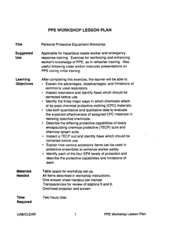
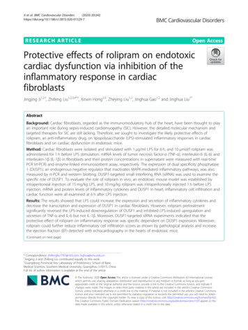
Ji et al. BMC Cardiovascular Disorders(2020) 20:242centrifuged at 250 g for 5 min. The supernatant was discarded, and the precipitate was resuspended in PBS andcells were filtered through a 70 μm nylon mesh (ThermoFisher, USA). Erythrocytes were removed by RBClysis Buffer (ThermoFisher, USA). Cells were stained by7-AAD, antibodies specific to Cd45-FITC and Ly6gAPC-Cy7 for 30 min. The cells were washed twice bycold PBS and subjected to flow cytometry (BD FACSCalibur) measurement. Data were analysed by FlowJo. 7AAD negative cells were regarded as the alive cells.Leukocyte cell ratio was calculated by Cd45 positivecells/all alive cells. Neutrophil ratio was calculated byLy6g positive cells/Cd45 positive cells.Page 5 of 12ResultsLPS increased the expression and secretion ofinflammatory cytokines in cardiac fibroblastsAfter cardiac fibroblasts were stimulated with 1 μg/mlLPS for 6 h, the mRNA levels and supernatant concentrations of inflammatory mediators were measured. Asshown in Fig. 1, the mRNA levels of TNF-ɑ, IL-6 andIL-1β were greatly upregulated, and the concentrationsof TNF-ɑ and IL-6 in supernatant also increased significantly after LPS stimulation (Fig. 1d and e). However, although the mRNA expression of IL-1β increasedconsiderably after LPS stimulation, no significant difference or even a moderate increase was observed in IL-1βconcentration in supernatant (Fig. 1f).Statistical analysisAll data were analyzed by SPSS 13.0 software (IBM,Armonk, New York, USA). After the homogeneity ofvariance test, one-way ANOVA followed by theNewman-Keuls test was performed for multigroup comparisons in the LPS-stimulated cardiac fibroblasts experiment. Two-way ANOVA was performed to evaluatethe effect of rolipram both in cardiac fibroblasts andendotoxemic mice. A value of P 0.05 was consideredindicative of statistical significance.Rolipram inhibited the LPS-induced inflammatoryresponse by upregulating DUSP1 expression in cardiacfibroblastsCardiac fibroblasts were treated with 1 μg/ml LPS for 6 hor 10 μmol/L rolipram for 1 h before LPS stimulation either alone or in combination. The mRNA levels (Fig. 1g)and protein expression (Fig. 1h) of DUSP1 in cardiac fibroblasts were measured 6 h after LPS stimuli.Fig. 1 Effects of LPS on the cardiac fibroblast inflammatory responses and DUSP1 expression. After cardiac fibroblasts were stimulated with 1 μg/ml LPS for 6 h, the mRNA levels and supernatant concentrations of inflammatory mediators were measured by qRT-PCR and ELISA, respectively.a-c and g, Relative fold change to control group of TNF-ɑ, IL-6, IL-1β and DUSP1 mRNA expression. d-f, Concentrations of TNF-ɑ, IL-6 and IL-1β inthe supernatant. h, DUSP1 protein expression in cardiac fibroblasts (original, uncropped blot was shown in supplement Fig. S1A). I, Proteinquantification of DUSP1. * represents P 0.05 compared with the control group
Ji et al. BMC Cardiovascular Disorders(2020) 20:242DUSP1 is a negative modulatory factor in the inflammatory response and may be upstream of LPS-inducedproinflammatory cytokine release in cardiac fibroblasts.Therefore, in our study, the expression of DUSP1 wasmeasured by qRT-PCR and western blotting after LPStreatment, and our results showed that both the mRNA(Fig. 1g) and protein expression (Fig. 1h and i) of DUSP1were decreased significantly after LPS stimulation (Fig. 1g-i), indicating that the downregulation of DUSP1 mightparticipate in the LPS-induced inflammatory response incardiac fibroblasts.A previous study found that rolipram could upregulatethe expression of DUSP1 in macrophages [22]. To investigate its effect on the LPS-induced inflammatory response in cardiac fibroblasts, rolipram was used beforeLPS administration. As shown in Fig. 2, pretreatmentwith 10 μmol/L rolipram significantly reversed the LPSinduced downregulation of DUSP1 at both the mRNAand protein levels (Fig. 2a-c).Rolipram significantly inhibited LPS-induced upregulation and secretion of TNF-α (Fig. 2d and g) and IL-6(Fig. 2e and h) in cardiac fibroblasts but not IL-1β secretion (Fig. 2i). After knocking down the expression ofDUSP1 by targeted siRNA, rolipram has no effect on theinflammatory cytokine secretion (Fig. 2j-l), indicatingthat the protective effect of rolipram on inflammatoryresponse was dependent on the DUSP1 expression.Rolipram inhibited the LPS-induced inflammatorymediator release by upregulating DUSP1 expression inthe hearts of endotoxic miceTo further evaluate the likely role of rolipram in vivo, weestablished an endotoxic mouse model by intraperitonealinjection of 15 mg/kg LPS for 6 h. In the LPS rolipramgroup, 10 mg/kg rolipram was injected intraperitoneally 1h before LPS injection. The hearts of the mice were dissociated, and the mRNA and protein levels of the proinflammatory molecules and DUSP1 were measured. Similarwith the in vitro results, LPS treatment significantly decreased the mRNA levels of DUSP1 in the heart (Fig. 3a).In addition, rolipram could increase the expression ofDUSP1 at both the mRNA and protein levels (Fig. 3a andb), similar with the results in cardiac fibroblasts. ThemRNA levels of TNF-α and IL-6 were decreased in theLPS rolipram group compared with those in the LPS vehicle group (Fig. 3c and d). However, no change in IL1β mRNA was observed in the hearts of endotoxic mice(Fig. 3e), in contrast with the cultured cardiac fibroblasts.The protein levels of TNF-α, IL-6 and IL-1β in heart tissue were also measured by ELISA. TNF-α and IL-6 levelswere increased significantly in the LPS group comparedwith those in the control group, and rolipram pretreatment decreased the LPS-induced production of TNF-αand IL-6 (Fig. 3f and g).Page 6 of 12Rolipram reduced the leukocyte infiltration and improvedheart dysfunction in endotoxic miceInflammatory cell infiltration was an important indicatorof the tissue inflammatory response, and our pathological results showed that inflammatory cell infiltrationwas present in the myocardial interstitium. However, thedegree of inflammatory cell infiltration in the roliprampretreatment group was significantly reduced (Fig. 4a).The inflammatory infiltration scores were also lower inthe LPS rolipram group than in the LPS vehiclegroup (Fig. 4b). In addition, flow cytometry analysisshowed that rolipram treatment could decrease theleukocyte ratios (Fig. 4c) and neutrophil ratios (Fig. 4d)in endotoxic mice heart. These results suggest that rolipram may inhibit the LPS-induced inflammatory response by upregulating DUSP1 expression in the heartsof endotoxic mice.Finally, echocardiography was used to measure the effects of rolipram on endotoxin-induced cardiac dysfunction in mice. M-mode at the level of the papillarymuscles was used to assess changes in left ventricularEF. Representative M-mode images of the left ventricularshort axis view are shown in Fig. 4e. Compared with theNS vehicle group, the EF was decreased in the LPS vehicle group, suggesting that LPS could induce cardiacdysfunction. However, EF was higher in the LPS rolipram group than in the LPS vehicle group, indicatingthat rolipram may improve the heart dysfunction induced by endotoxin.DiscussionThe present in vitro and in vivo study showed that LPScould induce inflammatory responses in cardiac fibroblasts with high levels of expression and secretion of inflammatory cytokines (TNF-α, IL-6, IL-1β) and couldincrease inflammatory cell infiltration in heart tissues, further inducing heart dysfunction, or SIC, in endotoxicmice. Moreover, LPS could also decrease the transcriptionand expression of DUSP1 (an endogenous negative regulator that inactivates MAPK-mediated inflammatory pathways) in cardiac fibroblasts and heart tissue. However,pretreatment with rolipram (an anti-inflammatory drugthat selectively increases DUSP1 expression) could significantly reverse LPS-induced downregulation of DUSP1 andinhibit LPS-induced upregulation and secretion of TNF-αand IL-6 to further reduce neutrophil cell infiltration andimprove heart dysfunction induced by endotoxin in mice.Sepsis is a syndrome of physiologic, pathologic, andbiochemical abnormalities induced by infection, whichmay have originated as pneumonia, peritonitis, urinarysystem infection, and the like [1]. Overall, 40–50% ofcases of sepsis are complicated by SIC, and the mortalityof the patients with SIC can exceed 50% [32]. However,the exact mechanism of SIC remains unclear. One
Ji et al. BMC Cardiovascular Disorders(2020) 20:242Page 7 of 12Fig. 2 Effects of rolipram on the LPS-induced DUSP1 expression and inflammatory response in cardiac fibroblasts. Cells were pretreated with10 μmol/L rolipram for 1 h before LPS stimulation. DUSP1 mRNA and protein expression, the mRNA expression and supernatant proteinconcentrations of inflammatory mediators in cardiac fibroblasts were measured after LPS stimuli for 6 h. For the RNAi experiment, cardiacfibroblasts were treated by small interfering RNA for 6 h, followed by normal culture for 48 h, then cells were pretreated with 10 μmol/L rolipramfor 1 h before LPS stimulation. The expression of DUSP1 and the TNF-ɑ, IL-6 concentrations in the supernatant of cardiac fibroblasts weremeasured. a, Relative fold of changes to control of DUSP1 mRNA; b and c, DUSP1 expression detection and quantification (original, uncroppedblot was shown in supplement Fig. S1B). d-f, Relative fold of changes to control of TNF-ɑ, IL-6 and IL-1β mRNA expression. g-i, Concentrations ofTNF-ɑ, IL-6 and IL-1β in the supernatant. j. The expression of DUSP1 after RNAi (original, uncropped blot was shown in supplement Fig. S1C). kand l. The TNF-ɑ and IL-6 concentrations in the supernatant after the DUSP1 knockdown* represents P 0.05explanation for SIC is the systemic inflammatory response. Since the viewpoint of “circulating myocardialdepressant factor” was put forward, an increasing number of researchers have found that endotoxin and inflammatory mediators affect cardiac depression [33]. Insepsis, SIC was more common in Gram-negative infection induced endotoxemia than that in Gram-positive infection, for the exotoxin from Gram-positive bacteriausually resulted in local symptom [34, 35]. Therefore, weestablished an endotoxemia model by LPS treatment. Inour study, we found that the concentrations of TNF-ɑand IL-6 were significantly increased in the heart tissueof endotoxic mice, which might be involved in the occurrence of cardiac dysfunction.Heart cell components include cardiomyocyte, fibroblast, immune cell, endothelial cell, stromal cell [36] . Inflammatory responses not only existed in the immunecells, but also the cardiac cells. Yao et al found that constitutive activation of inflammasome were found in cardiomyocyte, which contributed to the atrial fibrillation
Ji et al. BMC Cardiovascular Disorders(2020) 20:242Page 8 of 12Fig. 3 Effect of rolipram on the inflammatory response in the hearts of endotoxic mice. E
1985, Parrillo et al proposed that myocardial depressant substances existed in septic patients and that these de-pressant substances were the pathophysiologic factors that induced cardiomyopathy during sepsis [11]. Subse-quently, some studies found that MDF was likely to be an en